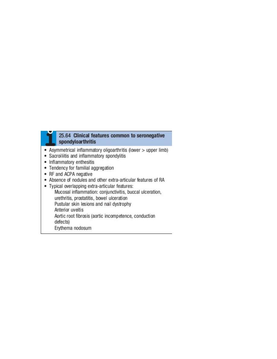
SERONEGATIVE SPONDYLOARTHROPATHIES
These comprise a group of related inflammatory joint
diseases, which show considerable overlap in their clinical features and a shared
immunogenetic association with the HLAB27 antigen . They include:
• ankylosing spondylitis
• axial spondyloarthritis
• reactive arthritis, including Reiter’s syndrome
• psoriatic arthritis
• arthropathy associated with inflammatory bowel disease.
Ankylosing spondylitis
Ankylosing spondylitis (AS) is characterised by a chronic
inflammatory arthritis predominantly affecting the sacroiliac
joints and spine, which can progress to bony fusion of the
spine.
The onset is typically between the ages of 20 and 30, with a
male preponderance of about 3 : 1.

In Europe, more than 90% of those affected are HLAB27
positive.
Even if severe ankylosis develops, functional limitation may
not be marked as long as the spine is fused in an erect
posture.
Clinical features
The cardinal feature is low back pain and early morning
stiffness with radiation to the buttocks or posterior thighs.
Symptoms are exacerbated by inactivity and relieved by
movement.
The disease tends to ascend slowly, ultimately involving the
whole spine, although some patients present with
symptoms of the thoracic or cervical spine.
As the disease progresses, the spine becomes increasingly
rigid as ankylosis occurs.
Secondary osteoporosis of the vertebral bodies frequently
occurs, leading to an increased risk of vertebral fracture.
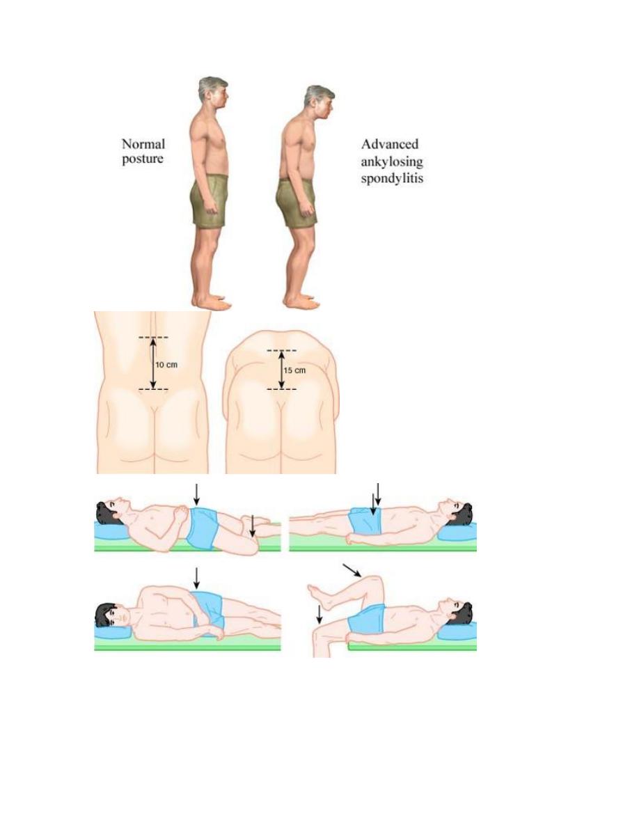
Early physical signs include a reduced range of lumbar spine
movements in all directions and pain on sacroiliac stressing.
As the disease progresses, stiffness increases throughout the
spine and chest expansion becomes restricted.
Spinal fusion varies in its extent and in most cases does not
cause a gross flexion deformity, but a few patients develop

marked kyphosis of the dorsal and cervical spine that may
interfere with forward vision.
Pleuritic chest pain aggravated by breathing is common and
results from costovertebral joint involvement.
Plantar fasciitis, Achilles tendinitis and tenderness over bony
prominences such as the iliac crest and greater trochanter
may all occur, reflecting inflammation at the sites of tendon
insertions (enthesitis).
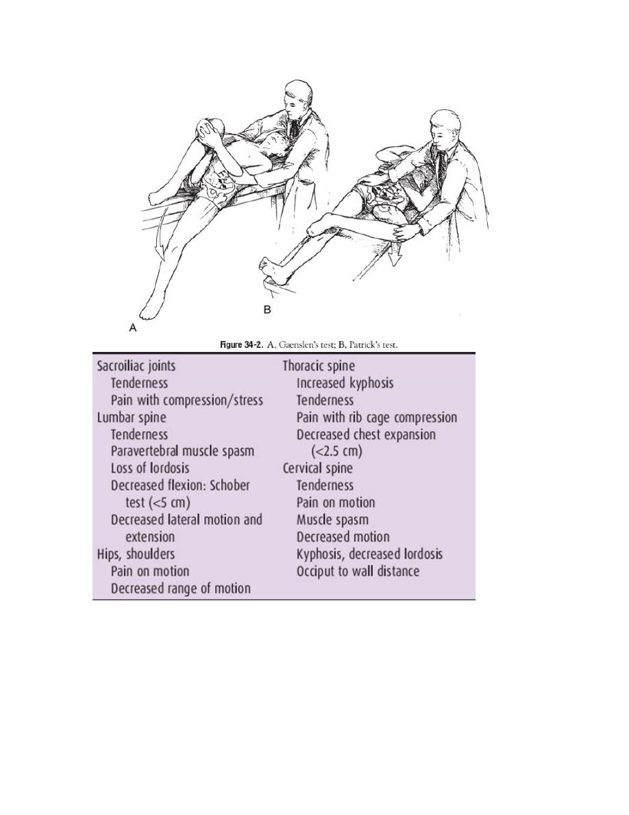
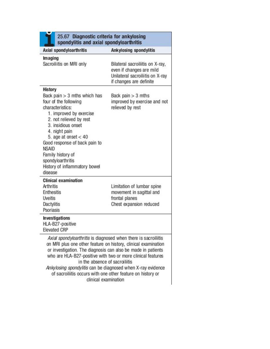
Describe six physical examination tests used to
assess sacroiliac joint tenderness or progression of
spinal disease in AS.
Occiput-to-wall test. Assesses loss of cervical range of
motion. Normally with the heels and scapulae touching the
wall, the occiput should also touch the wall. Any distance
from the occiput to the wall represents a forward stoop of
the neck secondary to cervical spine involvement with AS.
The tragus-to-wall test could also be used.
Chest expansion. Detects limited chest mobility. Measured
at the fourth intercostal space in men and just below the
breasts in women, normal chest expansion is approximately
5 cm. Chest expansion less than 2.5 cm is abnormal.
Schober test (modified).Detects limitation of forward
flexion of the lumbar spine. Place a mark at the level of the
posterior superior iliac spine (dimples of Venus) and another
10 cm above in the midline. With maximal forward spinal

flexion with locked knees, the measured distance should
increase from 10 cm to at least 15 cm .
Other spinal mobility tests will show that lateral flexion and
spinal rotation are also diminished, establishing that the
patient has a global loss of spinal mobility.
Pelvic compression. With the patient lying on one side,
compression of the pelvis should elicit sacroiliac joint pain.
Gaenslen’s test. With the patient supine, a leg is allowed to
drop over the side of the examination table while the
patient draws the other leg toward the chest. This test
should elicit sacroiliac joint pain on the side of the dropped
leg .
Patrick’s test.With the patient’s heel placed on the opposite
knee, downward pressure on the flexed knee with the hip
now in flexion, abduction, and external rotation (FABER)
should elicit sacroiliac joint tenderness.
Clinical features
Up to 40% of patients also have peripheral arthritis. This is
usually asymmetrical, affecting large joints such as the hips,
knees, ankles and shoulders.
In about 10% of cases, involvement of a peripheral joint
may antedate spinal symptoms.
In a further 10%, symptoms begin in childhood, as in the
syndrome of oligoarticular juvenile idiopathic arthritis.
Fatigue is a major complaint and may result from both
chronic interruption of sleep due to pain, and chronic
systemic inflammation with direct effects of inflammatory
cytokines on the brain.
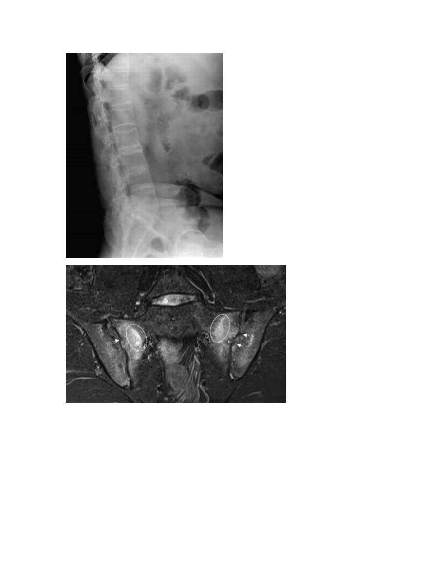
Investigations
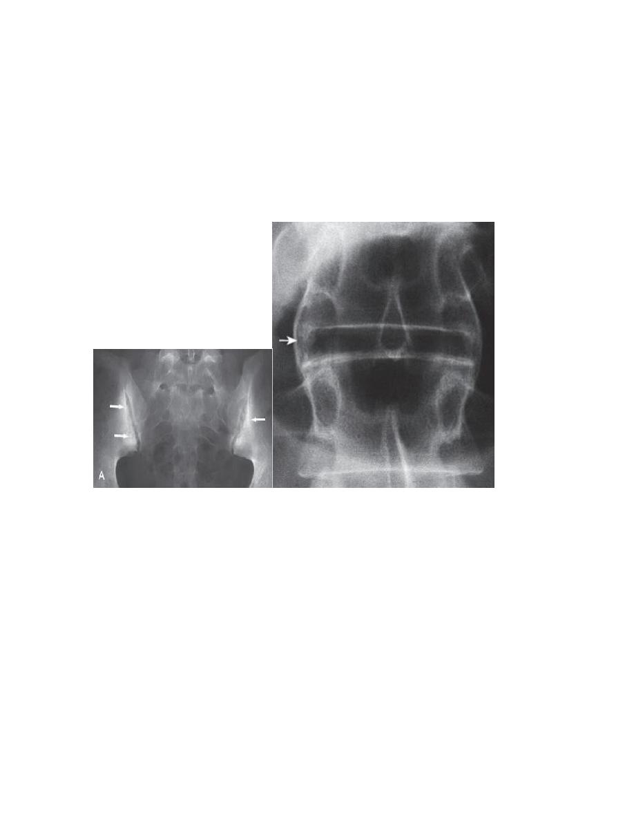
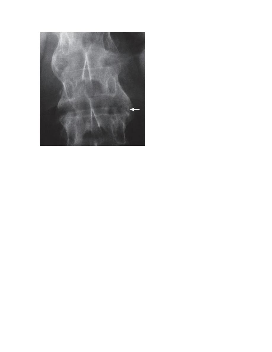
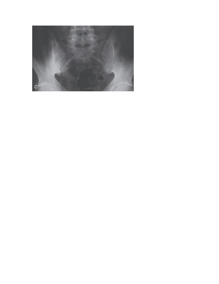
In established AS, radiographs of the sacroiliac joint show
irregularity and loss of cortical margins, widening of the joint
space and subsequently sclerosis, joint space narrowing and
fusion.
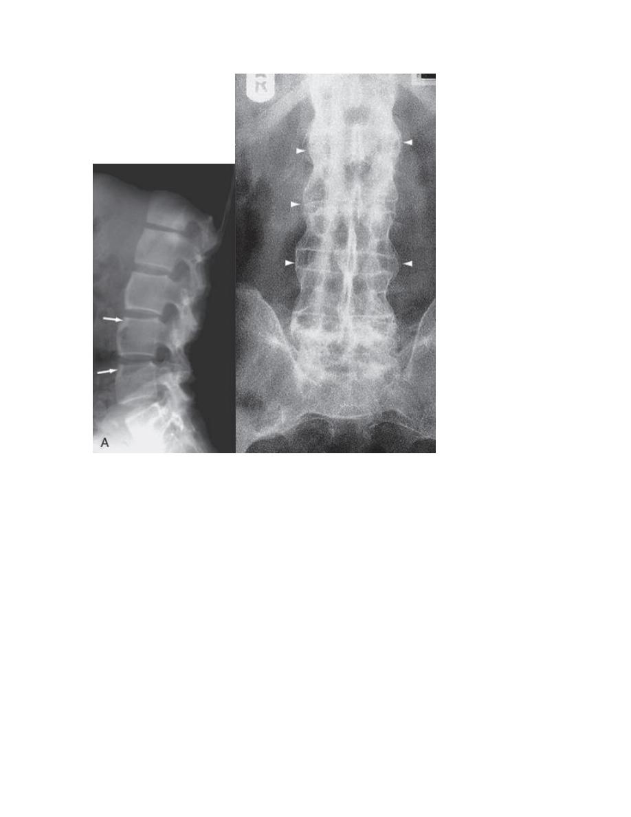
Lateral thoracolumbar spine Xrays may show anterior
‘squaring’ of vertebrae due to erosion and sclerosis of the
anterior corners and periostitis of the waist.
Bridging syndesmophytes may also be seen. These are areas
of calcification that follow the outermost fibres of the
annulus
In advanced disease, ossification of the anterior longitudinal
ligament and facet joint fusion may also be visible. The
combination of these features may result in the typical
‘bamboo’ spine.
Erosive changes may be seen in the symphysis pubis, the
ischial tuberosities and peripheral joints.
Osteoporosis and atlantoaxial dislocation can occur as late
features.
Patients with early disease can have normal Xrays, and if
clinical suspicion is high, MRI should be performed. This is
much more sensitive for detection of early sacroiliitis than X
ray and can also detect inflammatory changes in the
lumbar spine.
The ESR and CRP are usually raised in active disease but may
be normal.
Testing for HLAB27 can be helpful, especially in patients with
back pain suggestive of an inflammatory cause, when other
investigations have yielded equivocal results.
Autoantibodies such as RF, ACPA and ANA are negative.

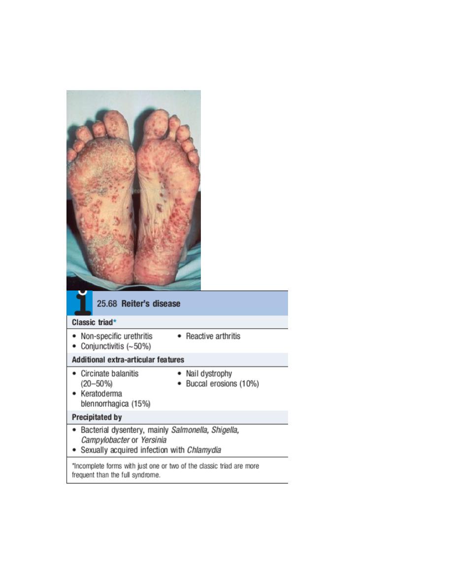
Reactive arthritis
Reactive arthritis (previously known as Reiter’s disease) is
predominantly a disease of young men, with a male
preponderance of 15 : 1.
It is the most common cause of inflammatory arthritis in
men aged 16–35 but may occur at any age.
Reactive arthritis may present with triad of non-specific
urethritis, reactive arthritis,and conjunctivitis, but many
patients present with arthritis only.
Clinical features
The onset is typically acute, with an inflammatory
oligoarthritis that is asymmetrical and targets lower limb
joints, such as the knees, ankles, midtarsal and MTP joints.
It occasionally presents with single joint involvement and
no clear history of an infectious trigger.
There may be considerable systemic disturbance, with fever
and weight loss.
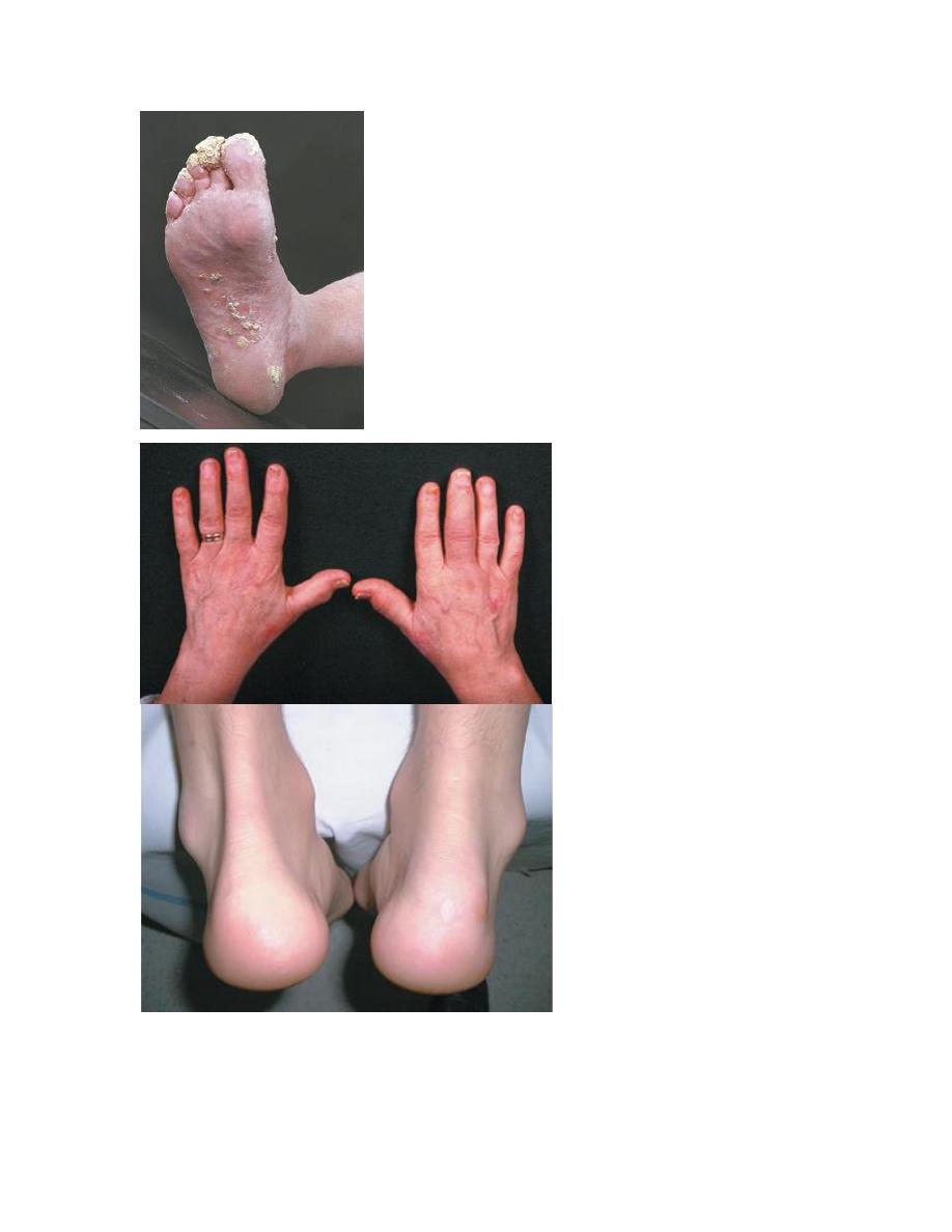
Achilles tendinitis or plantar fasciitis may also be present.
The first attack of arthritis is usually selflimiting, but
recurrent or chronic arthritis develops in more than 60% of
patients, and about 10% still have active disease 20 years
after the initial presentation.
Low back pain and stiffness are common and 15–20% of
patients develop sacroiliitis.
Investigations
The diagnosis is usually made clinically but joint aspiration
may be required to exclude crystal arthritis and articular
infection.
Synovial fluid is leucocyterich and may contain
multinucleated macrophages (Reiter’s cells).
ESR and CRP are raised during an acute attack.
Urethritis may be confirmed in the ‘twoglass test’ by
demonstration of mucoid threads in the firstvoid specimen
that clear in the second.
High vaginal swabs may reveal Chlamydia on culture.
Except for postSalmonella arthritis, stool cultures are
usually negative by the time the arthritis presents, but
serum agglutinin tests may help confirm previous dysentery.
RF, ACPA and ANA are negative.
In chronic or recurrent disease, Xrays show periarticular
osteoporosis, joint space narrowing and proliferative
erosions.
Another characteristic feature is periostitis, especially of
metatarsals, phalanges and pelvis, and large, ‘fluffy’
calcaneal spurs.
In contrast to AS, radiographic sacroiliitis is often
asymmetrical and sometimes unilateral, and
syndesmophytes are predominantly coarse and
asymmetrical, often extending beyond the contours of the
annulus (‘nonmarginal’)
Xray changes in the peripheral joints and spine are identical
to those in psoriasis.
Psoriatic arthritis
Psoriatic arthritis (PsA) occurs in 7–20% of patients with
psoriasis and in up to 0.6% of the general population.

The onset is usually between 25 and 40 years of age.
Most patients (70%) have preexisting psoriasis but in 20%
the arthritis predates the occurrence of skin disease.
Occasionally, the arthritis and psoriasis develop
synchronously.
Clinical features
The presentation is with pain and swelling affecting the
joints and entheses.
Several patterns of joint involvement are recognised but
the course is generally one of intermittent exacerbation
followed by varying periods of complete or nearcomplete
remission.
Destructive arthritis and disability are uncommon, except in
the case of arthritis mutilans.
PATTERNS OF PsA
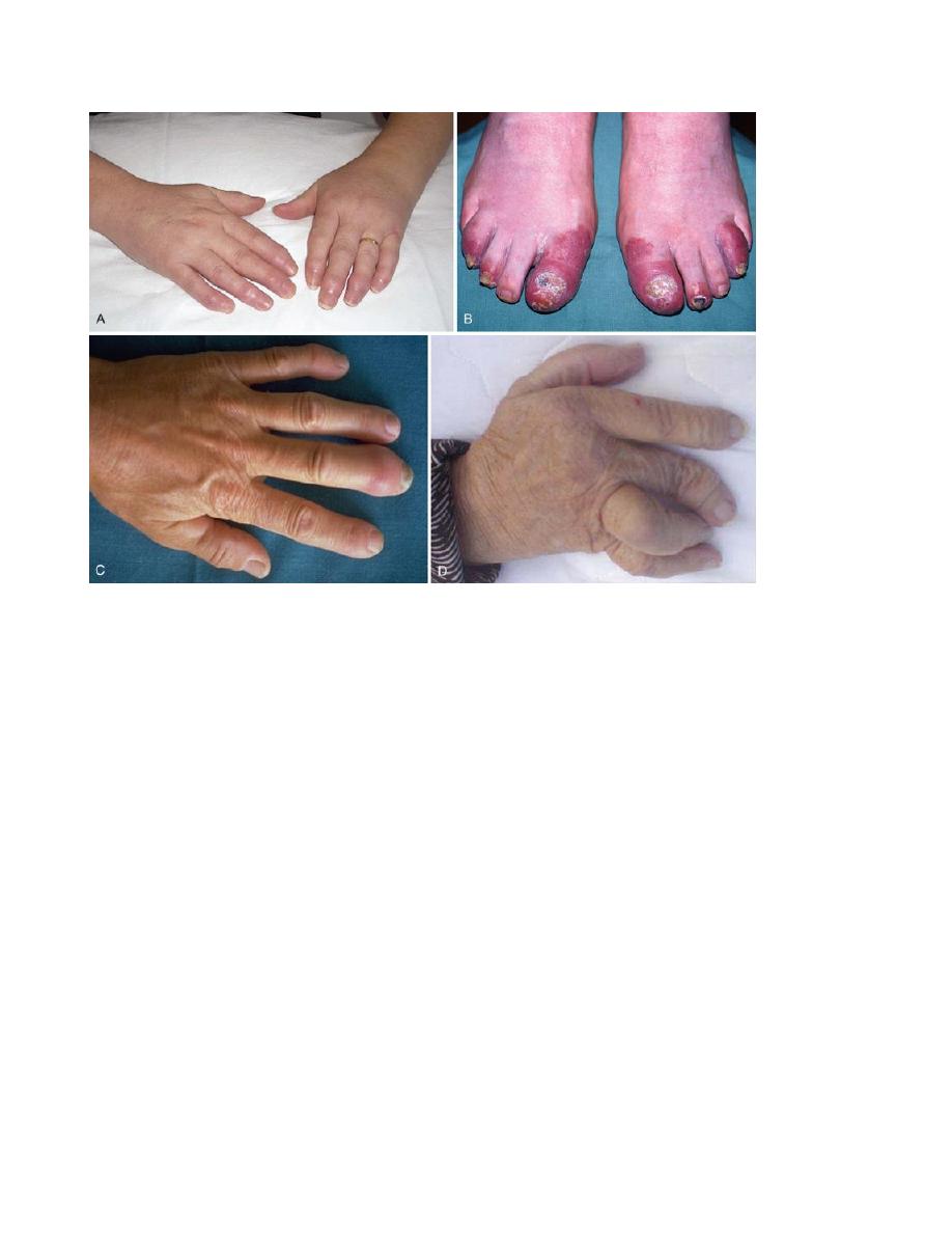
Asymmetrical inflammatory oligoarthritis affects about 40%
of patients and often presents abruptly with a combination
of synovitis and adjacent periarticular inflammation.
This occurs most characteristically in the hands and feet,
when synovitis of a finger or toe is coupled with
tenosynovitis, enthesitis and inflammation of intervening
tissue to give a ‘sausage digit’ or dactylitis

Large joints, such as the knee and ankle, may also be
involved, sometimes with very large effusions.
Symmetrical polyarthritis occurs in about 25% of cases.
It predominates in women and may strongly resemble RA,
with symmetrical involvement of small and large joints in
both upper and lower limbs.
Nodules and other extraarticular features of RA are absent
and arthritis is generally less extensive and more benign.
Much of the hand deformity often results from tenosynovitis
and soft tissue contractures.
Distal IPJ arthritisis an uncommon but characteristic pattern
affecting men more often than women. It targets finger DIP
joints and surrounding periarticular tissues, almost
invariably with accompanying nail dystrophy.
Psoriatic spondylitis presents a similar clinical picture to AS
but with less severe involvement. It may occur alone or with
any of the other clinical patterns described above and is
typically unilateral or asymmetric in severity.
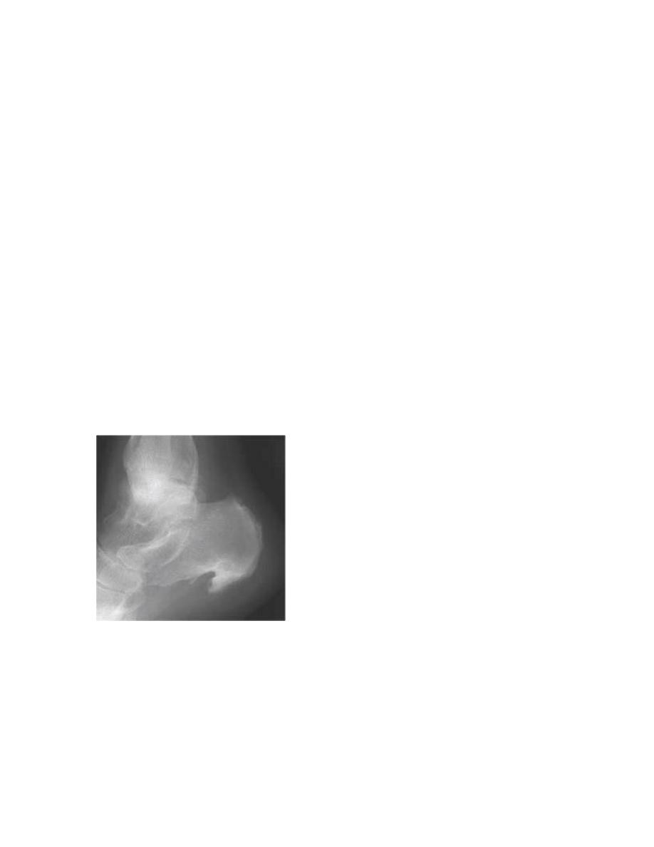
Arthritis mutilansis a deforming erosive arthritis targeting
the fingers and toes that occurs in 5% of cases of PsA.
Prominent cartilage and bone destruction results in marked
instability. The encasing skin appears invaginated and
‘telescoped’ and the finger can be pulled back to its
original length.
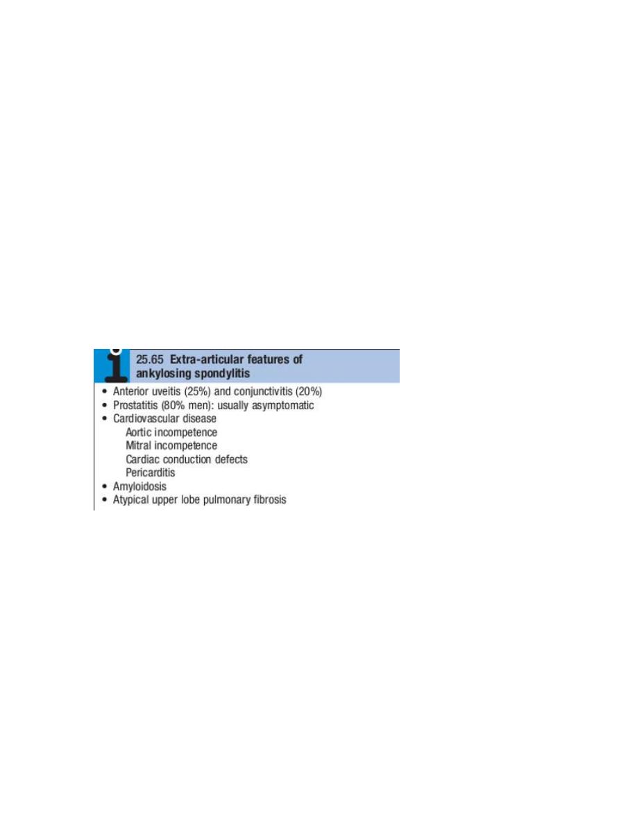
Extra-articular
features
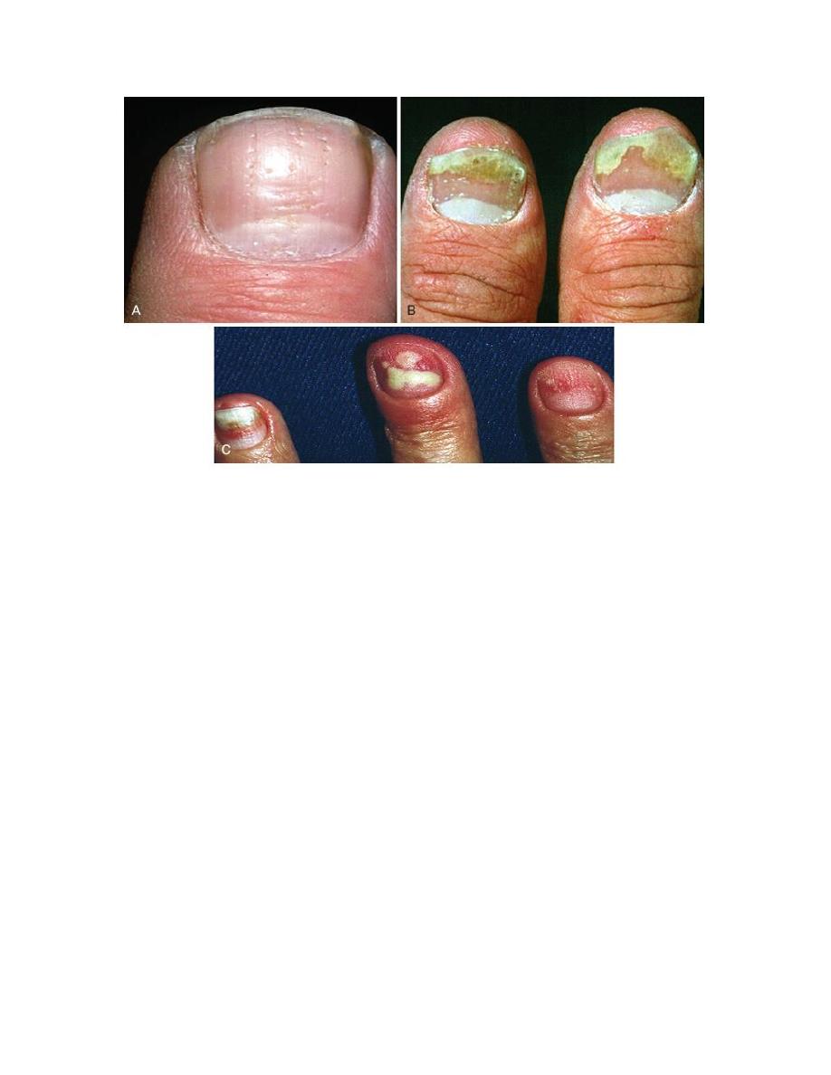
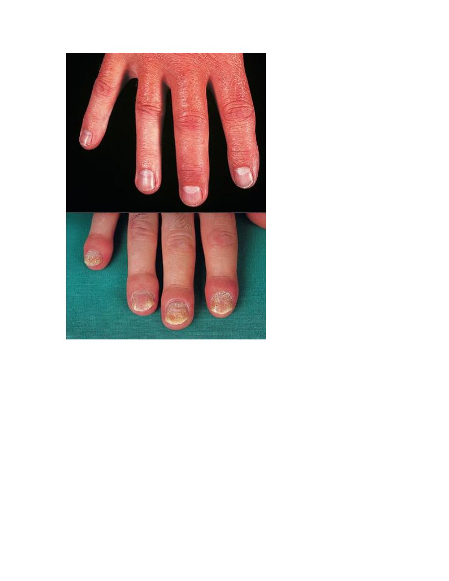
Nail changes include pitting, onycholysis, subungual
hyperkeratosis and horizontal ridging. They are found in 85%
of those with PsA and only 30% of those with uncomplicated
psoriasis, and can occur in the absence of skin disease.

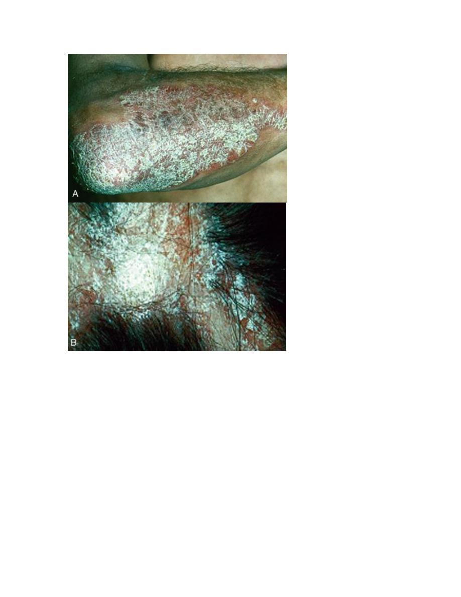
The characteristic rash of psoriasis may be widespread, or
confined to the scalp, natal cleft and umbilicus, where it is
easily overlooked.
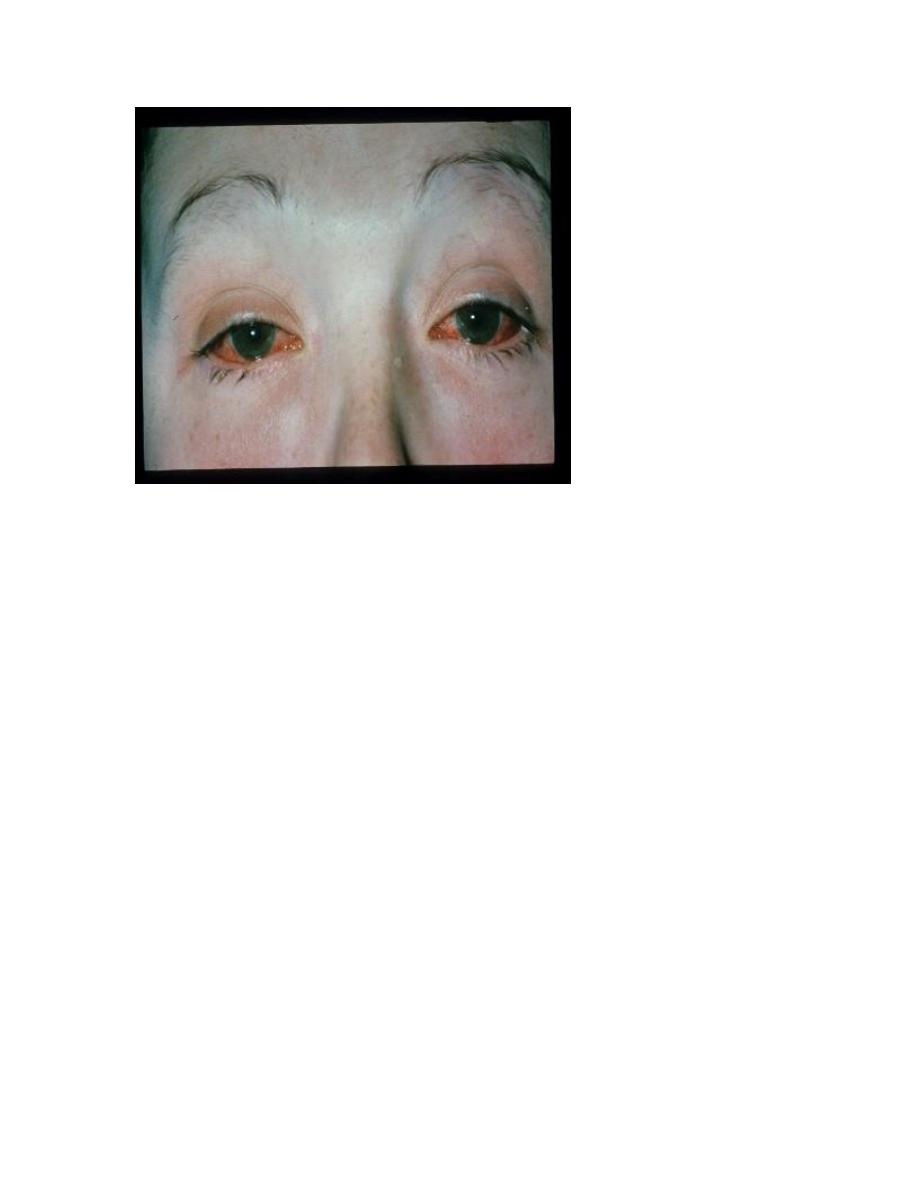
Conjunctivitis can occur, whereas uveitis is mainly confined
to HLAB27positive individuals with sacroiliitis and
spondylitis.
Investigations
The diagnosis is made on clinical grounds.
Autoantibodies are generally negative and acute phase
reactants, such as ESR and CRP, are raised in only a
proportion of patients with active disease.
Xrays may be normal or show erosive change with joint
space narrowing. Features that favour PsA over RA include
the characteristic distribution of proliferative erosions with
marked new bone formation, absence of periarticular
osteoporosis and osteosclerosis.
Imaging of the axial skeleton often reveals features similar
to those in chronic reactive arthritis, with coarse,
asymmetrical, nonmarginal syndesmophytes and
asymmetrical sacroiliitis.
MRI and ultrasound with power Doppler are increasingly
employed to detect synovial inflammation and inflammation
at the entheses.
Enteropathic arthritis
An acute inflammatory oligoarthritis occurs in around 10%
of patients with ulcerative colitis and 20% of those with
Crohn’s disease. It predominantly affects the large lower
limb joints (knees, ankles, hips) but wrists and small joints of
the hands and feet can also be involved. The arthritis usually
coincides with exacerbations of the underlying bowel
disease, and sometimes is accompanied by aphthous mouth
ulcers, iritis and erythema nodosum.
It improves with effective treatment of the bowel disease,
and can be cured by total colectomy in patients with
ulcerative colitis.
Patients with inflammatory bowel disease may also develop
sacroiliitis (16%) and AS (6%), which are clinically and
radiologically identical to classic AS. These can predate or
follow the onset of bowel disease and there is no correlation
between activity of the spondylitis and bowel disease.

The arthritis often remits with treatment of the bowel
disease but DMARD and biological treatment is occasionally
required.