JABIR IBN HAYYAN MEDICAL UNIVERSITYCOLLEGE OF MEDICINE DEPARTMENT OF HUMAN ANATOMY
Histology of Endocrine SystemLecture By;
Dr. Hayder ALKifaee
20/4/2017
Endocrine Glands& Organs Containing Endocrine Cells
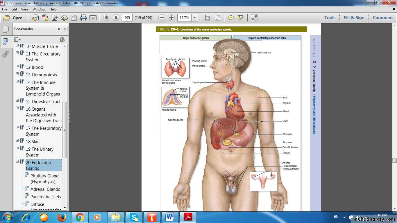
Primary Endocrine Organs
• Pituitary Gland (Hypophysis Cerebri)• Pineal Gland (Epiphysis Cerebri)
• Thyroid Gland
• Parathyroid Gland
• Adrenal Gland
Secondary Endocrine Organs
• Kidney• Testes
• Ovaries
• Pancreas
• Stomach
• Intestines
• Thymus
• Heart
• Placenta
Typical Endocrine Glands
• Pituitary Gland (Hypophysis Cerebri)
• Pineal Gland (Epiphysis Cerebri)
• Thyroid Gland
• Parathyroid Gland
• Adrenal Gland
Scattered Endocrine Masses
Islets of LangerhansCorpus luteum
Interstitial cells of Leydig
Placental lactogen secreting cells
Chorionic gonadotropin secreting cells
Juxtaglomerular cells
Isolated Endocrine Cells (Diffuse Neuroendocrine System)
Called APUD cells – Amine Precursor Uptake & Decarboxylation cells.Present within the lining epithelium of the digestive and respiratory system.
Pituitary Gland(Hypophysis Cerebri)
Dual Origin of Pituitary
Partly from the developing brain (Neurohypophyseal bud )Partly from the developing oral cavity (Hypophyseal ( Rathke ) pouch )
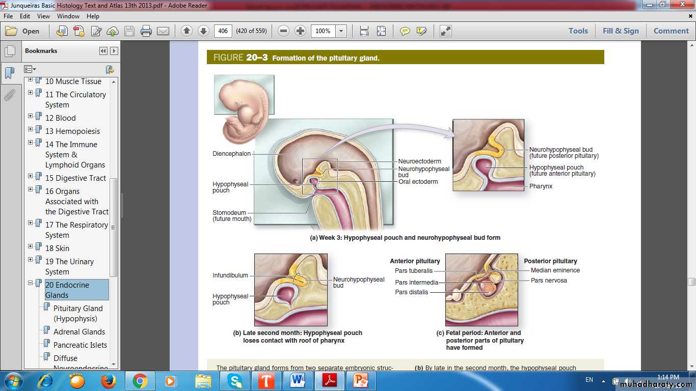
Formation of Pituitary Gland
Dual Origin of Pituitary Gland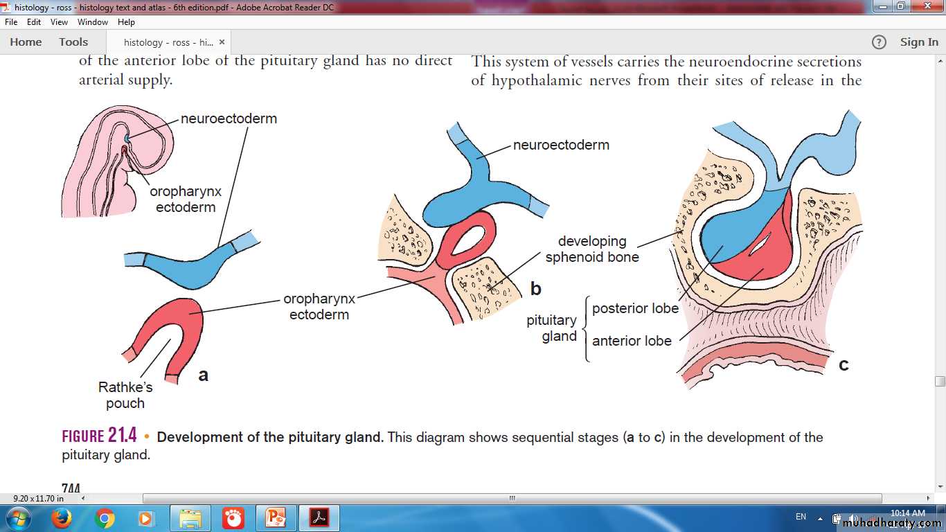
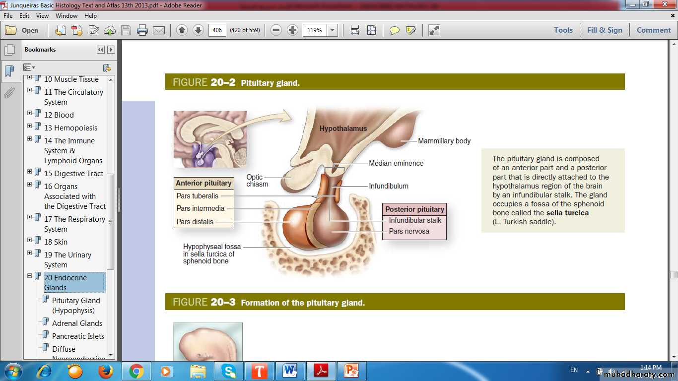
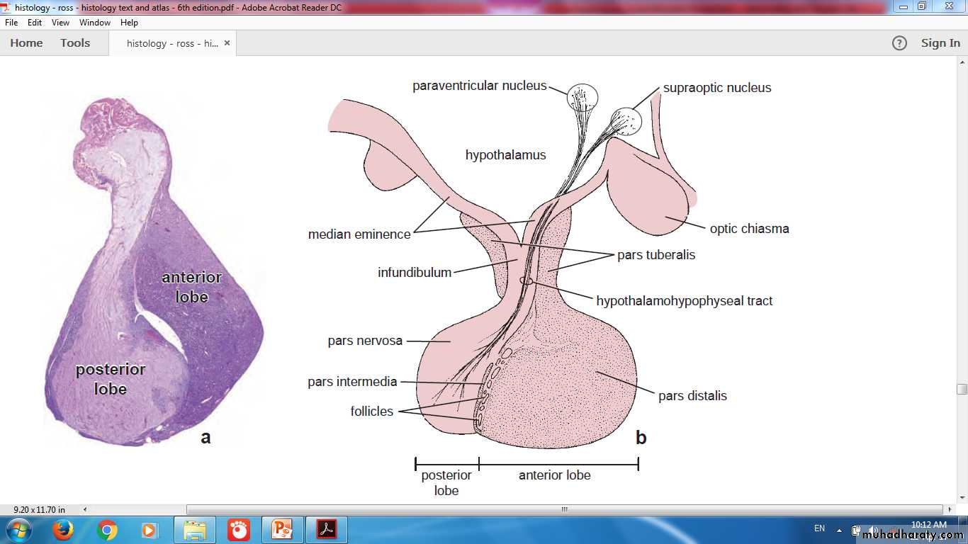
Pituitary Gland
Pituitary Gland
AdenohypophysisPars distalis
Pars tuberalis
Pars intermedia
Neurohypophysis
Pars nervosaStalk (Infundibulum)
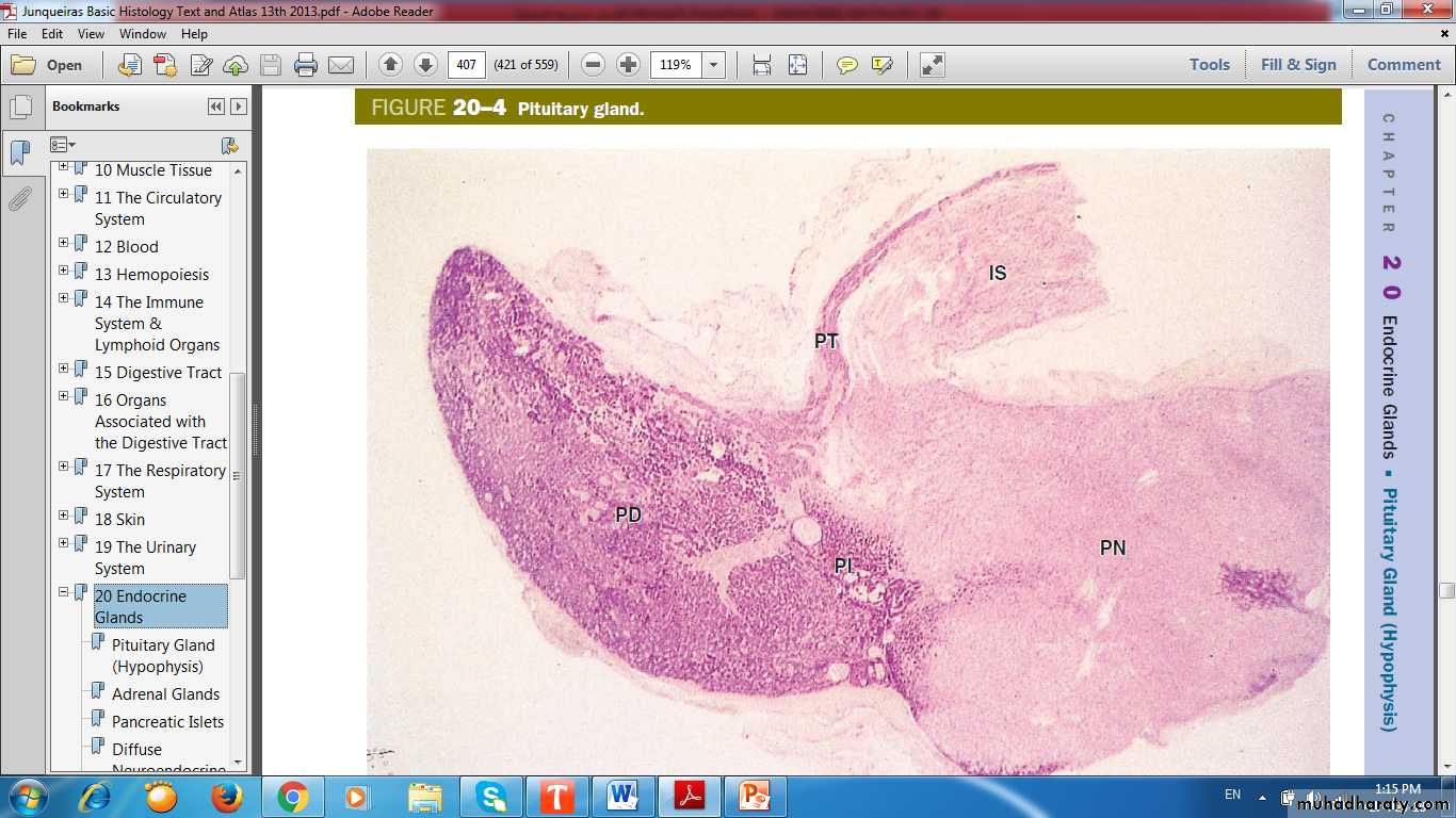
Pituitary Gland
infundibular StalkPars Nervosa
Pars Tuberalis
Pars Intermedia
Pars Distalis
Adenohypophysis
Neurohypophysis
Pituitary Gland
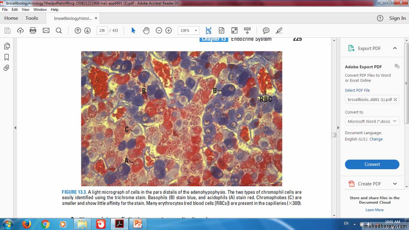
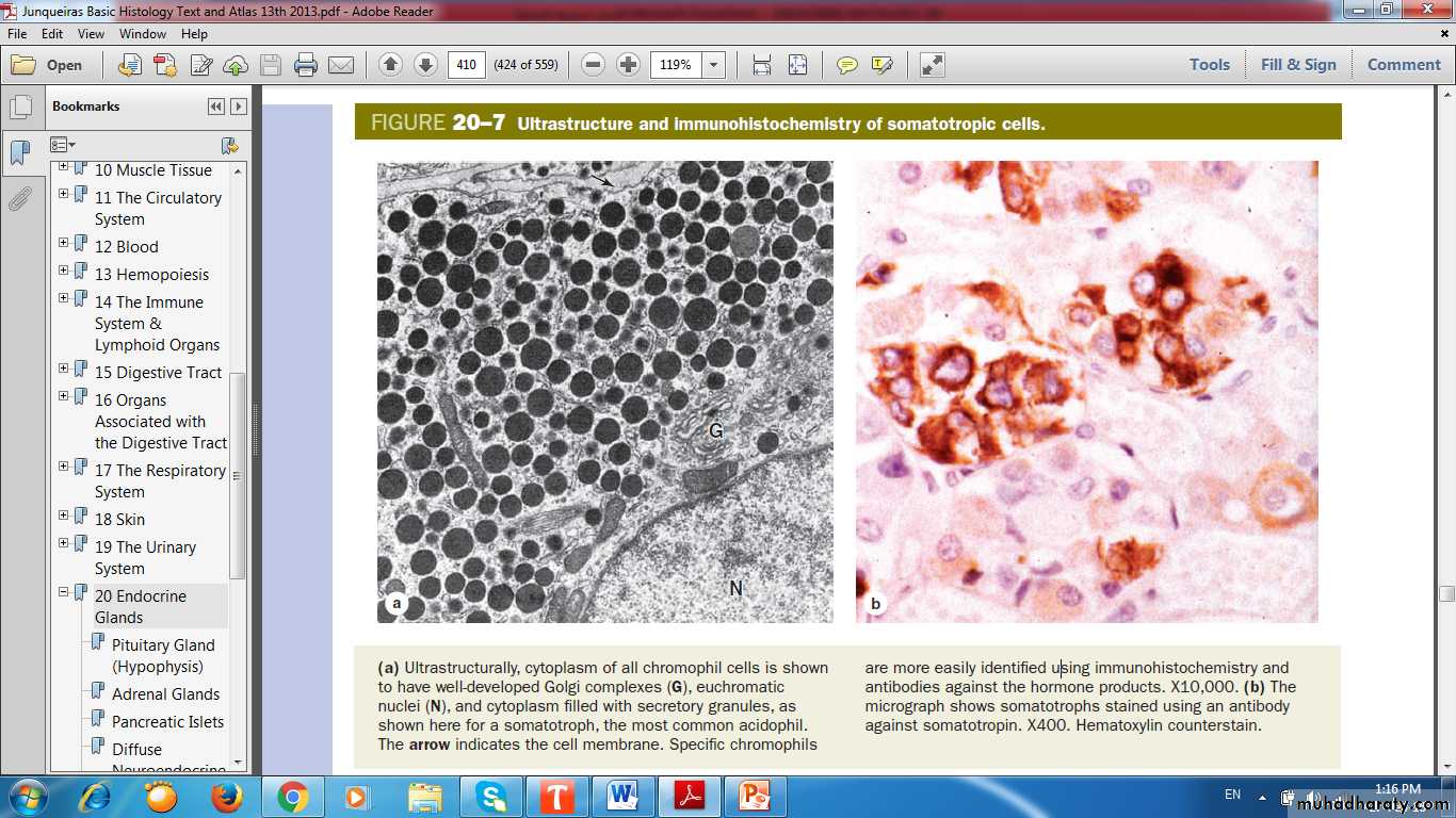
Pars distalisChromophils
Acidophils
Basophils
Chromophobes
Stain weakly
Few or no secretory granules
Stem & progenitor cells
Degranulated cells
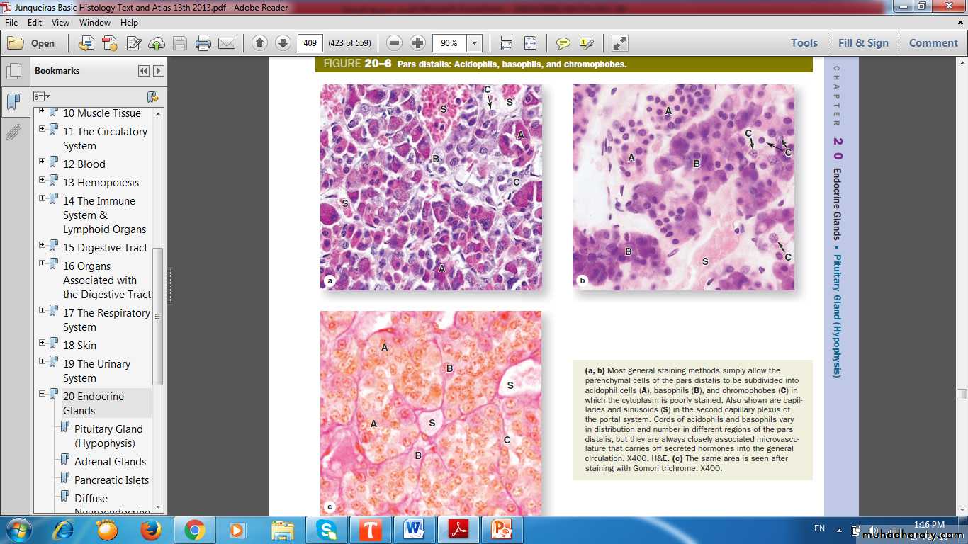
Pars Distalis: Acidophil, Basophil & Chromophobe
ChromophilsAcidophils
Somatotrophs
Mammotrophs
Basophils
Gonadotrophs
Thyrotrophs
Corticotrophs
Acidophil, Basophil & Chromophobe
Somatotropic Cells
UltrastructureImmunohistochemistry
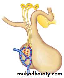
Control of the Pituitary
Hypothalamic-Hypophyseal TractHypothalamic-Hypophyseal Portal System
Negative Feedback Loops
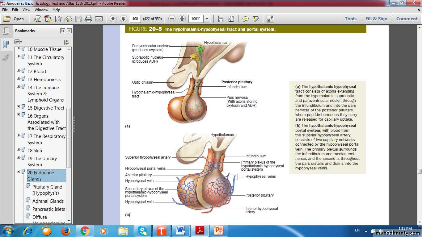
Hypothalamic-Hypophyseal Tract
Hypothalamic-Hypophyseal Tract
Hypothalamic-Hypophyseal Portal System
Hypothalamic Control of pituitary function
Hypothalamic-Hypophyseal Portal SystemHypothalamo-Hypophyseal Tract
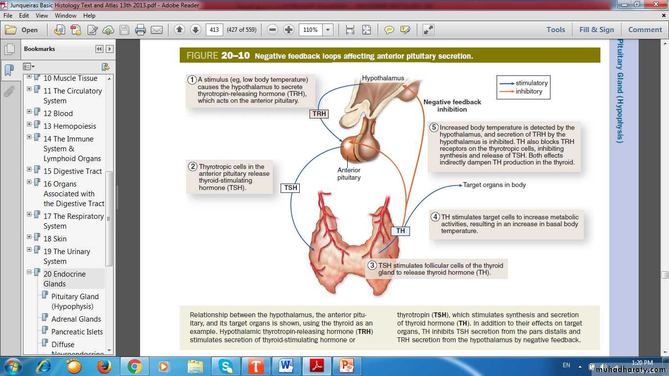
Negative Feedback Loops Affecting Anterior Pituitary Secretion
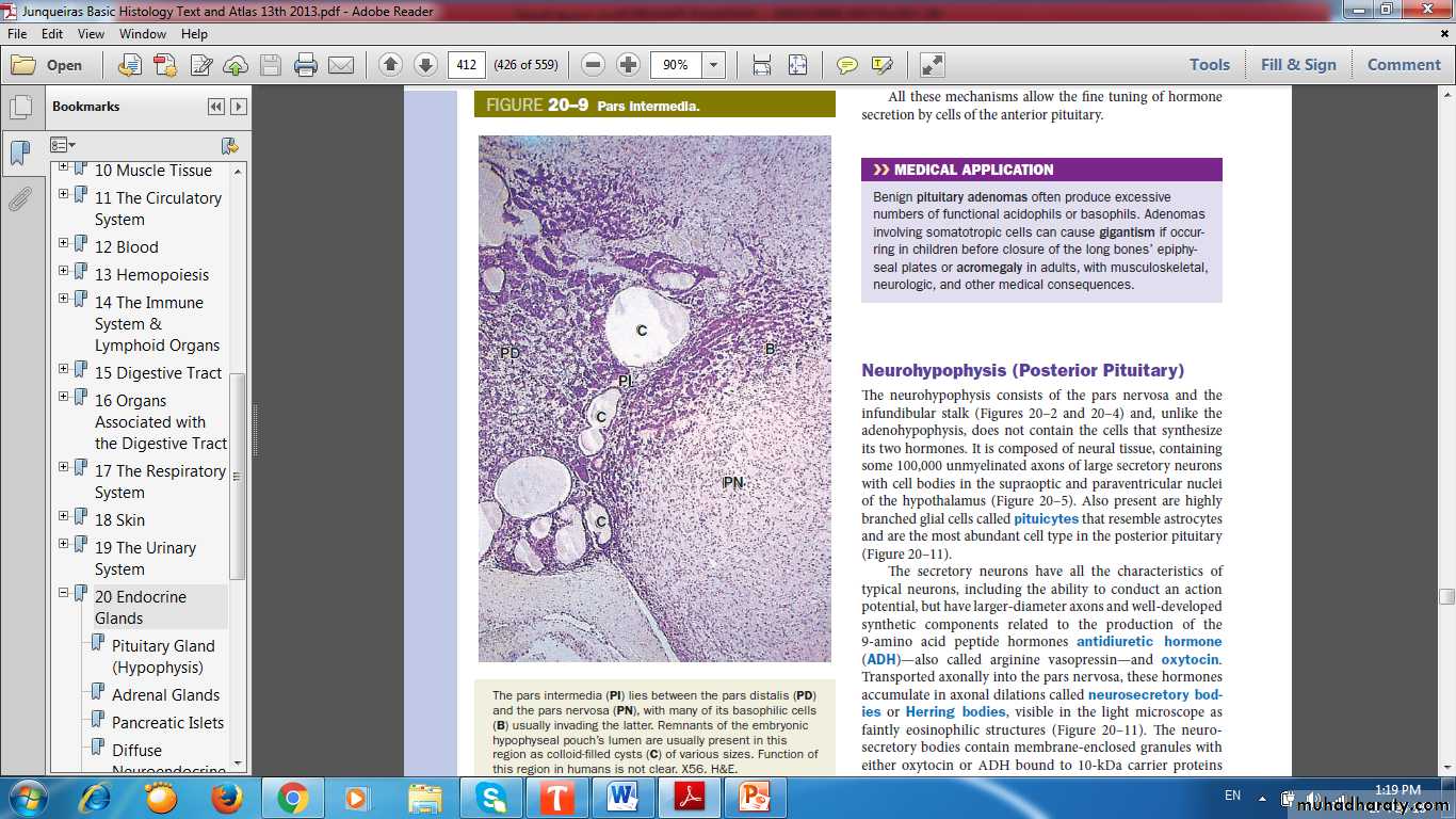
Pars Intermedia
Pars NervosaPars Distalis
Cyst
Basophilic cells
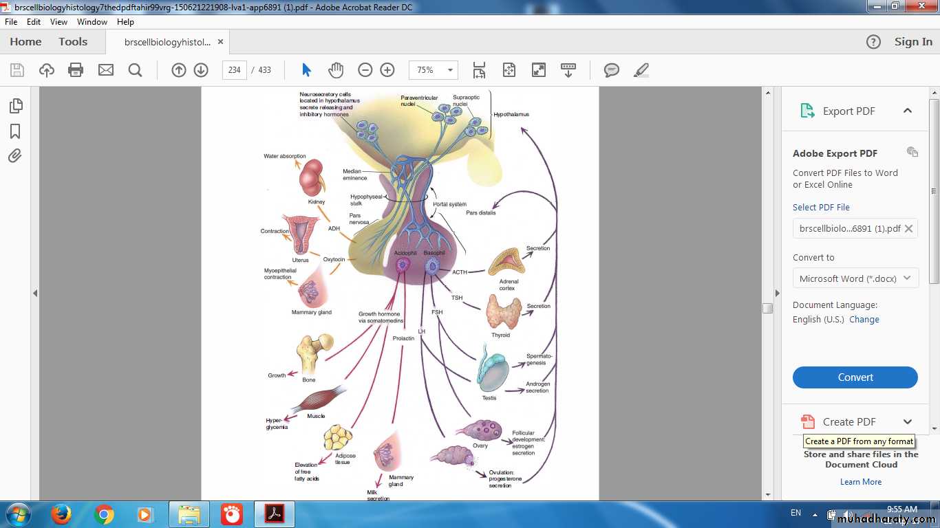
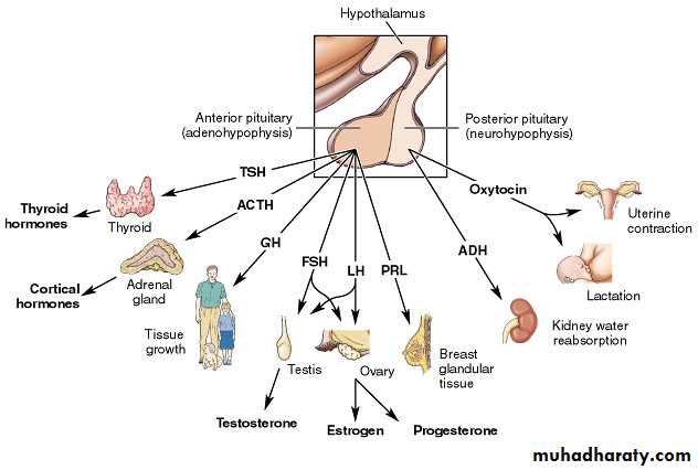
Hormones of Pituitary
Adenohypophysis• Growth hormone
• Prolactin
• FSH
• LH
• ACTH
• TSH
Neurohypophysis
• ADH
• Oxytocin
•
Gigantism & Dwarfism

Acromegaly
Pars Nervosa
• Neurosecretory Bodies• Unmylinated axons
• Pituicytes
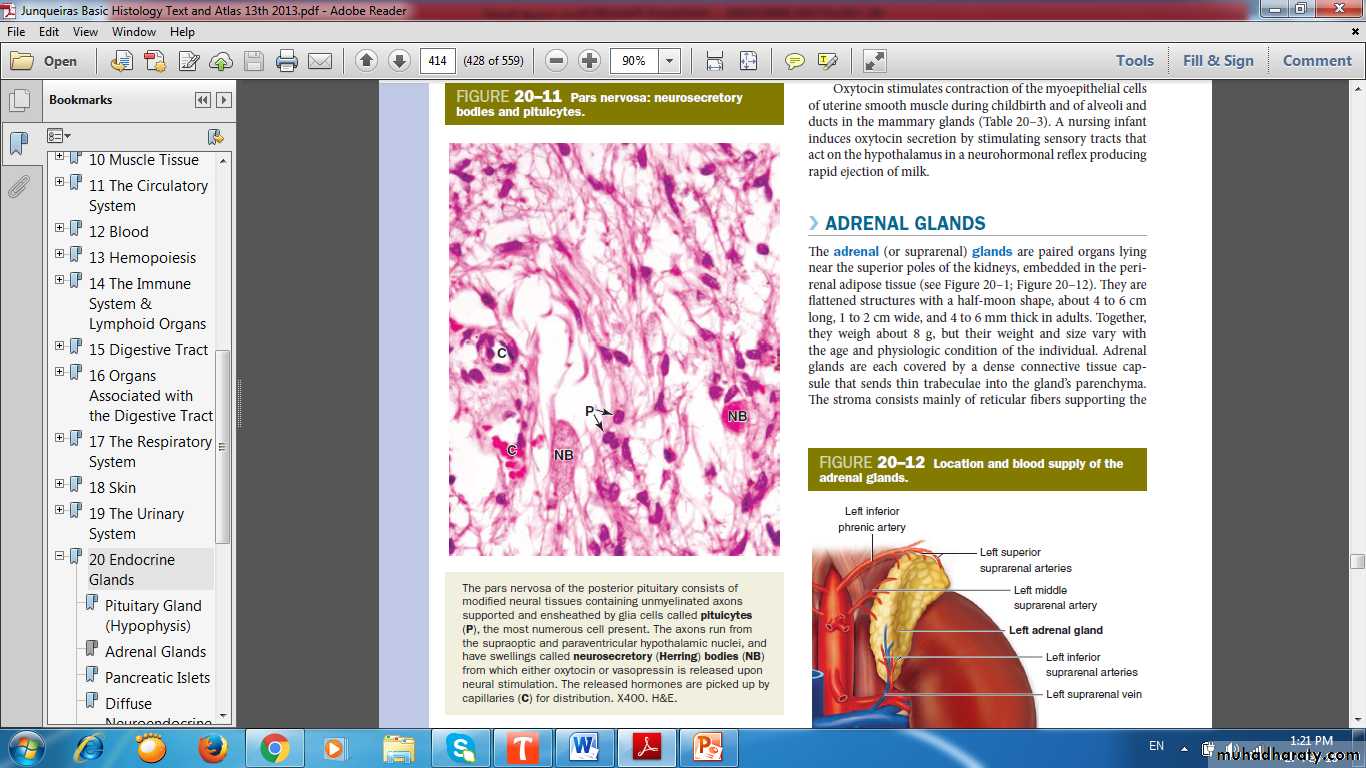
Neurosecretory Bodies
PituicytesCapillary
Hormones of Pituitary
Adrenal Gland
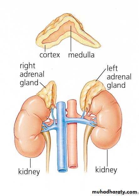
Adrenal Gland
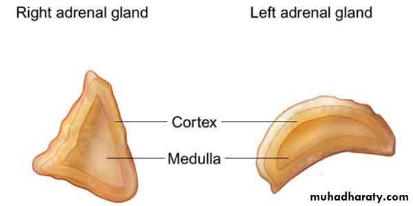
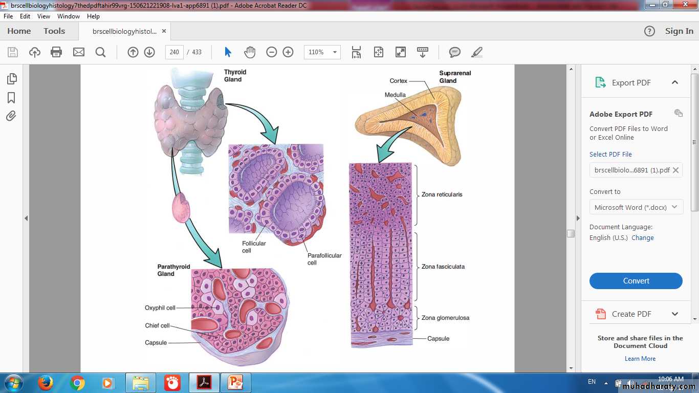
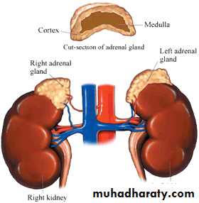
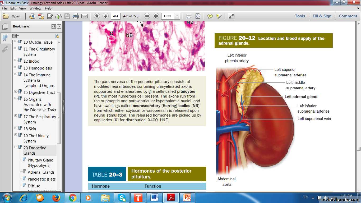
Adrenal Gland
Adrenal Gland
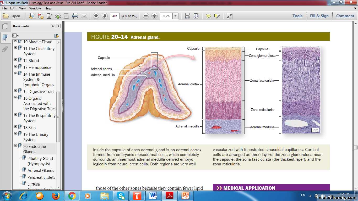
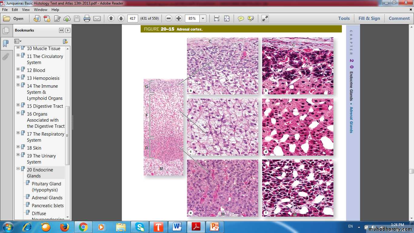
Adrenal CortexOrigin from mesoderm
Features of steroid- secreting cells
Three concentric zones:
Zona Glomerulosa
Zona Fasciculata
Zona Reticularis
Adrenal Cortex
Zona Glomerulosa (15%): rounded or arched cords of columnar or pyramidal cellsZona Fasciculata (65-80%): long cords of large polyhedral cells, one or two cells thick
Zona Reticularis (10%): smaller cells in a network of irregular cords
Adrenal Cortex Zones & Function
Zona Glomerulosa:Aldosterone
Zona Fasciculata:
CortisolZona Reticularis:
Androgen
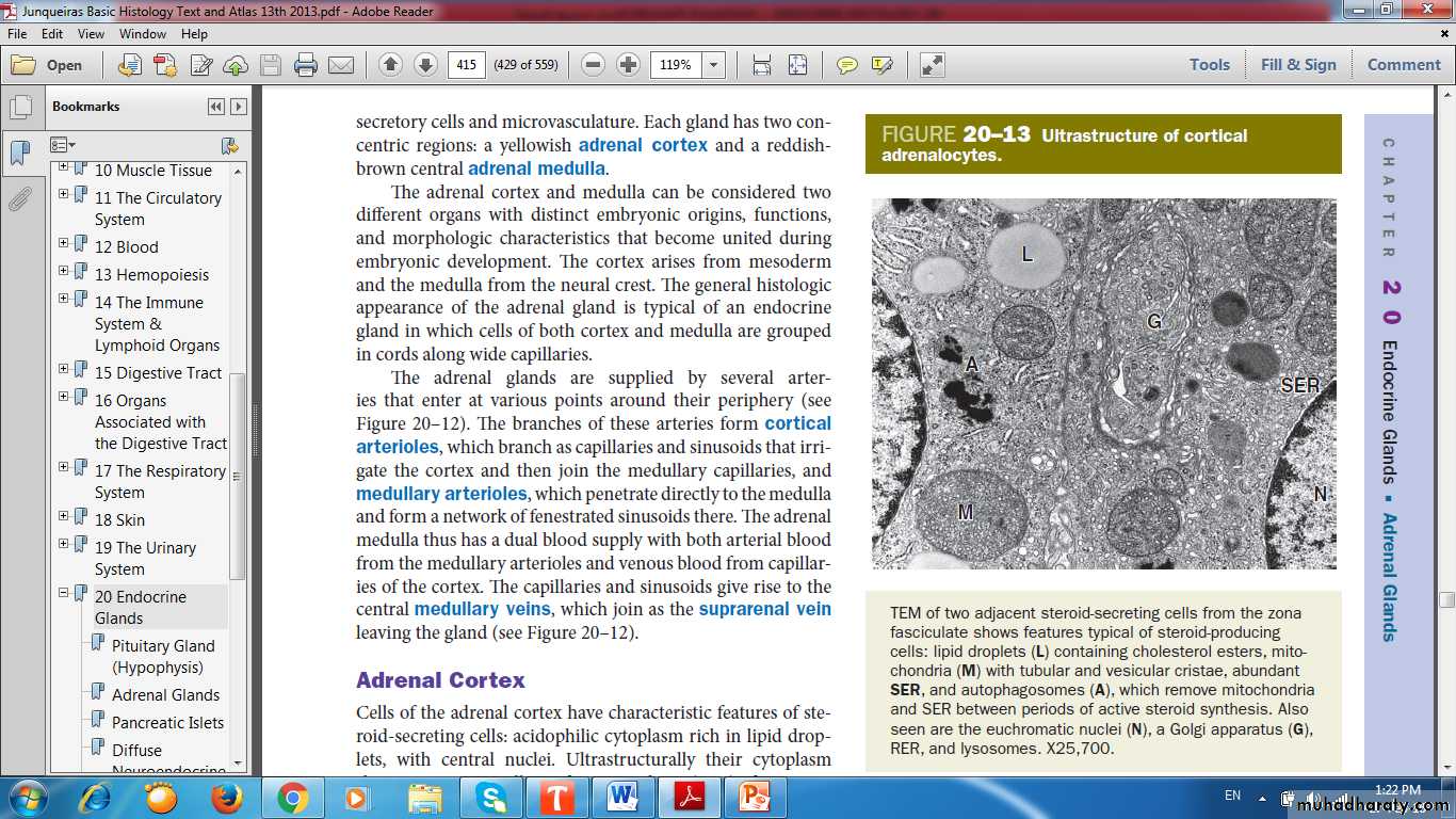
Ultrastructure of Cortical Adrenalocytes
Lipid dropletMitochondria
Smooth Endoplasmic Reticulum
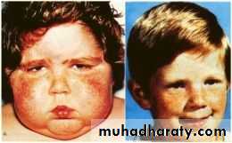
Cushing Syndrome
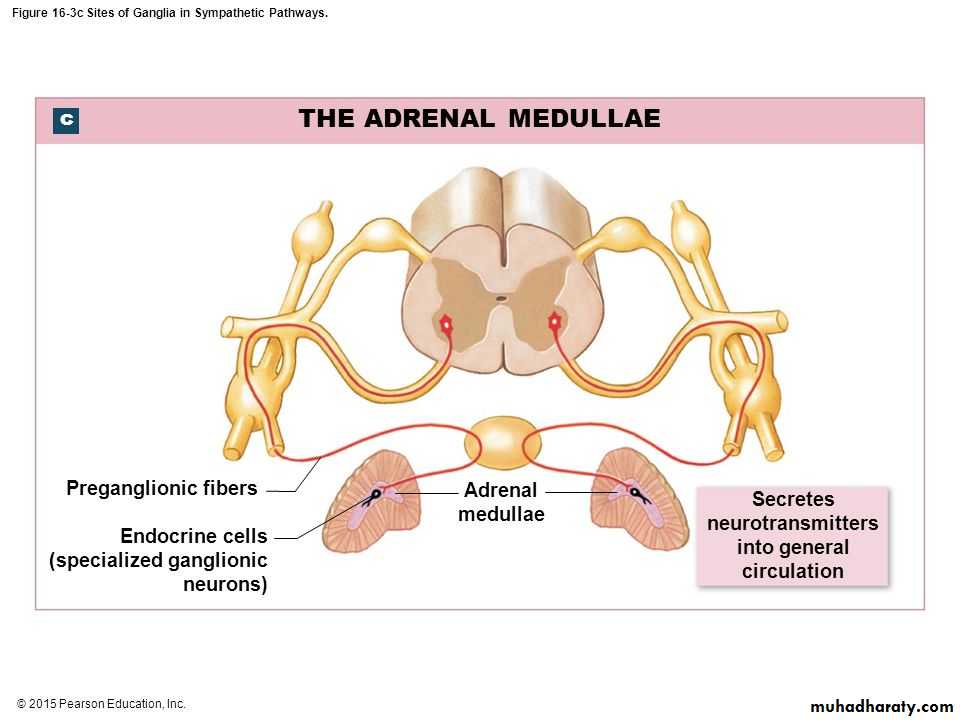
Adrenal Medulla
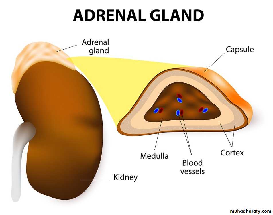
Adrenal Medulla
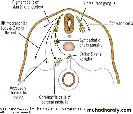
Origin from neural crestReddish brown in color
Cells called Chromaffin Cells
Profuse supply of sinusoidal capillaries
Adrenal Medulla Origin from Neural Crest
Chromaffin Cells
Modified neurons, lacking axons and dendritesSpecialized as secretory cells
Contain many electron-dense granules for storage and secretion of catecholamines
large, pale-staining polyhedral cells arranged in cords or clumps
Modified Neurons, Lacking Axons & Dendrites
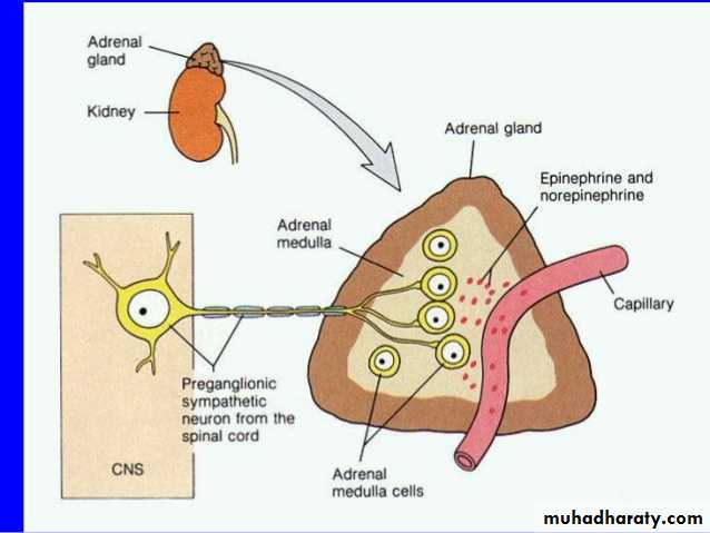
Innervated by Preganglionic Nerve, which Trigger Hormone Release
Specialized as Secretory Cells
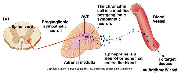
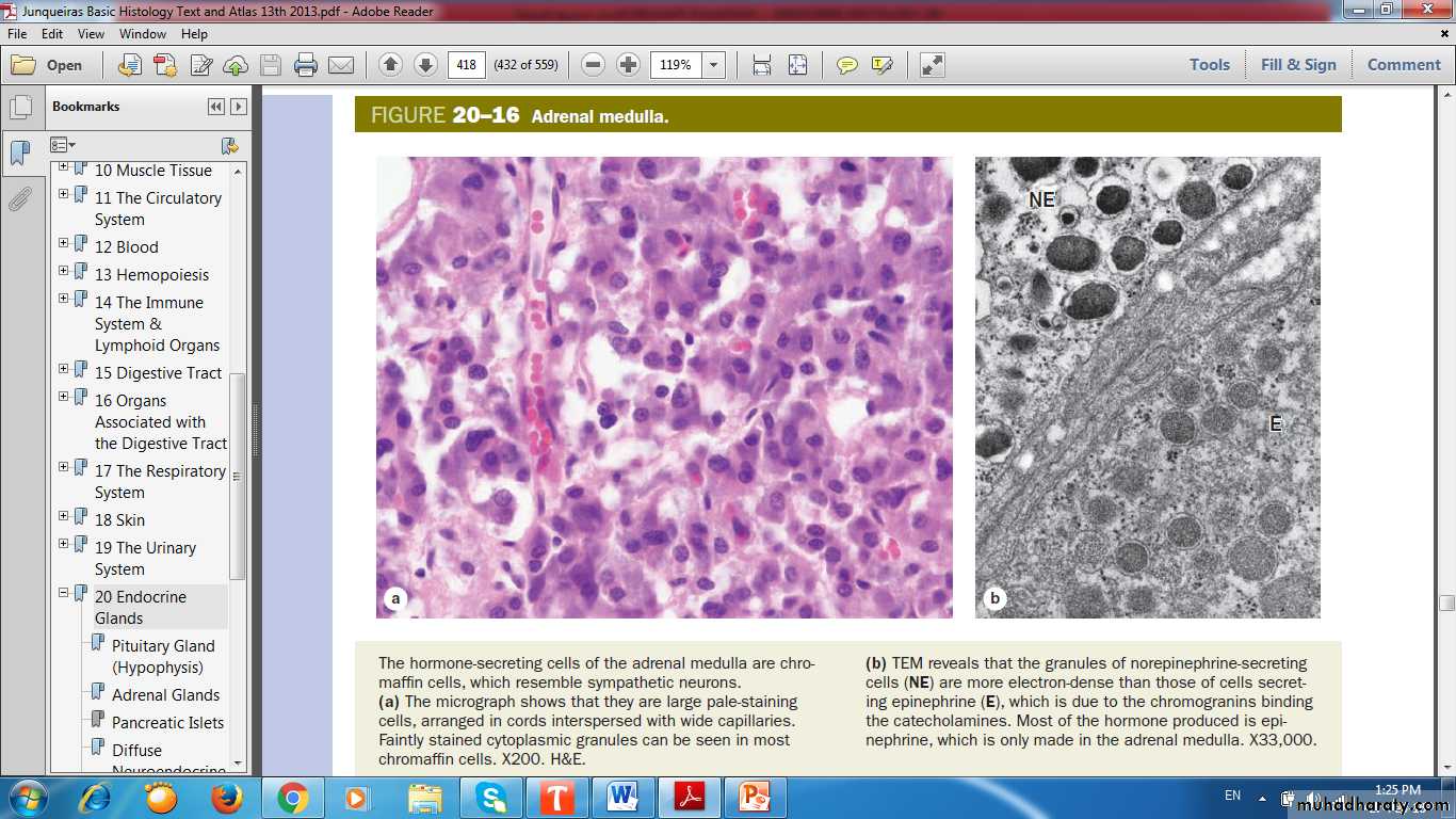
Chromaffin Cells are large, pale-staining polyhedral cells arranged in cords or clumps
NorEpinephreinEpinehrin
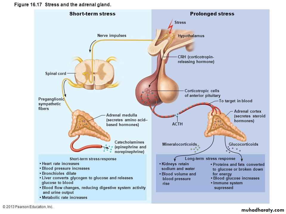
Stress & Adrenal Gland
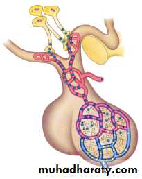
Thyroid Gland
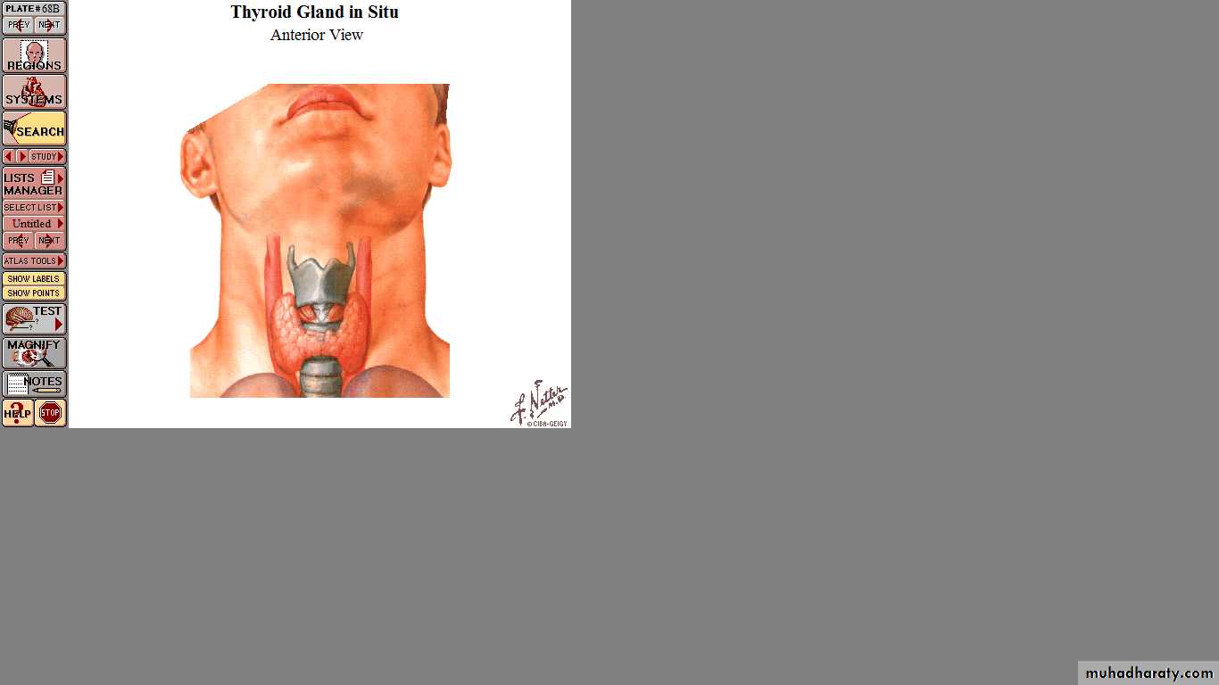
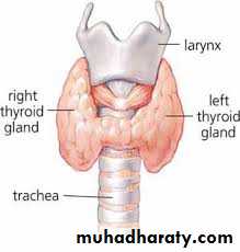
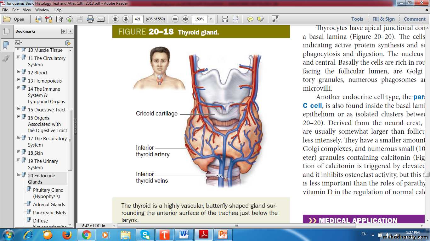
Thyroid Gland
Thyroid GlandOrigin: foregut endoderm
Hormones – (T3, T4 & Calcitonin)
Thyroid follicles
Thyroglobulin
Parafollicular cell, or C cell
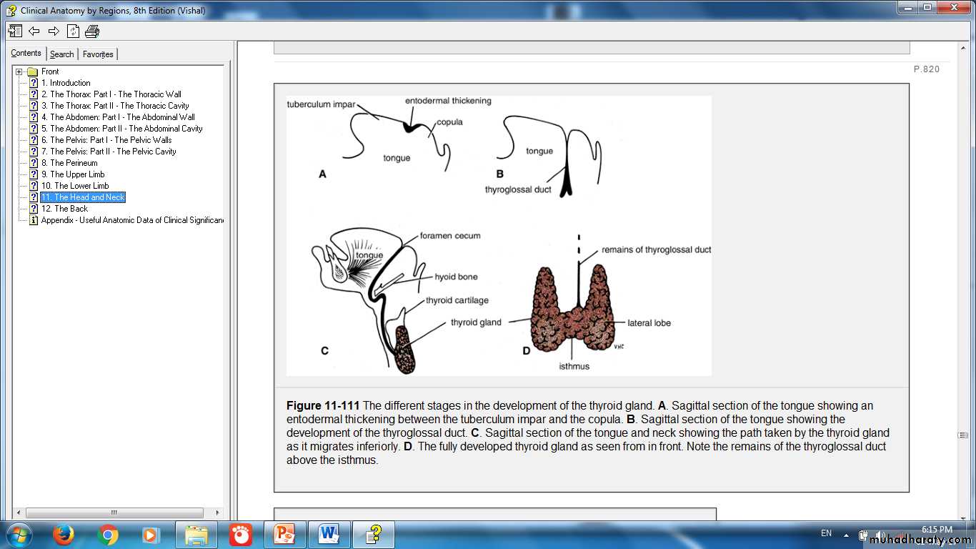
Origin of Thyroid: from foregut endoderm near the base of the tongue

Thyroid Follicular Cells & Parafollicular Cells
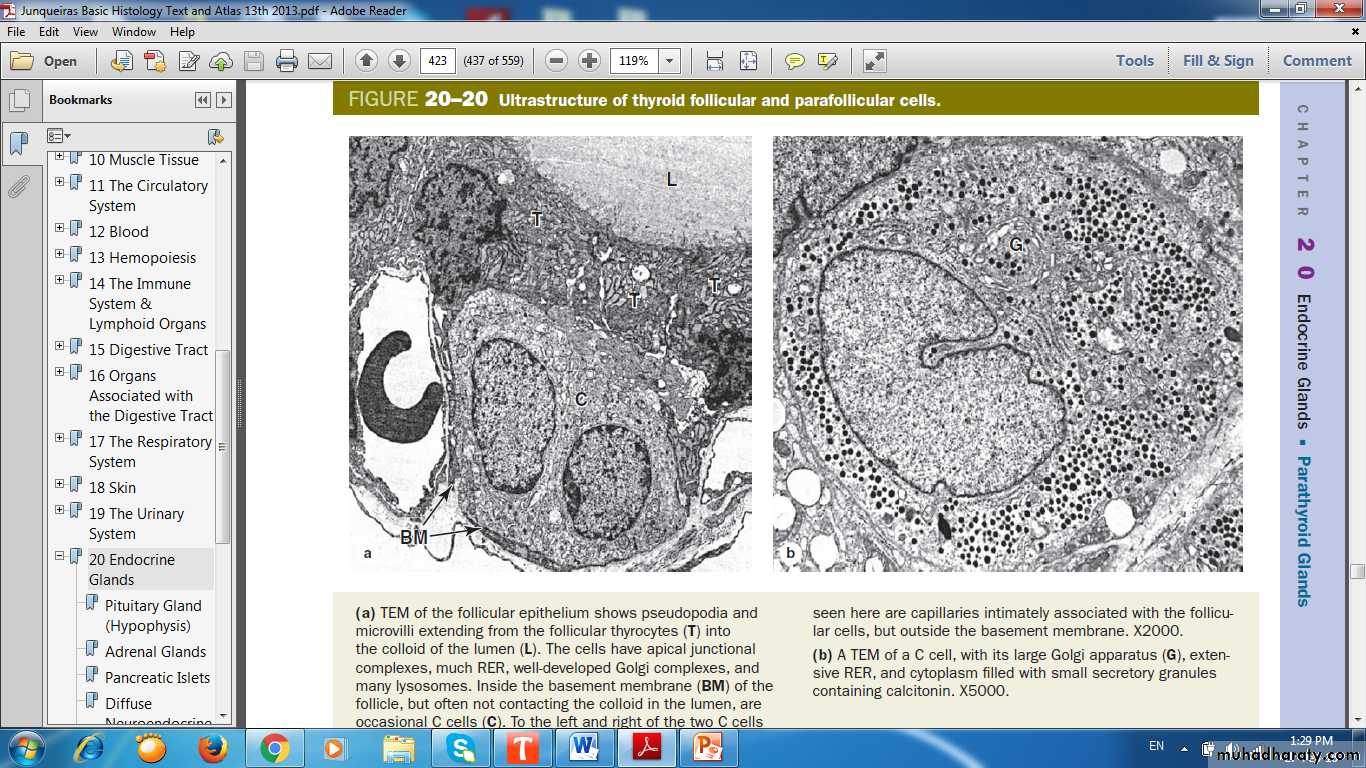
Ultrastructure of Thyroid Follicular Cells & Parafollicular (C) Cells
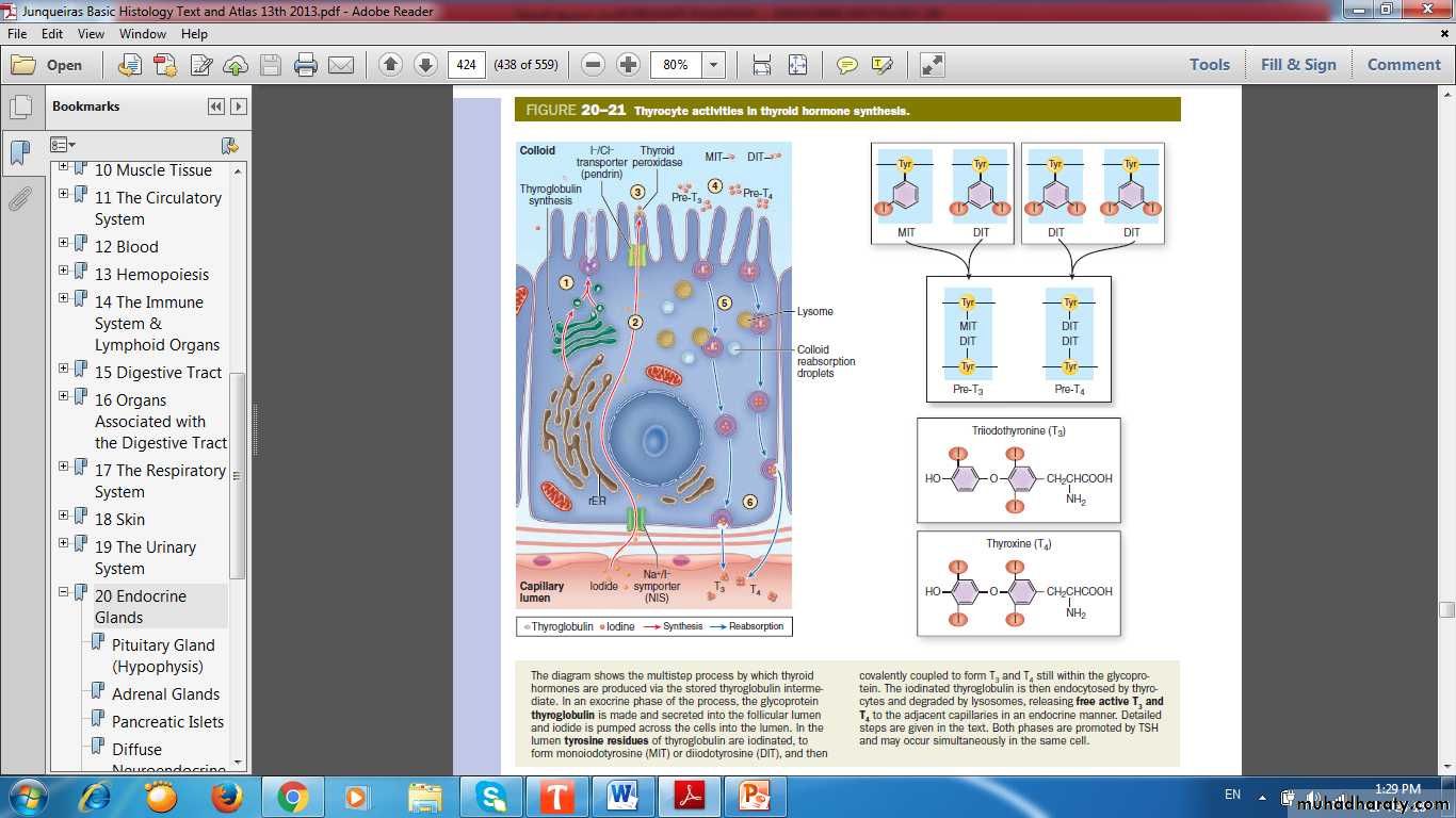
Thyrocytes Activities in Thyroid Hormone Synthesis
Hyperthyroidism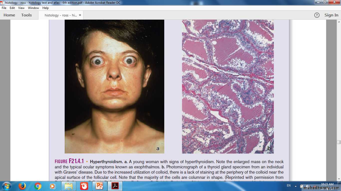
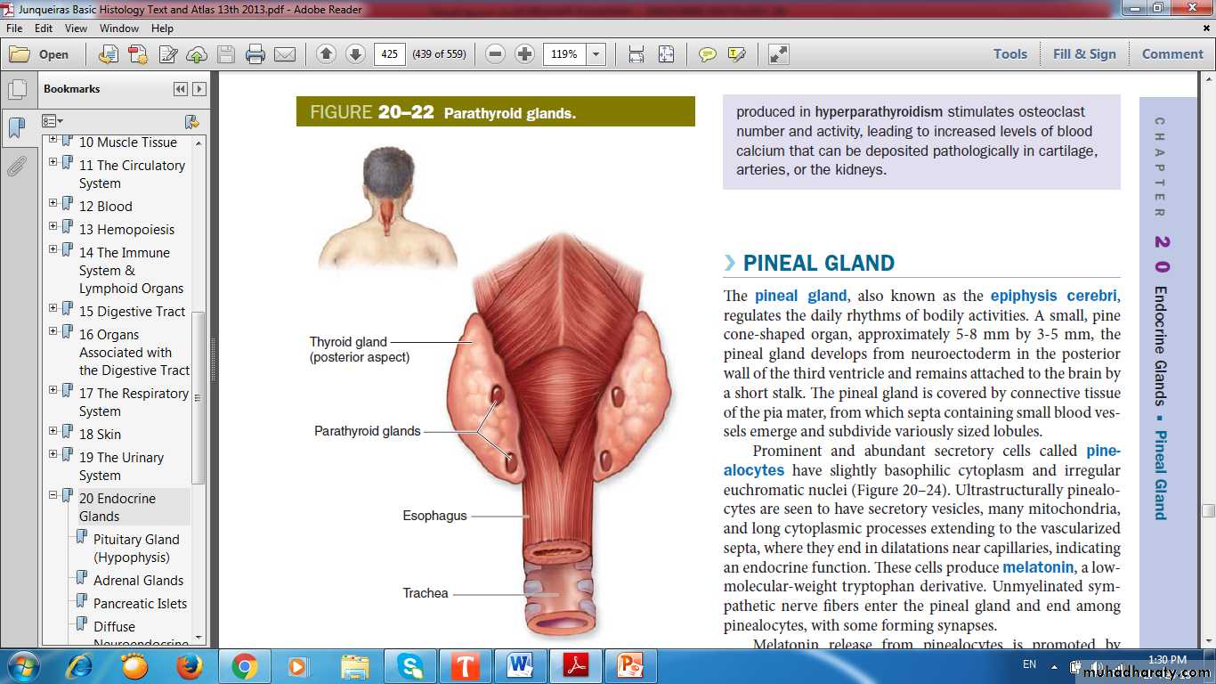
Parathyroid Gland
Parathyroid GlandFour small ovoid masses
Location
Origin
Principal (chief) cells
Parathyroid hormone (PTH)
Oxyphil cells
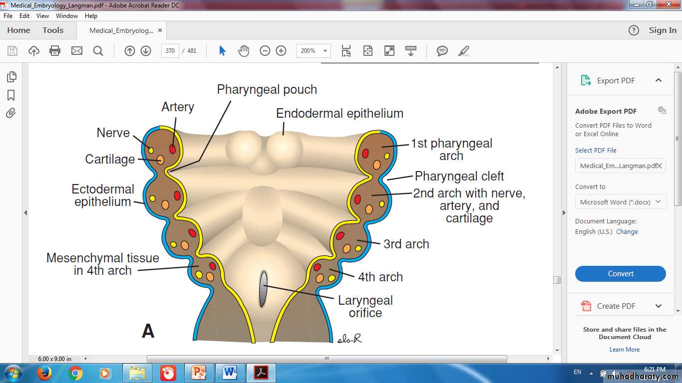
Pharyngeal Arches, Clefts & Pouches
Origin of Parathyroid Glands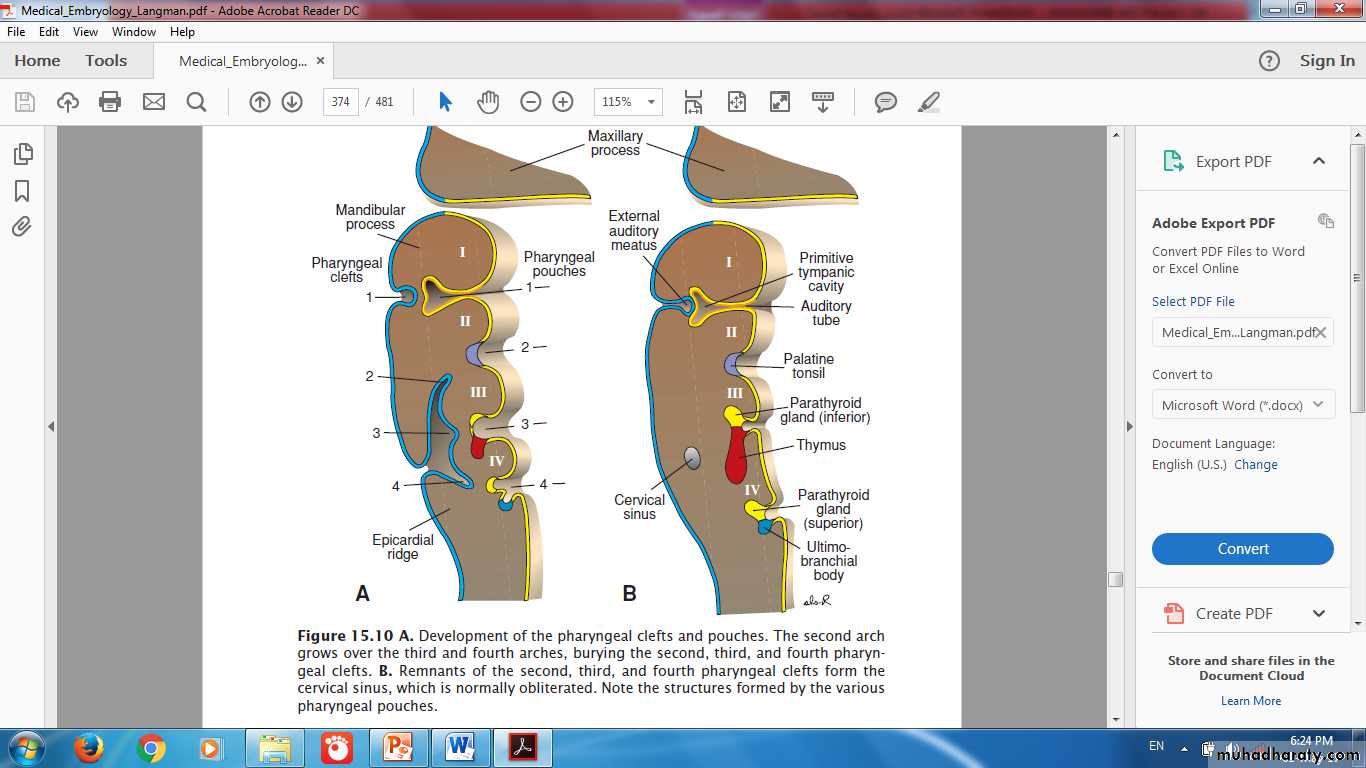
Thymus & Inferior Parathyroid Gland
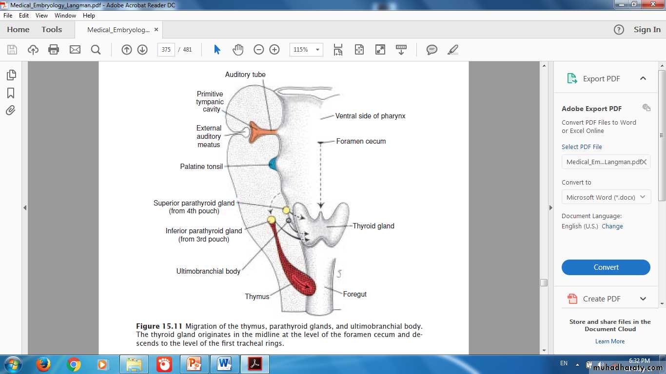
Thyroid & Parathyroid Glands
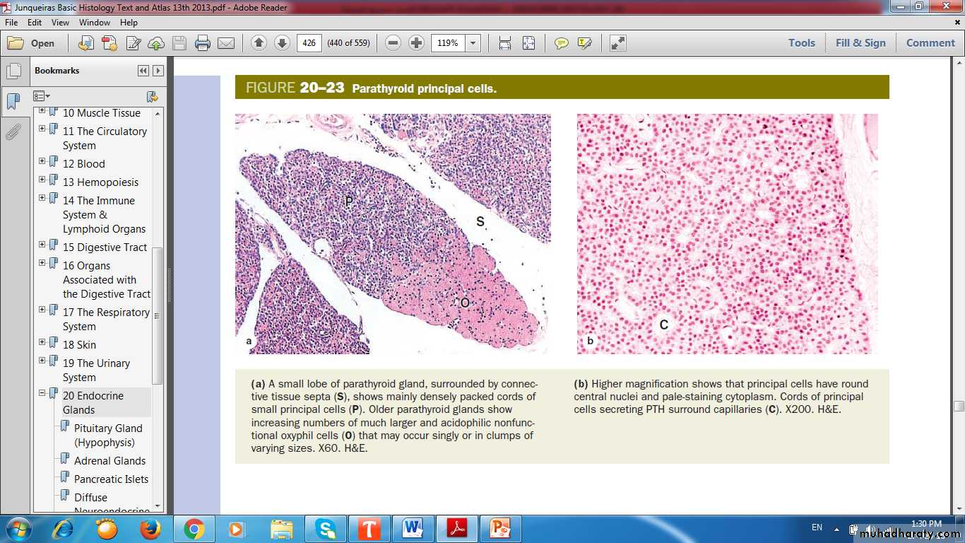
Parathyroid Principle Cells
Pineal Gland (Epiphysis Cerebri)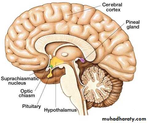
Pineal Gland
ShapeOrigin
Cells : Pinealocytes
Hormone: Melatonin
Glial cells: Modified astrocytes
Corpora arenacea (brain sand)
Neuroendocrine Transducer
Pineal Gland
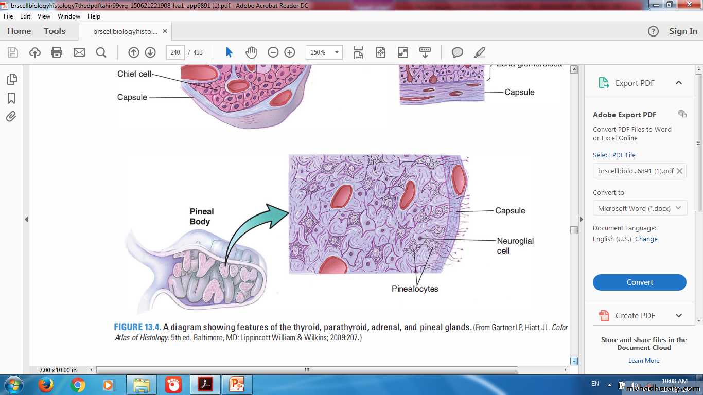
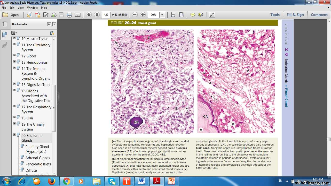
Pineal Gland
Corpora ArenaceaSepta
Astrocyte
Pinealocytes