1
First stage
Biology
Lec / 9
Abeer H. Alkhafaf
Muscle tissue
Muscle cells are structurally and functionally specialized for contraction, which
contain two types of special protein filaments called myofilaments; including thin
filaments containing actin and thick filaments containing myosin. Nearly all muscle
cells are of mesodermal origin.
Muscle tissues are groups of muscle cells organized
by connective tissue. It permits movement of the body, maintains posture, producing
body heat and circulates blood throughout the body.
The cytoplasm of muscle cells is called sarcoplasm and the smooth ER is called
sarcoplasmic reticulum. The cell membrane or plasmalemma is the sarcolemma.
Three types of muscle tissue can be distinguished on the basis of morphologic and
functional characteristics, and each type of muscle tissue has a structure adapted to
its physiologic role :-
.g.
intestines and blood vessels).
2
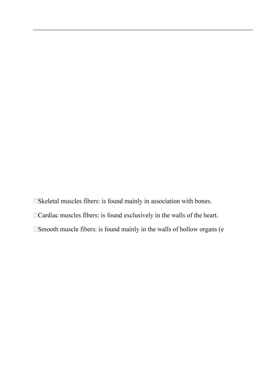
Skeletal muscle
- They are striated and voluntary muscles, they are attached to the skeleton, and so
their contraction moves the skeleton.
- Skeletal muscle is composed of muscle fibers which are bundles of very long,
cylindrical multinucleated cells that show cross-striations.
- The long oval nuclei are usually found at the periphery of the cell under the cell
membrane. This characteristic nuclear location is helpful in distinguishing skeletal
muscle from cardiac and smooth muscle, both of which have centrally located
nuclei.
3
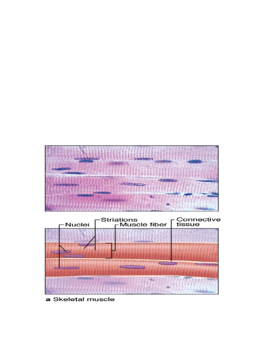
Organization of The connective tissue
The muscle fibers that make up the various types of muscle are arranged in regular
bundles surrounded by C.T. arranged around and in between muscles fibers and
bundles. C.T. carries blood vessels, lymphatic, and nerve fiber to muscle.
The connective tissues are arranged as:-
1- Epimysium: the external sheath of dense C.T. surrounding the entire muscle.
2- Perimysium: its thin septa of C.T. extend inward, surrounding the bundles of
fibers muscle or fascicles within the muscle.
3- Endomysium: each muscle fiber is itself surrounded by more delicate C.T.
Organization of skeletal muscle
4
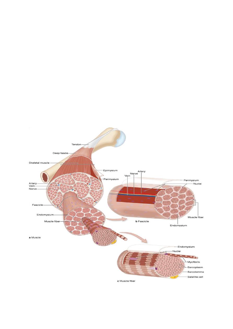
Skeletal muscle fibers
- The muscle fiber is long and cylindrical in shape. The nuclei are multiple, long and
peripheral. The cytoplasm (sarcoplasm) show alternative dark and light striations.
- The dark band is called anisotropic or A- band.
- The light band is called isotropic or I - band.
- In the middle of the dark A band there is a pale region called H-zone.
- Bisecting the H zone is the M line, a region where lateral connections are made
between adjacent thick filaments.
- In the middle of the light band there is a dark line called Z- line.
- The distance between two successive Z lines is called Sarcomere which is the
functional unit for muscle contraction.
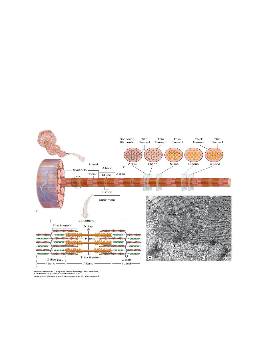
5
Myofibrils:
- The cytoplasm or sarcoplasm is full of parallel myofibrils, which are formed of
two types of fine myofilaments:
* Thick myosin filaments in the A-bands the central portion of sarcomere.
* Thin actin filaments in the both I & A bands.
- The arrangements of actin and myosin filaments give the myofibrils their striation.
- Changes in the amount of overlap between Thick and Thin filaments allows for
contraction and relaxation of muscle fibers.
Structure of Myofibril
6
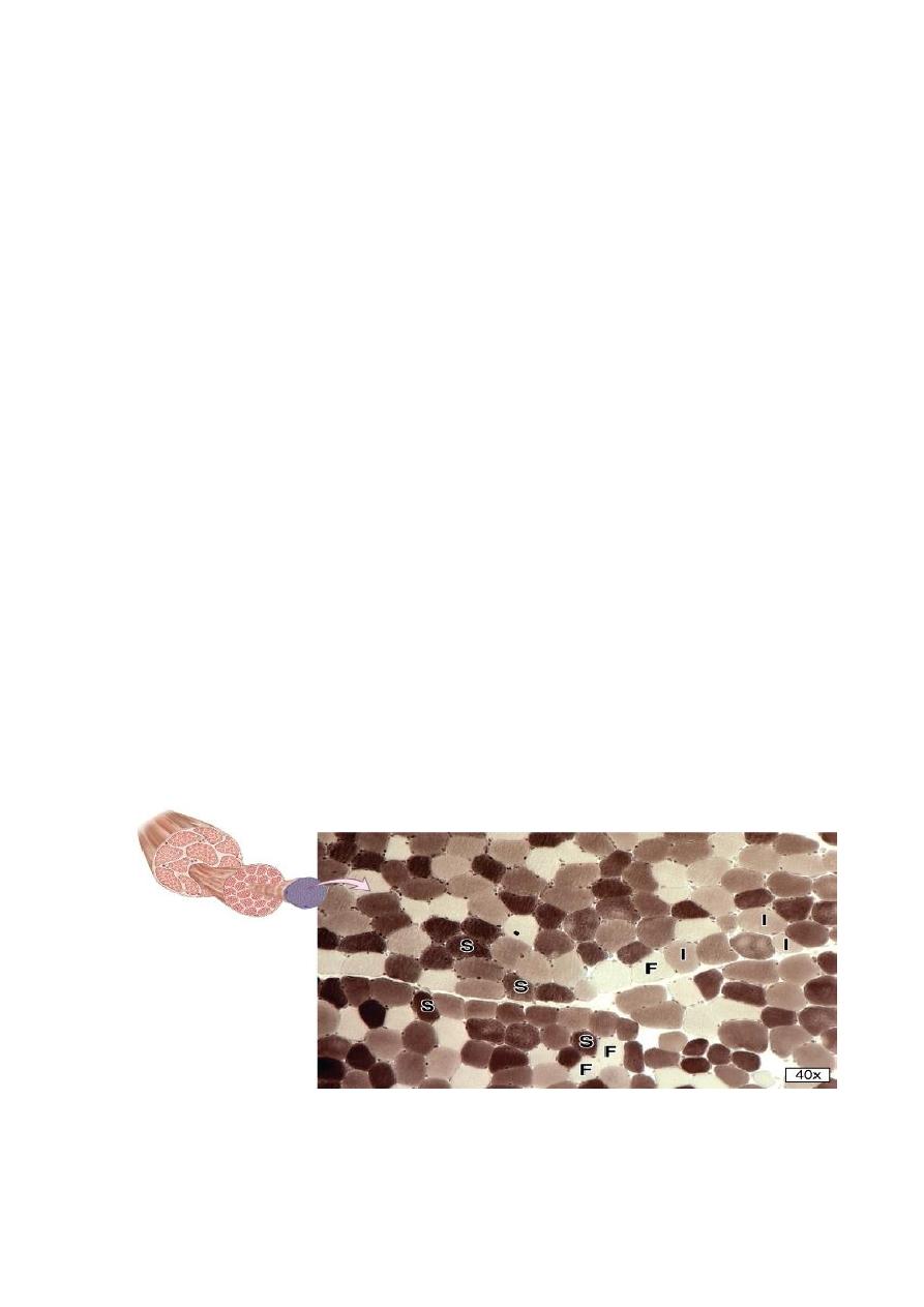
Types of skeletal muscle fibers
There are three types of skeletal muscle fibers differ in myoglobin content, number
of mitochondria, and speed of contraction.
1- Red
muscle
fibers:
- Contain many mitochondria and abundant myoglobin ( a protein with iron groups
that bind O
2
and produce a dark red color ).
- Red fibers are slow and continuous contraction over prolonged periods, as required
for example in the postural muscles of the back.
2- Intermediate fibers:
- Contain many mitochondria and much myoglobin and have considerable glycogen.
- They are intermediate between the other fiber types both in color and in energy
metabolism.
- They are adapted for rapid contractions and short bursts of
activity, such as those
required for athletics.
3- White fibers
- Contain fewer mitochondria and less myoglobin, but abundant glycogen, making
them very pale in color.
- They are adapted for rapid contractions
but cannot sustain contraction for long
periods
such as the muscles that move the eyes and digits.
Skeletal muscle fiber types
7
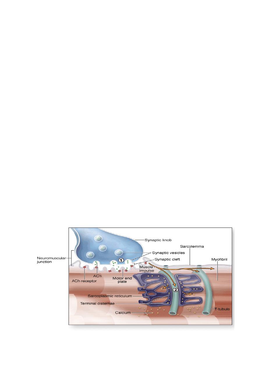
Sarcoplasmic Reticulum & Transverse Tubule ( T Tubule )
Sarcoplasmic reticulum is Smooth endoplasmic reticulum ( SER ) specialized to
sequester calcium ions. The sarcoplasmic reticulum specifically regulates calcium
flow, which is necessary for rapid contraction which depends on the availability of
Ca
2+
ions and relaxation which is related to an absence of Ca
2+
.
It consists of an anastomosing complex of membrane-limited tubules and cisternae
that ensheathe each myofibril.
Skeletal muscle fibers have a system of Transverse (T) tubules these fingerlike
invagination of the sarcolemma penetrates the muscle fiber and comes to lie close to
the surface of the myofibrils near the A-I band boundaries of each sarcomere.
On each side of the T tubule, lies an expansion of the sarcoplasmic reticulum
termed a terminal cisternae. A complex of 2 terminal cisternae and an intervening
T tubule constitutes a Triad, triads are important in initiating muscle contraction.
Contraction is the result of an increase in the amount of overlap between the
filaments caused by the sliding of thin and thick filaments past one another; this
means that during contraction, neither the thick nor thin filaments changes their
length.
Contraction is induced by an action potential produced at a synapse, the
neuromuscular junction, between the muscle fiber and a terminus of a motor axon.
8
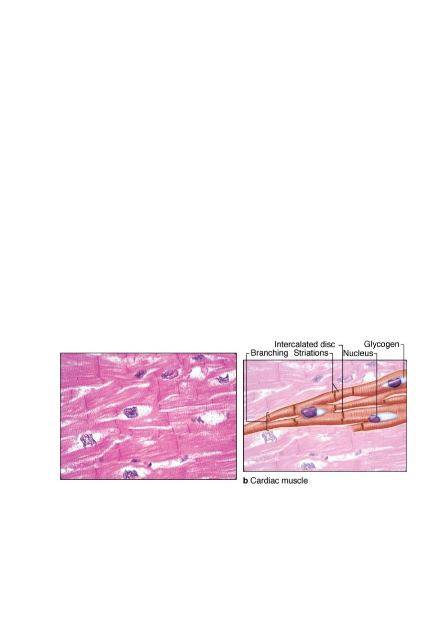
Cardiac Muscle
- Cardiac muscle is striated involuntary muscle, forms the majority of heart tissue.
- The sarcoplasm contains single, oval, prominent and central nucleus.
- The sarcoplasm near the nuclei contains many mitochondria, glycogen granules
and some lipofuscin pigment.
- The muscle fibers are branched but shorter than skeletal muscle fibers. Joined
together by intercalated discs.
- Intercalated disks they appear as dark transverse lines between the muscle fibers
and represent specialized junctional complexes. Intercalated disks prevent
detachment of the cardiac muscle fibers from one another during contraction and
provide electrotonic coupling between adjacent cardiac muscle fibers and pass the
stimulus for contraction from cell to cell.
- The structure and function of the contractile proteins in cardiac cells are essentially
the same as in skeletal muscle.
- Sarcoplasmic reticnlum and T tubule system are irregularly arranged in cardiac
muscle fibers.
Cardiac Muscle
9
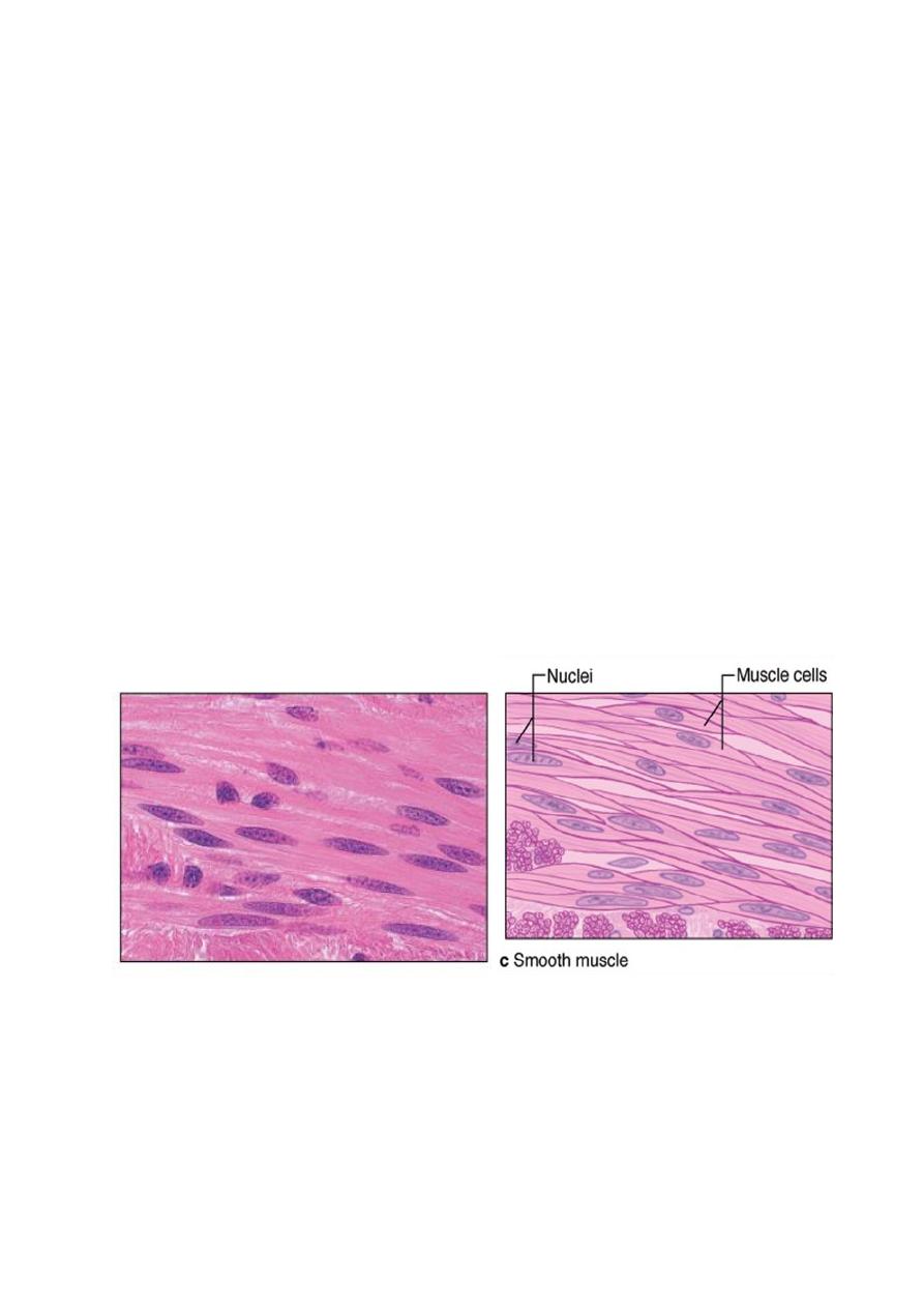
Smooth Muscle
- Smooth muscle is an involuntary, controlled directly by the autonomic nervous
system; it is smooth because there is no striation
- Smooth muscle fibers are elongated, spindle shaped cells, and nonstriated cells
with a single central nucleus, the narrow part of one cell lie adjacent to the broad
parts of neighboring cells.
- Concentrated near the nucleus are mitochondria, polyribosomes, cisternae of
rough ER, and the Golgi apparatus.
- Different internal organization of actin and myosin filament. The myosin filaments
of smooth muscle are less stable and less regular than those in striated muscle cells
and thin filaments attached to dense bodies, which are transmit contractions from
cell to cell.
- Smooth muscle is found within the walls of blood vessels. It is also found in
lymphatic vessels, bladder, uterus, male and female reproductive tracts,
gastrointestinal tract, respiratory tract and iris of the eye.
- A poorly organized sarcoplasmic reticulum is present in smooth muscle cells, but
T tubules are not present.
Smooth Muscle