Department of Surgery & Obstetrics
College of Veterinary MedicineUniversity of Mosul

Haematoma
Department of Surgery & ObstetricsCollege of Veterinary Medicine
University of Mosul
Haematoma:
An accumulation of free blood anywhere in the body, that has partially clotted to form a semi-solid mass. Haematomas may be caused by injury or may occur spontaneously as a result of a bleeding or clotting disorder. In some sites, as within the skull, enlarging haematomas may be very dangerous. Infected haematomas may form abscessesOR:
Accumulation of blood under subcutaneous tissue and skin due to rupture of blood vessels
Department of Surgery & Obstetrics
College of Veterinary Medicine
University of Mosul
On manipulation it is found
SymptomsIt appear suddenly
Characterized by the formation of a uniformly fluctuating enlargement.
The liquid dose not completely fill the space
It can displaced from one part to other by manipulation,
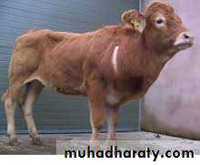
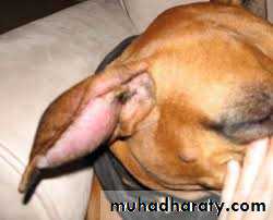
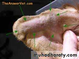
Common site of lesion in the thigh, buttocks or forearm in large animals, in the dog ear hematoma
Department of Surgery & Obstetrics
College of Veterinary MedicineUniversity of Mosul
Treatment:
Cold application and astringent application to arrest hemorrhage
2- Not open before 3 Week
3- Excision and remove after organized hematoma, put draine
Department of Surgery & Obstetrics
College of Veterinary MedicineUniversity of Mosul
Treatment:
Cold application and astringent application to arrest hemorrhage2- Not open before 3 Week
3- Excision and remove after organized hematoma, put draine
Hematoma occurs because the wall of a blood vessel wall, artery, vein or capillary, has been damaged and blood has leaked into tissues where it does not belong. The hematoma may be tiny, with just a dot of blood or it can be large and cause significant swelling be inflammatory or fluctuating and crepitating due to presence of liquid and coagulated blood.
Minor injuries occur routinely and the body is usually able to repair the damaged vessel wall by activating the blood clotting cascade and forming fibrin patches. Sometimes the repair fails if the damage is extensive and the large defect allows for continued bleeding. As well, if there is great pressure within the blood vessel, for example a major artery, the blood will continue to leak and the hematoma
• Types of haematomas:
Hematomas are often described based upon their location.1. Cranial haematoma: Haematomas can be formed inside the cranium or brain following to the damage to the blood vessels due to trauma or increased blood pressure. They can be
• Epidural hematomas: between the skull and duramater
• Subdural hematomas : between the duramater and arachnoid mater
• Intra cerebral hematomas This type of haematoma is very difficult to manage in case of farm animals as involves more cost and is complex in nature..
2. Orthopaedic haematoma: Injuries and fractures are often associated with hematoma formation in and around bones. Bones are very vascular structures since the marrow is where blood cells are made. Fractures are always associated with hematomas at the fracture site. Fractures of long bones such as the thigh (femur) and upper arm (humerus) can be associated with a significant amount of bleeding, sometimes up to one unit of blood or 10% of the body's blood supply.
This is the haematoma in the pelvic cavity. This results from the tearing of the blood vessels during forceful parturition or any injury to the pelvic cavity. This is common in the high producing big breeds of cattle and buffalo.
4. Intramuscular haematomas
Intramuscular hematomas formed between the muscular layers. Some muscles are surrounded by tough bands of tissues. If enough bleeding occurs, the pressure within these compartments can increase to the point that a‘ compartment syndrome‘ can occur. In this situation, the blood supply of the muscle is compromised and the muscle and other structures such as nerves can be permanently damaged. This is most commonly seen in the lower leg and forearm.5. Subcutaneous haematomas.
These occur due to trauma or injuries to the superficial blood vessels under the skin characterized by formation of haematomas in the subcutaneous layer.
3. Pelvic haematoma
7. Aural or ear haematomas.This is most common type of haematoma occurring in the animals. This occurs if an injury causes bleeding to the outside helix or cartilage structure of the ear. Often called boxer's, wrestler's ear, or cauliflower ear, blood gets trapped between the thin layer of skin and the cartilage itself. Since the ear cartilage gets its blood supply directly from the overlying skin, a hematoma can decrease blood flow causing parts of the cartilage to shrivel and die. Hematomas within the ear flaps (“aural haematomas”) occur when head shaking breaks a blood vessel within the ear flap.
6. Intra-abdominal haematomas
Regardless of how the blood gets into the abdomen, Haematomas may occur in solid organs such as the liver, spleen,or kidney.They may occur within the walls of the bowel, including the small Intestine (duodenum, jejunum, ileum) or the large intestine(colon). Haematomas may also form within the lining of the abdomen called the peritoneumor behind the peritoneum in the retroperitoneal space (retro=behind).The ear flap may partially or completely swell with blood. The swelling maybe so large and painful that the opening of the ear canal is occluded. The extra weight of the ear flap may be uncomfortable and may lead to a permanent change in the carriage of the ears. This conditions more common in dogs but can occur cats as well. The ear flap will feel fluctuant and fluid-filled, like a water balloon
Department of Surgery & Obstetrics
College of Veterinary MedicineUniversity of Mosul
• OTHER COMMON HAEMATOMAS OCCURRING IN ANIMALS Cattle:
• Teat/ Udder haematoma: caused by rubbing against hard ground, biting of calf during suckling.
• Mammary Vein Haematoma: Cattle can sustain huge hematomas cranial to the udder, probably resulting from trauma to the subcutaneous abdominal vein. Possible mechanisms that cause the trauma include lacerations, getting up and down, or fighting other cattle.
Department of Surgery & Obstetrics
College of Veterinary MedicineUniversity of Mosul
• Vaginal haematoma:
Fetus passage may damage the internal pudendal artery, resulting in formation of a large hematoma lateral to the vaginal wall. In rare cases, this condition may be bilateral. In most instances, these haematomata resolve spontaneously; sometimes they become infected and persist as abscesses. Haematomata of the vagina may protrude from the vulva. Hematoma of the vulva is usually obvious. Both may be readily drained after allowing 3 days for haemostasis.• Penile haematoma: A hematoma results from sudden or forceful bending of the erect penis. During the peak of erection, blood pressure within the corpus cavernosum penis rises to astronomical levels. Deviation of the penis at this point (by sudden movement of the cow or by thrusting of the bull against the thigh of the cow before intromissions achieved, results in rupture of the tunica albuginea and hemorrhage. The hematoma may be exacerbated by repeated mating attempts by the bull. The site of the hematoma is usually distal to the distal curve of the sigmoid flexure ..
Department of Surgery & Obstetrics
College of Veterinary MedicineUniversity of Mosul
• Horse:
Shoulder haematoma: injury to the spur vein or evternal thoracic vein by the rider is common cause of haematoma in horse• Dog/ Cat:
Aural haematoma: Haematoma of the ear flap. It is very common condition in dogs which have got very long pendulous ears. Blood and serum accumulate in between conchal cartilage and the skin of the skin of the ear. It results from prior contusion.Dogs, cats and pigs suffer.
Vaginal mucus membrane haematoma: Caused due to injury during copulation, common in dogs but is seen in all species. Testicular haematoma: improper ligation of the arteries during castration of dog or pic and improper surgical technique without proper haemostatic technique may result in the extravasion of the blood into the testicular cavity causing large haematomas.Department of Surgery & Obstetrics
College of Veterinary MedicineUniversity of Mosul
ETIOLOGY
1-Trauma It is the most common cause of haematoma in animals. Trauma during various management operations and the animal itself causes the rupture of the delicate blood vessels giving rise to the extravasion of blood when a blood vessel is damaged blood leaks into the surrounding tissue; this blood tends to coagulate or clot. The greater the amount of bleeding that occurs, the larger the amount of clot formation.
• Trauma includes
- Rubbing against hard objects during sitting or accidentally causing udder haematoma in dairy cattle.• Injury to teat during suckling milk by the claves also result in teat haematoma.
• Deviation of the penis at the time of peak erection (by sudden movement of the cow or by thrusting of the bull against the thigh of the cow before intromission is achieved, results in rupture of the tunica albuginea and hemorrhage resulting the haematoma formation.
- Damage to the internal pudendal artery during foetal passage causes haematoma of the vagina. The trauma during copulation also results in vaginal haematoma.).
-Pets that paw at their ears or shake their heads vigorously, especially those with large ears, can cause a hematoma. This pawing and shaking can be due to irritants around the face and ears, or irritants in the external ear canals like infections or foreign bodies(foxtails
• Damage to the spur vein or external thoracic vein in case of horses by horse riders.
Department of Surgery & ObstetricsCollege of Veterinary Medicine
University of Mosul
2. Itching produced due to allergic response may cause severe rubbing and
scratching of the ears resulting to haematoma.3. Ear mites Otodectes cynoticsis one of the very important cause of haematomas
in dog. S. Scabieiandpediculosis (lice) may cause haematomas in pigs.4. Autoimmune disease Sometimes autoimmune disease of the pinnal tissue has found to cause haematomas of the
Department of Surgery & Obstetrics
College of Veterinary MedicineUniversity of Mosul
• CLINICAL SIGNS:
In case of aural haematoma:• Repeated shaking of the head,
• rubbing of ears and pain of the ear.
• Ear may appear as ovoid or round tendered, swelling, fluctuating on pressure.
• Swelling is either in the inner or outer surface of the ear.
• The ear flap may partially or completely swell with blood. The swelling may be so large and Painful that the opening of the ear canal is occluded
• The head is carried at an angle keeping the affected side lowermost.
• Discolouration of the skin, reddish to bluish red.
• Hematomas of the bladder wall (cystic hematoma) cause hematuria in neonatal foals
Department of Surgery & Obstetrics
College of Veterinary Medicine
University of Mosul
CLINICAL SIGNS:
• In case of penile haematoma, when the initial haematoma is over 15 cm in diameter, more extensive damage to the penile adnexa (telescoping fascia) results, making restrictive adhesion formation more likely.• Haematomata of the vagina may protrude from the vulva as bluish coloured sac like structure. Hematoma of the vulva is usually obvious.
• Secondary bacterial complications may lead the condition more painful and there maybe accumulation of pus.
• Swollen and oedematous testis with fluctuates uniformly on palpation with doughy consistency
• Puncture with needle (aspiration) will reveal oozing of blood and serum from the swelled part
Department of Surgery & Obstetrics
College of Veterinary MedicineUniversity of Mosul
TREATMENT
The treatment of haematoma can be grouped into two categories:1.Medical treatment
2.Surgical treatmentDepartment of Surgery & Obstetrics
College of Veterinary MedicineUniversity of Mosul
Medical treatment:
Haematomas should not be insised as far as possible. Time should be given ( 2-3 wks) for resorption. If no resolution of haematoma occurs, then only incision is indicated.
This is also called conservative therapy where bya needle is inserted to drain out the fluid called aspiration and the area is bandaged tightly (esp inaural haematoma) to prevent the fluid from forming again.