Introduction to
Lamenessr
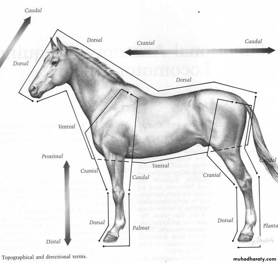
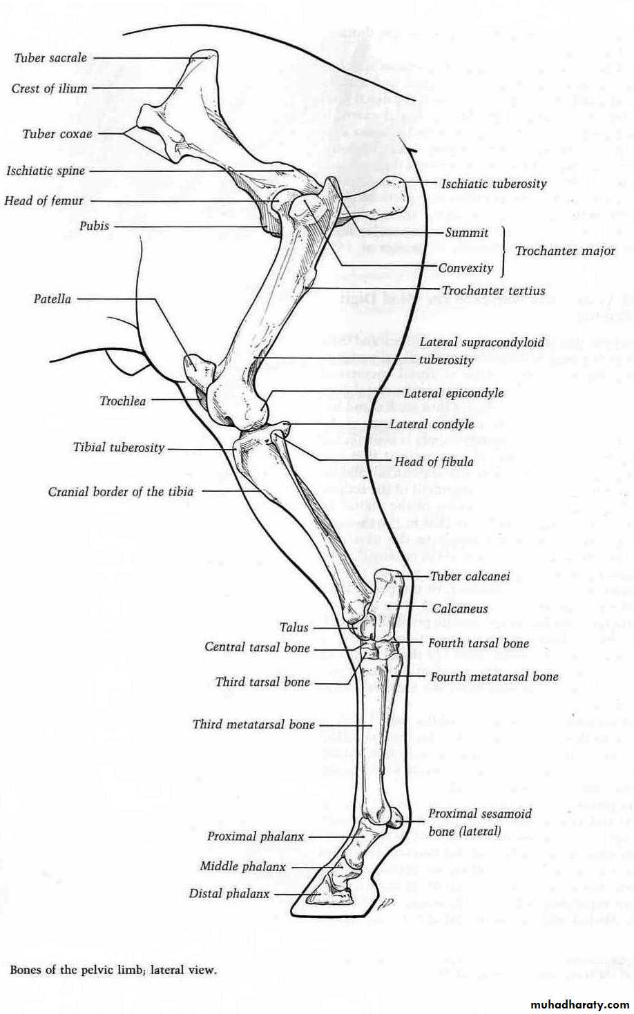
Center Gravity:-
Where the mass of the horse is centered?It will vary with horses shape. It is most commonly located in the middle of the rib cage just caudal to the line separating the cranial and middle third of the body.
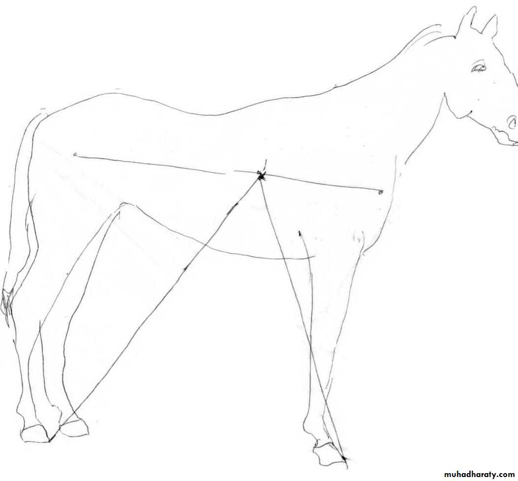
Middle of rib cage
Because the center of gravity is located more cranially, the forelimbs bear 60-65% of the body weight. This is causes an increased stress in the forelimbs resulting in an increased incidence of lameness in these limbs.
Lameness in Horses
Definition:- It is an indication of a structural or functional disorder in one or more limbs that is manifested during progression or in the standing position.Lameness can be caused by:-
Trauma
Congenital or aquired anomalies
Infection
Metabolic disorder
Circulatory and nervous disorders, or any combinations of these.
The diagnosis of lameness requires a detailed knowledge of anatomy and physiology of limbs movement.
Classification of lameness
1- Supporting limb lameness:- This is evidenced or appeared when the horse supporting weight on the foot or when the horse lands on it. Causes mostly due to bones injury or injury to joints collateral ligaments or motor nerves.2- Swinging lameness:- This is evident when the limb is in motion.
Pathologic changes involving joint capsules, muscles, tendons, tendon sheaths or bursas are considered to be the cause.3- Mixed lameness:- This is evident both when the limb is moving and when it is supporting weight. Causes can involve any combination of the structures affected in swinging or in supporting limb lameness.
4-Complementary lameness:- Pain in the limb will cause uneven distribution of weight on another limb or limbs which can produce lameness in a previously affected limb or opposite limb.
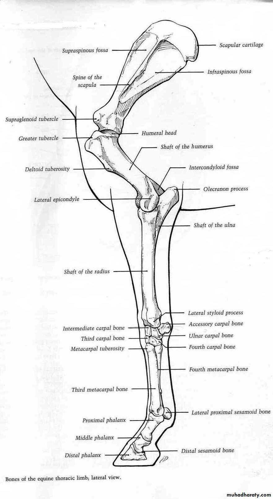
Faults in conformation of the fore limb
Base narrow:- In base narrow conformation , the distance between the center lines of the feet at their placement on the ground is less than the distance between the center line of the limbs at their origin in the chest when viewed from the front. This is found most often in horses having large chest and well developed pectoral muscles, such as quarter horses.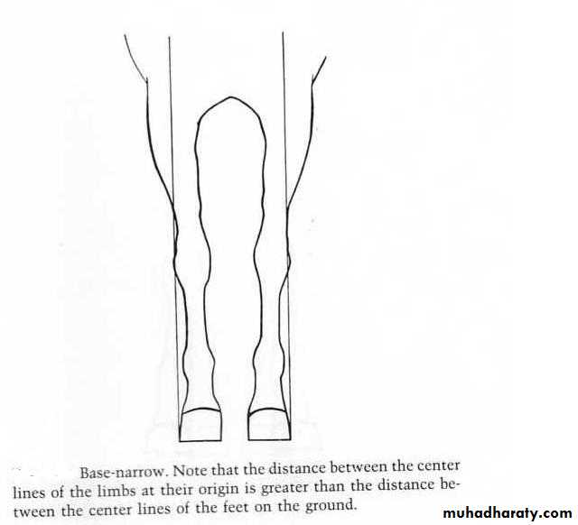
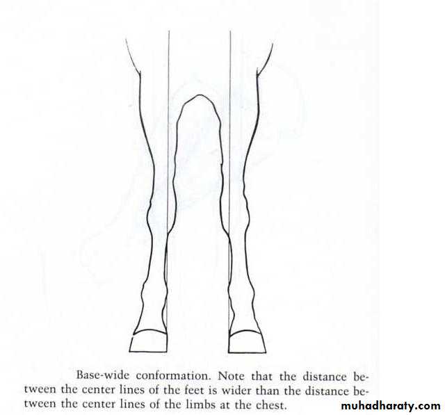
Base wide:- The distance between the center lines of the feet is wider than the distance between the center lines of the limbs at the chest.
Toe-in or pigeon –toed:- Toe-in is a position of the feet in which the toes point toward one another when viewed from the front. It is congenital and usually accompanied by a base-narrow and rarely with base-wide.
Toe-out or splay footed:- When viewed from the front , the toes point a way from one another. The condition is usually a congenital and may be accompanied by either base-wide or base narrow conformation.
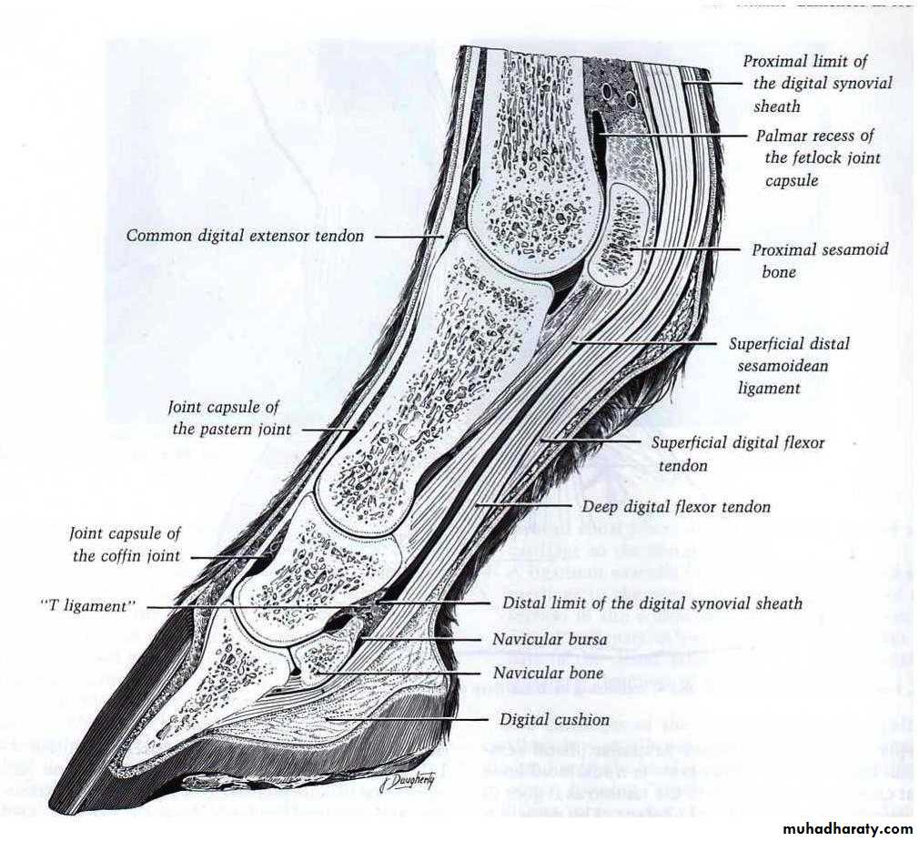
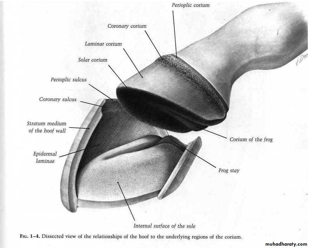
Foot (Hoof)
The foot consists of the epidermal hoof and all encloses, and the connective tissue corium which is derived from the dermis of the coronary band.The hoof includes the following tissues:-
digital cushion
distal phalanx(coffin bone)
most of the collateral cartilage of the 3rd phalanx
coffin joint
distal extremity of the 2nd phalanx
distal sesamoid (navicular bone) and navicular bursa
several ligaments, tendons of insertion of the common digital extensor and deep digital flexor muscles
blood vessels and nerves
The hoof extends from the ground proximad to the coronary border, where the priople joins the epidermis of the skin with the hoof at the coronet.
The layers of hoof wall
Stratum tectorium:- which is a thin horny layer extending from the priople.Stratum medium:- which form the bulk of wall which consisting mainly of horn tubules and interlobular horn.
Stratum internum(stratum lamellatumStr):- containing about 600 primary epidermal laminae(lamellae) and approximately 100 microscopic secondary laminae branch at an each primary lamina.
Nutrition of the hoof wall is from the coria:-
prioplic coriumcoronary corium
corium of the wall
corium of the sole
corium of the frog
Affections of the hoof
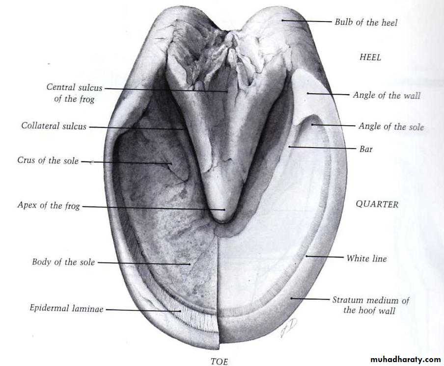
Corns and Bruised sole:-A corn is an involvement of the sensitive and insensitive tissues of the sole at the angle formed by the bar. Corns occur most frequently on the inner angle of the front feet are rarely found in the hind feet. This is due to the fact that the front feet beer more weight than the hind feet.
Etiology:-
Corns are usually due to improper shoeing.
When shoes are left on the feet too long , the heels of the shoe are forced inside the wall and cause pressure on the sole at the angle of the wall and bar.
Improper trimming of the feet making the heels too low , increases pressure at the angle of the wall.
A long, weak fetlock and a narrow foot may cause corns to appear at the bars, while in a wide foot corns are more likely to occur in the sole.
Trauma to the sole from rocks and other objects.
Horses have been affected previously with laminitis are more susceptible to this disease.
3 types of corn lesions may be evident:-
Dry corn: - In this case hemorrhage within the inner surface of the horn resulting from bruising of sensitive tissue usually causes red stains.Moist corn: - This is caused by severe injury that results in serum accumulation beneath injured horn.
Suppurating corn: - The corn becomes infected resulting in necrosis of the sensitive laminae.
Pathologic changes due to bruised sole are similar to those caused by corns but occur in toe or quarter regions of the sole rather than at the angle of the wall or bar.
Bruised sole also may be of dry, moist or suppurating type.
Clinical sings:-
The horse will show varying degrees of lameness depending upon the severity of the bruise or corn, while the attitude of the lameness will vary according to the location of the bruise or corn.Hoof tester will reveal the location of the pathologic changes.
A cleaning of the sole from the bottom of the foot with a hoof knife will reveal red stains in the sole indicating a bruised region.
In some cases this region may show a bluish discoloration, especially if a sole abscess is developing.
If the corn is present at the inside heel, the horse will tend to place more weight on the outside of the foot because of the pain.
6-In some cases, the horse will tend to bear most of the weight on the toe and will rest the foot with the knee forward to decrease heel pressure.
Treatment:-
In cases in which shoeing is the cause, removal of the shoe may be all that is necessary.To prevent shoes from causing corns the heels of the shoe should always extend well back and should fit full on the wall at the quarters and heels.
Removal of some of the tissue over the corn helps relieve pressure, but sensitive tissue should not be exposed.
The horse should be rested and should not reshod until symptoms disappear.
In case of suppurating corn the sole over the region should be removed until drainage of the sensitive tissues is established.
The foot should be soaked daily in an antiseptic or in solution of mag. Sulph. , after which Tr. Of iodine is applied.
The foot should be bandaged and protected from contamination.
Prognosis:-
Is always guarded, since some cases tend to become chronic which finally cause osteitis of the distal phalanx.Canker:-
It is a chronic hypertrophy of the horn producing tissues of the foot, which may involve any one or all of the feet, it is most often found in the hind feet, and it is a rare condition.
Etiology:-
The chief etiologic agent is related to unhygienic stabling, so it develops in horses that stand in mud or in bedding that is soaked with urine and feces.It also appear in horses whose feet do not receive regular attention.
3-The specific cause is thought to be an infectious process agent but it is unknown.
Clinical sings:-
Lameness usually is not present in early stages of the disease and may not be detected until well advanced.The foot usually has a fetid odor and the horn tissue of the frog looses easily and when removed, reveals a foul smelling, swollen corium covered with a caseous white exudates.
The disease may extend to the sole or even to the wall of the foot.
Diagnosis:-
It can be made by the appearance of the foot and by the offensive odor.Treatment:-
All loose horn and affected tissues should be removed and an antiseptic, astringent dressing applied like 5% of picric acid should be applied under bandage.
Caustic agent, such as a mixture of copper sulphate and zinc sulphate crystals are sometimes used.
Canker sometimes treated successfully with penicilline used at a rate of 3 million units per day until improvement was shown.
Prognosis:- It is guarded to unfavorable
Thrush:-A degenerative condition of the frog involving the central and lateral sulci, is characterized by the presence of a black necrotic material in the affected areas. The infection may penetrate the horny tissues and involve the sensitive structures.
Etiology:-
The predisposing causes of thrush are unhygienic conditions, dirty uncleaned feet.Lack of frog pressure resulting from poor shoeing or poor foot trimming.
Many organisms are probably involved, but spherophorous necrophorous appear to be the most important one.
Clinical sings:-
There is an increased amount of moisture and a black discharge in the sulci of the frog. This discharge which varies in the quantity has a very offensive odor.When the affected sulci are cleaned, it will be found that they are deeper than normal and may extend into the sensitive tissues of the foot, causing the horse to flinch when they are cleaned.
In severe cases that have penetrated into the sensitive structures of the foot, the horse may be lame.
Generally, the hind limbs are more frequently affected.
Treatment:-
Cleanliness, removal of the cause and return the frog and hoof to normal conformation and condition.
The foot should be cleaned daily and the cleft of the frog packed with a proper medication such as equal parts of phenol and iodine, Tr. Of iodine and 10% formalin.
Another treatment consists of packing the sulci with cotton soaked in 10-15% sodium sulphapyridine solution.
Degenerated frog tissue should be removed.
Prognosis:-
It is favorable if the disease diagnosed early, and it is guarded if the sensitive structures are involved.Hoof wall cracks(sand cracks):-
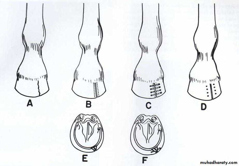
These are cracks in the wall of the hoof, starting at the bearing surface of the wall and extending to a variable distance up the hoof wall, or cracks originating at the coronary band, as a result of a defect in the band and extending downward. These cracks can located on the toe(toe crack) or on the quarter(quarter crack), or on the heel(heel crack), and may occur in either the front or hind feet. Quarter cracks and heel cracks are usually the most severe because they often involve the sensitive laminae.
Etiology:-
Excessive growth of the hoof wall.Ingury to the coronary band producing a weak and deformed hoof wall, will lead to cracks originating at the coronary band.
Weakening of the hoof wall due to excessive drying or excessively thin walls also causes hoof cracks.
Clinical sings:-
1-Lameness may not be present, but it will become evident if the crack extends into the sensitive tissues allowing infection to gain access to these structures.2-An exudates under the cracks or suppurative inflammation of the laminae may be present depending upon the size of the opening into the sensitive tissues.
3- Traumatic lesions found above the coronary band when the crack is due to injury of the coronary band.
Diagnosis:-
Presence of the crack or cracks, which is easily identified and is classified according to its location
Hoof tester can be used to identify if there is a pain associated with the crack in the hoof wall.
Bleeding from the hoof wall crack after exercise indicates that the crack has extended down to the sensitive laminae.
Pus will exude from the infected hoof when pressure is applied.
Treatment:-
It depends on the location of the crack.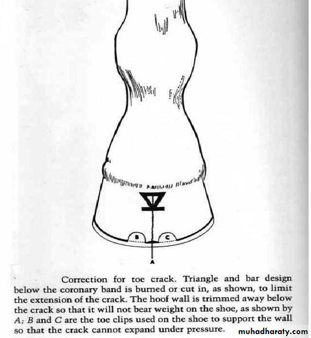
Toe crack
The hoof wall on the bearing surface should be lowered about 1 inch on either side of the crack, this will aids in preventing expansion of the crack.If the crack does not extend into the coronary band, a pattern of a triangle and bar design is grooved or burned below the coronary band, this will limit the crack to progress upward.
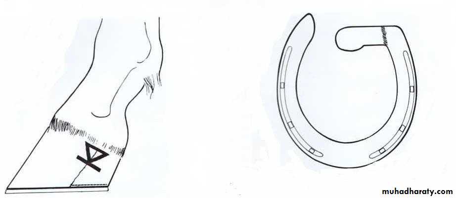
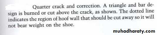
Quarter crack
The bearing surface of the hoof wall should be lowered in back of the crack.A half bar shoe should be applied, with the bar on the heel of the affected side and this bar should press against the frog, so it will make frog to bear the weight that normally would be taken by the hoof wall.
The same pattern of a triangle and bar design is applied as in toe crack treatment.
Ground surface view of a half – bar shoe used in treating quarter crack and heel cracks.
N.B.
Another treatment for the wall cracks is by drilling in the sides of the crack to form many holes on either side of the crack, then thread the holes by umbilical cotton tape or stainless steel wire.Any wall cracks can be treated by use of special materials to fill the crack like Epoxy glues, Fiber glass or special hoof- repair material in combination with the first treatment.