Puncture wounds of the white line
(Gravel)It is happened as an opening in the white line that permits infection to invade the sensitive structures. Because there is no drainage, inflammation follows the line of least resistance and drainage occurs at the coronary band.
Etiology:-
A puncture wound or crack in the white line may occur in feet that are too dry.Chronic laminitis with its associated seedy toe.
Clinical sings:--
Lameness will usually appear before drainage at the coronet band occurs.Careful examination of the white line and sole will reveal black spots, which should be propped to there depth, where sensitive laminae will found.
After crack is propped pus will often exude from the wound.
When the condition has been present for sometime drainage at the coronary band will be noted.
Systemic reaction to the infection varies, but in most cases infection remain localized.
Diagnosis:-
Careful examination of the foot with hoof tester.Most cases can be diagnosed before coronary band region breaks and drains.
Careful observation of the way the horse sets his foot on the ground, this will help in localization the region of penetration.
Treatment:-
Establishing proper drainage for the infection.
The foot may be soaked in Mag . sulphate salts.
Applying iodine to drainage area and bandaging the foot until healing.
Seedy toe:-
This condition is interfere with Gravel in the clinical sings, diagnosis and treatment.It is characterized by separation of the sensitive and insensitive laminae at the toe region of the sole. Enough separation of the white line may occur to allow infection to penetrate the sensitive laminae.
Etiology:-
The condition is usually present in chronic laminitis.Quittor(Necrosis of the collateral cartilage):-
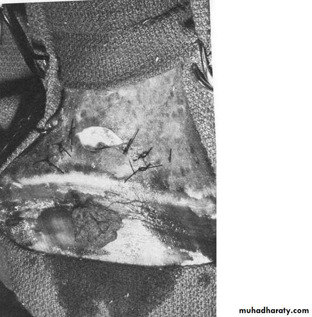
It is a chronic purulent inflammation of a collateral cartilage of the distal phalanx characterized by necrosis of the cartilage and sinus drainage at or above the coronary band. It is most common in fore limb.Etiology:-
Injury near the coronary band over the region of the collateral cartilages that producing sub coronary abscess and finally quitter.Penetrating wound through the sole, where infection is transmitted to the collateral cartilage.
Wire cuts or bruises that damage the cartilage and reduce circulation in the region.
Clinical sings:-
1-The condition may occur over either the medial or lateral collateral cartilage.
2- Swelling , heat and pain over the coronary band.3-Chronic suppurative sinus tracts that tend to heal and then break open at intervals is characteristic sign of quitter
4- Lameness occurs in acute stages that may show remission when the lesion appear to be .
healing.
5- Permanent damage and deformity of the foot may result causing persistent lameness.
Diagnosis:-
Depending on the clinical sings.Using probe to differentiate it from shallow abscess.
Radiographs can be helpful to show the involvement of the middle and distal phalanx.
Treatment:-
Medical treatment such as injection of caustics e.g. silver nitrate 20% or using of enzymes but it appear of lest effect.The treatment of choice is surgical excision of the necrotic cartilage. The surgical technique as follows:-
The region is clipped, the hoof is trimmed and rasped, scrubbed and placed in a povidone iodine soaked bandage for 24 hr.
Tourniquet is applied.
An elliptical or curved incision beginning just dorsal to the coronary band over the diseased collateral cartilage.
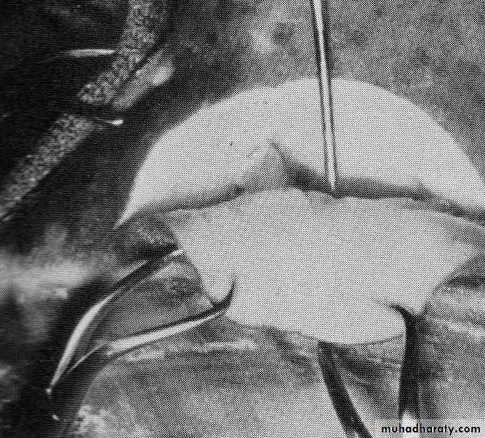
The flap is dissected distally to expose the collateral cartilage and a sterile probe is induced to identify the drainage tract.
Necrotic cartilage is recognized by its dark blue or reddish blue appearance.
All the necrotic tissue and cartilage is excised.
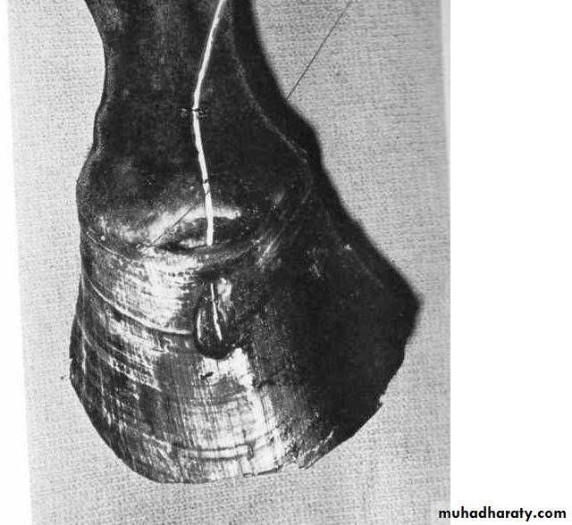
If necrotic cartilage extends below the coronary band a hole is drilled in the hoof wall over the ventral most limits of the necrotic cartilage to provide drainage.
A polyethylene tube is placed in the wound and is sutured to the limb proximally.
The remaining tract is then packed with an antiseptic soaked sponge, and leaving an opening at the top of skin flap.
The foot and sole can either be bandaged or placed in a protective boot.
One day following the operation the bandage is removed and the wound flushed with a 1% povidone iodine solution. This will repeated once daily until infection is gone.
After the tract has been debrided the skin is sutured leaving a small opening at the top. The tract is packed with gauze soaked in antiseptic solution.
A curved incision is made just proximal to the coronary band. A sterile probe was used to identify the tract.
A hole was made in the dorsal surface of the hoof wall to remove the involved tissue and provide good drainage.
Prognosis:-
For acute and sub acute cases is good with the treatment describedSide Bone:-
An ossification of the collateral cartilages, are usually found in the fore feet and are most common in horses having poor conformation.
Etiology:-
Concussion of the quarters of the foot causing trauma to cartilages is probably the cause of most cases.Hereditary predisposing factors through poor conformation e.g. horses that are base narrow are susceptible to develop to develop lateral side bone, while horses that are base wide develop medial side bone.
Poor shoeing may cause increased concussion resulting in side bone. And using shoes with long heel calks.
Clinical sings:-
Lameness resulting from side bones is rare, usually being present only when cartilages are in the process of becoming ossified and when inflammation is present.Massive bone formation may cause mechanical interference with foot action.
If side bones are a cause of lameness, there will be heat and pain over one or both of the cartilages.
Hardening of the cartilages can be palpated.
Pressure over the region will cause the horse to flinch if the cartilage in the active stage of bone formation.
In some cases there will be a visible bulging of the quarter at the coronary band.
Diagnosis:-
Radiological examination will reveal bone formation in the cartilage.Palpation of the cartilage.
Treatment:-
3-4 vertical grooves along the hornified layers (do not penetrate the sensitive laminae) at the quarters of the hoof wall below the coronary band are made, this will permit expansion of the foot and relieves the pain.When fractured side bone cause acute lameness, only the small proximal chips can be removed, but the large pieces should not.
When chronic lameness is present a palmer (posterior) digital neurectomy can be done on the affected side.