Fractures
Definition of fractureIs dissolution of bony continuity with or without displacement of the fragments. (Break down of bone continuity)
Principles of fracture repair
1- The blood supply and to bone and fragments of bone must be preserved.
2- Accurate restoration of bone contour is essential particularly when joints are
involved.
3- The repair must be mechanically stable.
4- Technics that result in a minimal trauma should be used.
5- Controlled exercise should be allowed early.
Blood supply of bone
The vascular supply to a long bone consists of 3 systems:-1- Nutrient blood supply.
The nutrient artery usually enters a long bone through a foramen in its proximal half. It divides in the medullary cavity into ascending and descending branches.
2- Periosteal blood supply.
It has 2 types of vessels
A- Where muscles are attached to the fibrous layer of periosteum, the intramascular and periosteal circulations are closely related and capillaries that penetrate the cortex are continuous with the capillaries of the muscles.
B- Where there is no muscle attachment to the periosteum, vessels from neighboring soft tissues pass to the fibrous layer of the periosteum and form a network of vessels around the bone.
3- Metaphyseal-epiphyseal blood supply.
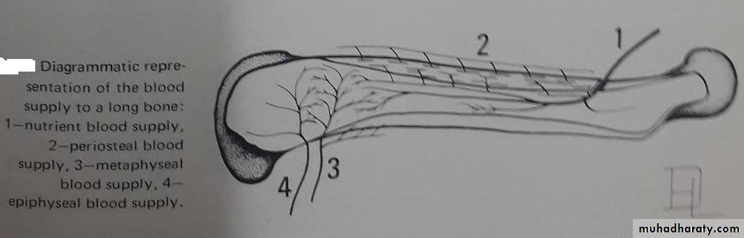
The metaphyseal and epiphyseal vessels are larger than the peiosteal vessels, and either penetrate the bone and anastomose with the nutrient artery or ramify over the bone cortex.
Types of fractures
1- Pathological fractures:- those fractures associated with rickets, malnutrition, or neoplasms.
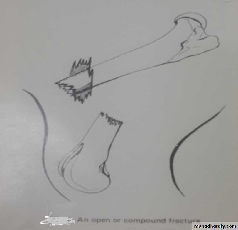
2- open and closed fractures:- A fracture is closed or simple when there is no communication between the fractured bone and the exterior of the body. It is classified as open or compound when there is an opening through the skin and underlying soft tissues leading to the fracture. Open fractures are subject to contamination or infection.
3- Complicated fracture: -
It is fracture associated with injury to nerve (like radial paralysis), artery or vein, opening of a joint, opening of a body cavity (like chest)
Patterns of fractures:-
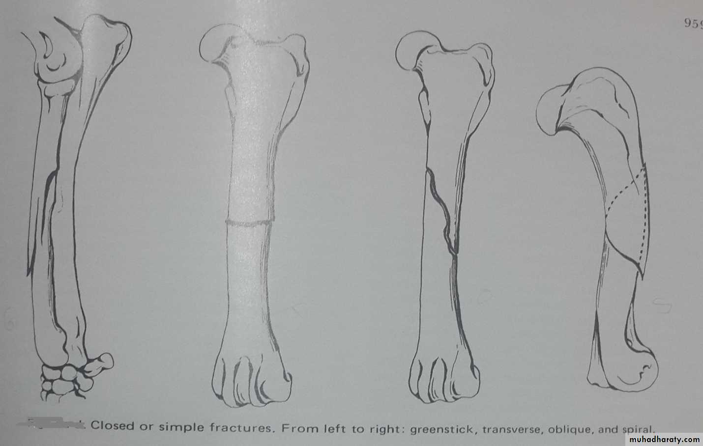
1- Greenstick fracture: with this fracture the bone is partially broken and bent. These fractures occur primarily in young animals whose bones are resilient. Displacement is minimal and healing is rapid.2- Transverse fracture: The bone is broken at a right angle to its long axis. After the fragments are apposed normal length of the bone is usually achieved.
3- Oblique fracture: The line of fracture is diagonal to the axis of the bone. The fragments tend to slip by each other unless they are held firmly together. With good reduction and rigid fixation healing is usually rapid because there ia a large area of contact between the fragments.
4- Spiral fracture: The line of fracture is curved. These fractures usually are caused by torsion. The bone fragments tend to overlap and may rotate unless firmly fixed.
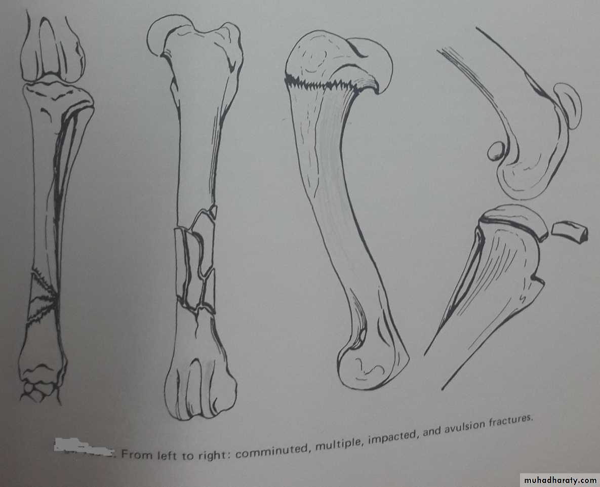
5- Comminuted fracture: Several fracture lines communicate at a common point.
6- Multiple fracture: The bone is splintered into 3 or more pieces and the fracture lines do not meet. These fractures are accompanied by severe injury to soft tissue and blood supply to the bone is impaired. Reduction can be difficult to achieve and maintain and healing can be prolonged.
7- Impacted fracture: The fragments of bone are driven together. Union is rapid but slight shortening and angulation may occur.
8- Avulsion fracture: A bone fragment usually at site of muscular attachment can be detached by forcible muscular contraction. When reduced promptly and fixed firmly union is rapid and complete function is regained.
Fracture healing:-
Prerequisites of bone healing demands:1- Good blood supply.
2- Accurate positioning of the fragments.
3- Adequate immobilization.
4- Early treatment.
The fracture healing involves the growth of new tissue around the fracture site. This tissue or known as callus forms a bridge that unites the fragments. Callus is best considered as nature’s way of stabilizing a fracture. The more unstable fracture the greater is callus formation, in rigid stabilization callus formation is minimal.
External callus is that which develops around the ends of the bone fragments. Internal callus forms in the medullary cavity and between the ends of the bone.
Stages of bone healing:-
1- The initial stage of bone healing is hematoma formation at the site of fracture due to laceration of the adjacent soft tissue, rupture of cortical and endosteal blood vessels, and blood flows into the fracture site and produces a hematoma.
2- Fibroblasts and capillary buds penetrate the hematoma which gradually becomes organized and converted to a mass of granulation tissue.
3- The hematoma between the end of the bone is quickly invaded by osteogenic cells from the medullary cavity an d endosteum.
4- Mesenchymal cells which is originated from the endothelial cells of blood vessels differentiated and forming collagenous tissue (fibroblasts), cartilage (chondroblasts) or bone (osteoblasts).
5- Shortly after the fracture occurs there is proliferation of osteogenic cells beneath undamaged periosteum which is progressively elevated from the bone on each side of the fracture.
6- Within the medullary cavity there is an area of necrotic bone adjacent to the fracture site. Distal to this the osteogenic of the endosteum an d marrow proliferate to become osteoblasts and osteoclasts and a network of bone trabeculae is formed. This network of bone is laid down toward the fracture site with simultaneous absorption of the necrotic bone by osteoclasts.
7- When the external callus and trabeculae of the internal callus bridge the site the fracture is united. When the fracture site is bridged by trabecular bone it is said to have healed.
8- The size of the callus decreases and the bony bridge becomes remodeld into lamellar bone by the action of osteoclasts until the normal contour of the bone is restored.