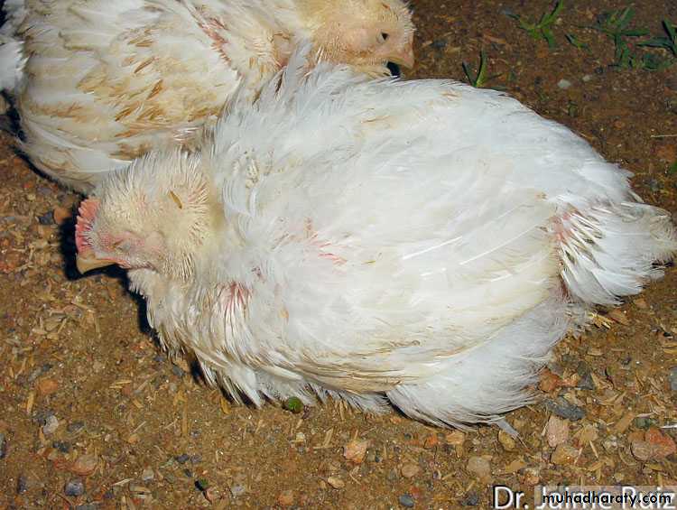
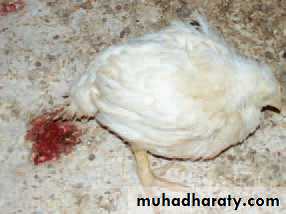
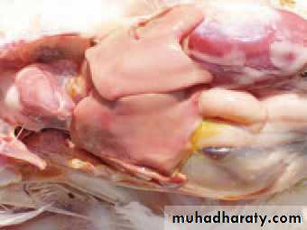
Picture 1
Depression , associated with severe respiratory disease(CRD)
Mycoplasma gallicepticum
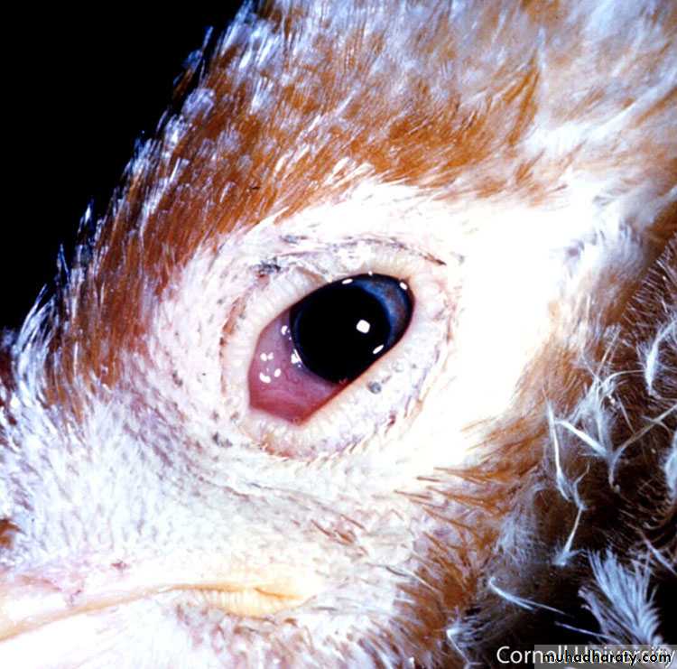
Picture 2
Conjunctivitis eyelid edema pre orbital swollen(CRD)
Mycoplasma gallicepticum
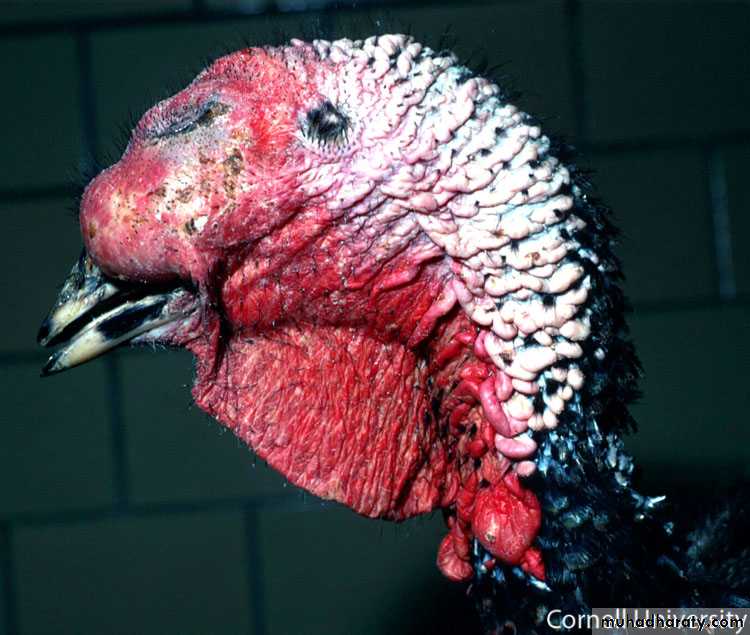
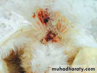
Picture 3
Conjunctivitis , severe swollen of the infraorbital sinuses combination of sinusitis and conjunctivitis may be come so severe that the bird can not open the eye
(CRD)
Mycoplasma gallicepticum
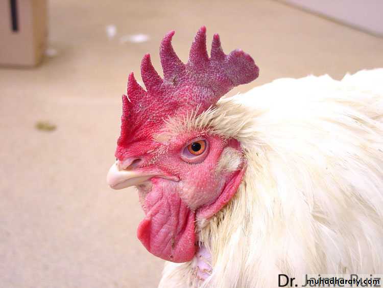
Picture 4
Swollen face and cyanosis of the comb(CRD)
Mycoplasma gallicepticum
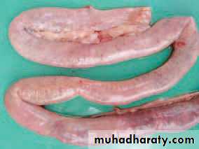
Picture 5
Mild trachitis , small amount of mucoid exudate in trachea lumen(CRD)
Mycoplasma gallicepticum
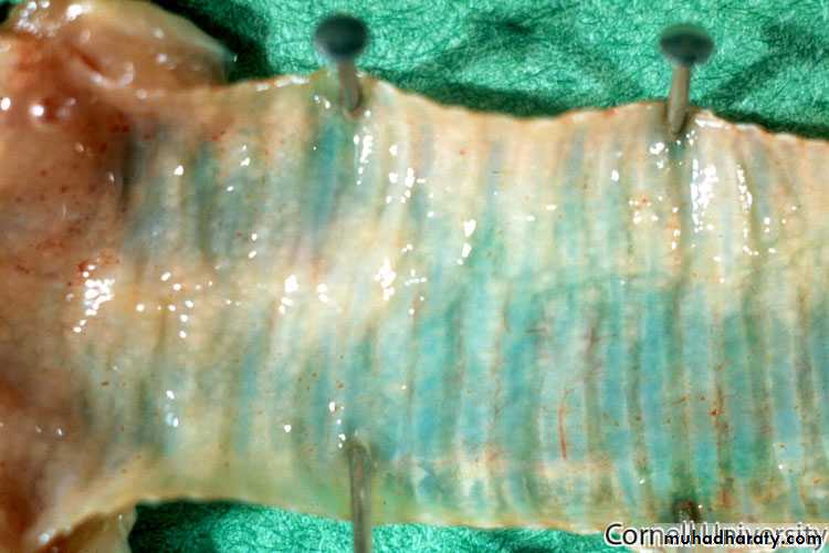
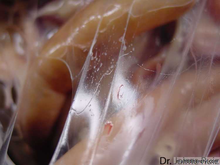
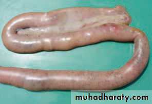
Picture 6
Organ :- trachea and bronchimucoid to gaseous exudate may be seen in nasal passage sinuses , trachea and bronchi
(CRD)
Mycoplasma gallicepticum
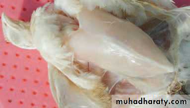
Picture 7
Organ – air sacclitisAir sacclitis with gaseous exudate
(CRD)
Mycoplasma gallicepticum
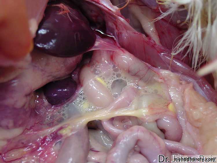
Picture 8
Organ – air sacclitis
Severe air saculitis , abundant foam and aggregate with gaseous exudate
(CRD)
Mycoplasma gallicepticum
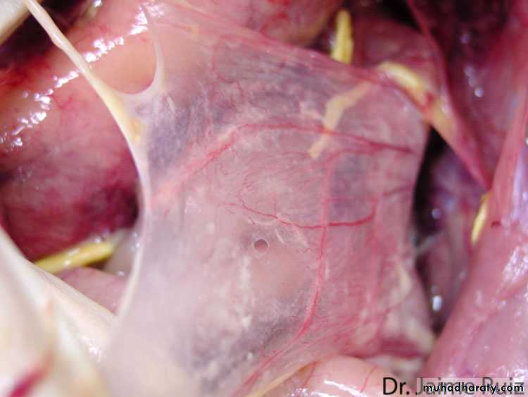
Picture 9
Organ – air sacNormal air sac are complete transudate this pic mild post vaccine reaction to mycoplasma that could be present in healthy bird

Mycoplasma gallicepticum
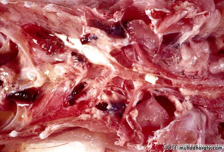
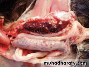
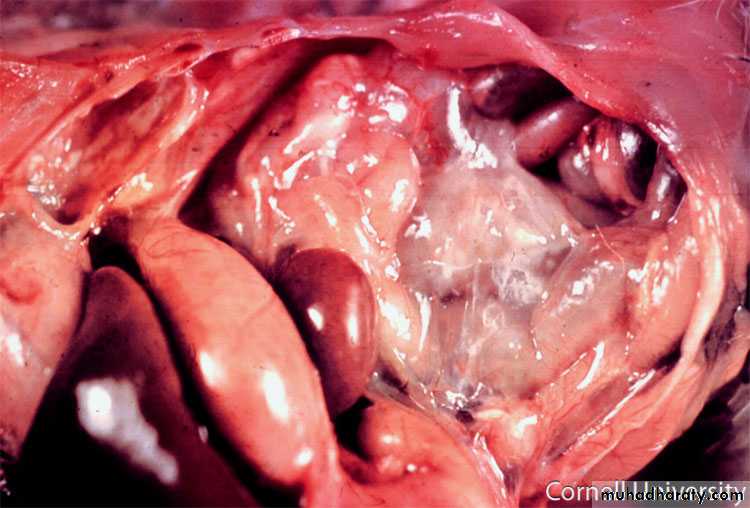
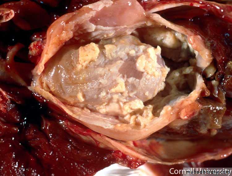
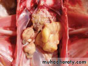
Picture 10Pericarditis peritonitis , peri hepatitis is frequently observed in bird CRB
(CRD)
Mycoplasma gallicepticum
1
CLINCAL SINGS – bloody diarrhea , roughly feather , anemia somnolence (sleepy) are observe
Coccidiosis
2
Organ – vent
The area around vent is stain with blood oocyst are distributed by fecesCoccidiosis
3
Organ – breast muscle
Lesion – dehydration and a high degree of anemia of body and viscera
Coccidiosis
4
Organ – coelomic cavity
Lesion – anemic appearance of internal organ
Coccidiosis


5
Organ – cecum or cecum coccidiosis (typhlitis)
A marked typhlitis is present and hemorrhageCoccidiosis
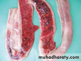
6
Organ – cecum
Lesion – cecum are filled with fresh or clotted bloodCoccidiosis
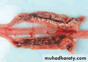
7
Organ – cecum
Lesion – later stage cecum contents become thick mixed with fibrous exudate and accrued cheese like appearance
Coccidiosis
8
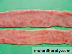
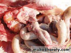
Organ- intestine
Lesion – hemorrhage with varies intestine in different part along intestine . The hemorrhage petechial and could be seen through the intestinal wall
Coccidiosis
9
Organ – cecum and intestine
Lesion – content mixed with fresh or clotted blood and mucous coat is mottled with multiple petechial hemorrhage
Coccidiosis
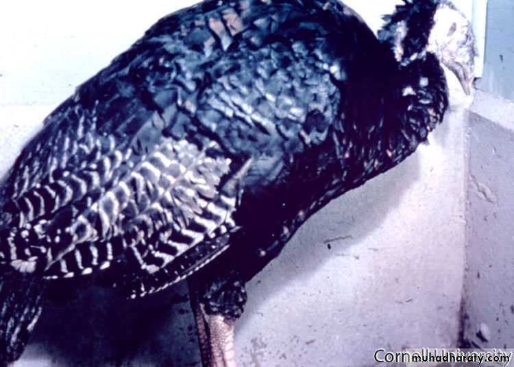
1
Species : turkey
Signs – depression , showing lethargy and weakness
Chlamydophila psittaci
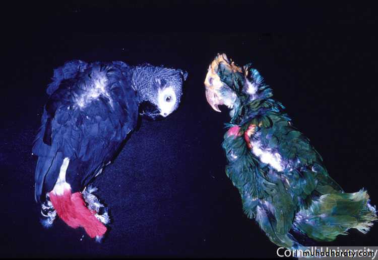
2
Species – parrot
Sign – prostration , fever
Diagnosis
Chlamydophila psittaci
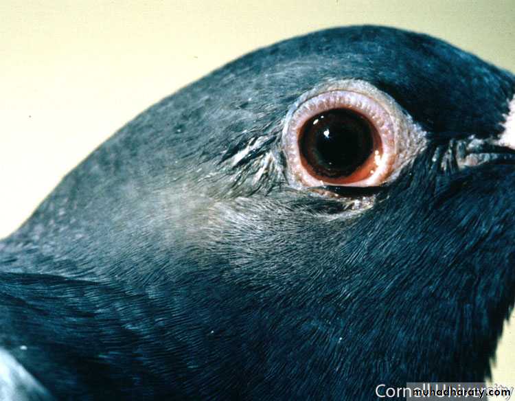
3
Organ – eye of pigeon
Lesion – mild acute conjunctivitis , sometime associated with purulent oculo- nasal discharge
Chlamydophila psittaci
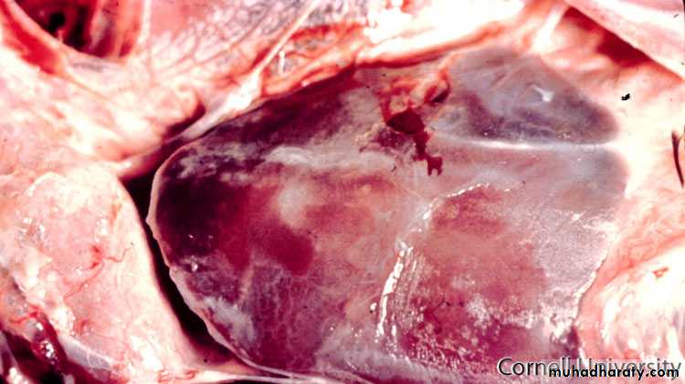
4
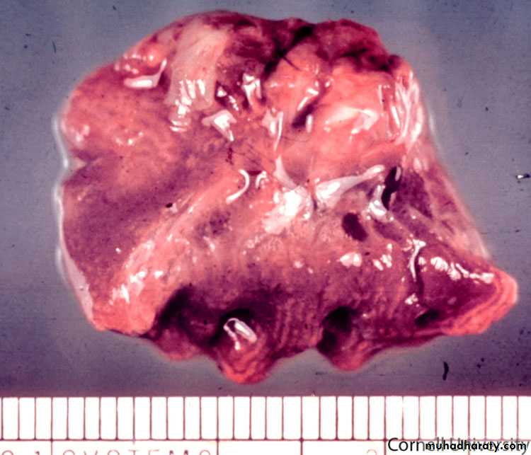
Organ – liver
Lesion – moderate acute fibrinous peri hepatitis the liver is often enlarged and covered with fibrin
Chlamydophila psittaci
5
Organ- air sac
Lesion – acute fibrinous air sacculitis
Chlamydophila psittaci
6
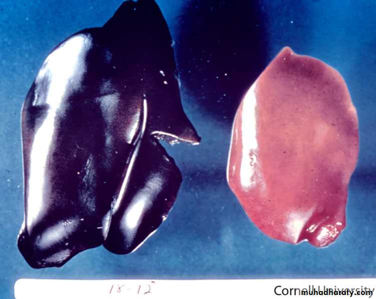
Organ – liver
Lesion – severe hepatomegaly and congestion the liver may be enlarged and discolored
Chlamydophila psittaci
7
Organ – liver
Lesion – cut surface of liver shown white multifocal area of necrosis of parenchyma
Chlamydophila psittaci
8
Organ – heart
Lesion – may be enlarged due to myocarditis and the epicardial surface may be covered with fibrin plaques
Chlamydophila psittaci
9
Organ – heart (cut surface)
Lesion – the epicardial (membrane of turkey thickened and covered with fibrinous exudate reline dry yellow exudate adhere to epicardium
Chlamydophila psittaci
10
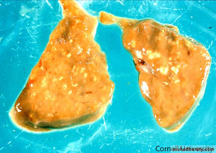
Organ – lung
Lesion – congestion and fibrinous exudate in pleural cavity
Chlamydophila psittaci
11
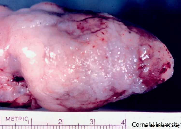
Organ – spleen
Lesion – enlarge most congestion and discoloration
Chlamydophila psittaci
Picture 1
Clinical – are drousy with eye closed . Roughed feather and grouped near source of heatS. typhimurium Paratyphoid
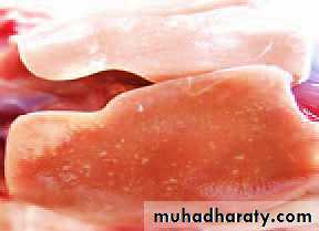
Picture 2
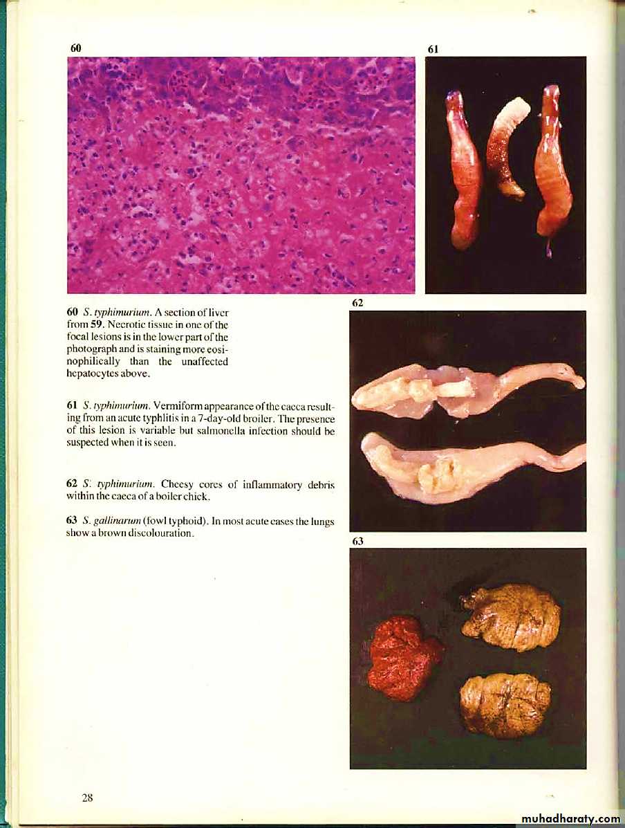
Organ – liverLesion –necrotic foci in the liver
S. typhimurium Paratyphoid
Picture 3
Organ – liverLesion-Multifocal necrotic hepatic in the liver
S. typhimurium Paratyphoid
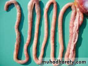
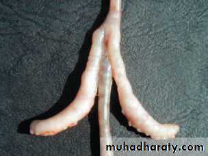
Picture 4
organ- - cecumLesion – vermiform of the cecum result from an acute typhlitis in 7 day old broiler
S. typhimurium Paratyphoid
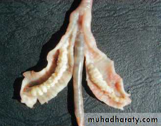
Picture 5
Organ – cecum inflammatory fibrinal exudate in cecum often forms casts with the shape of mucosal foldDiagnosis – typhemirum
S. typhimurium Paratyphoid
Picture 6
Organ- cecumLesion – necrohemorrhogic typhlitis
S. typhimurium Paratyphoid
Picture 1
Organ- eyeLesion – hponpyon- inflammatory cell accumulation in anterior chamber of eye this is fluid which is made of leukocyte diagnosis
Arizonosis s. arizonae
Picture 2
Organ – brain
Lesion – purulent meningoencephalitis
Arizonosis s. arizonae
Picture 3
Organ – air sacLesion – fibrinous air saculitis
Arizonosis s. arizonae
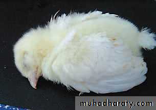
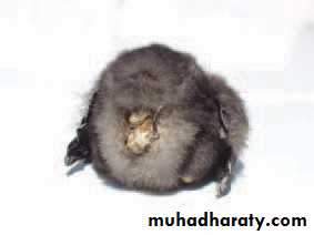
Pic.1species: chick(vent).Clinical signs: sleepy, depressed and their growth is retarded. The feather around vent is stained with diarrhea feces or pasted with dry feces.
S. pullorum
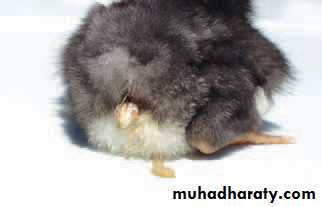
Pic. 2organ: legslesion: synovitis of hock joints
S. pullorum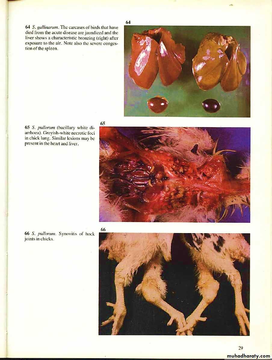
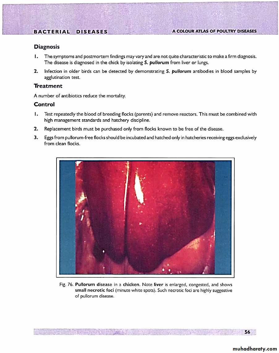
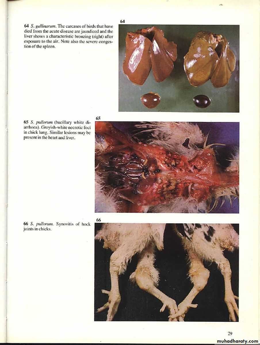
Pic. 3organ: liverlesion:liver is enlarged, congested and show small necrotic foci (minute white spots).
S. pullorum
Pic. 4 organ: liver lesion:multifocal necrotizing hepatitis.
S. pullorumPic. 5 organ: heartlesion:multifocal granulomatous myocarditis.
S. pullorumPic. 6organ: spleenlesion:chick multifocal necro-hemorrhagic splenetis.
S. pullorumPic. 7organ: ureterslesion:ureters are often filled with urates.
S. pullorum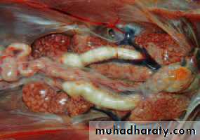
Pic. 8organ: cecumlesion:cecum (closed/open) fibrino-necrotic typhlitis.
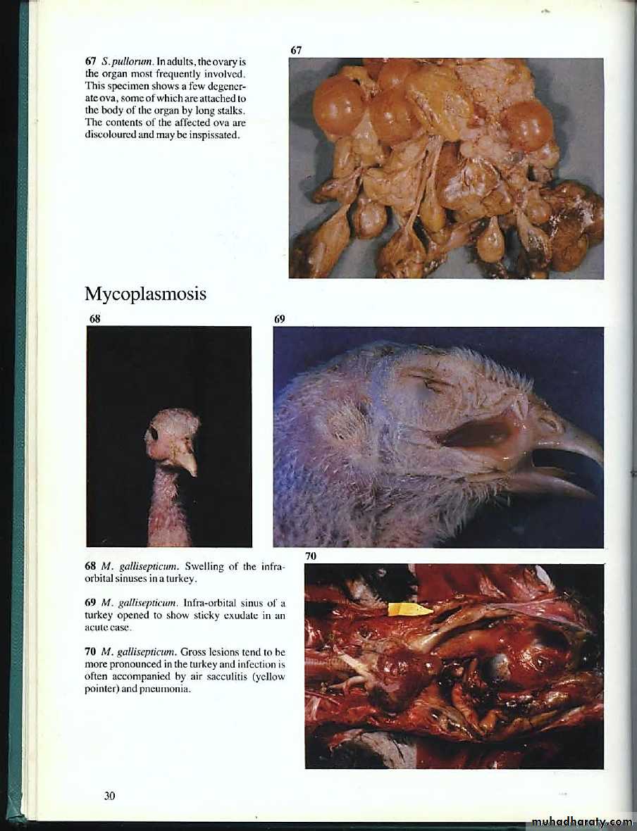
S. pullorumPic. 9organ: ovary (adult)lesion:this specimen show a few degenerate ova, some of which are attached to the body of the organ by long stalks. The content of the affected ova are discolored and may be inspissated (thickener).
S. pullorum
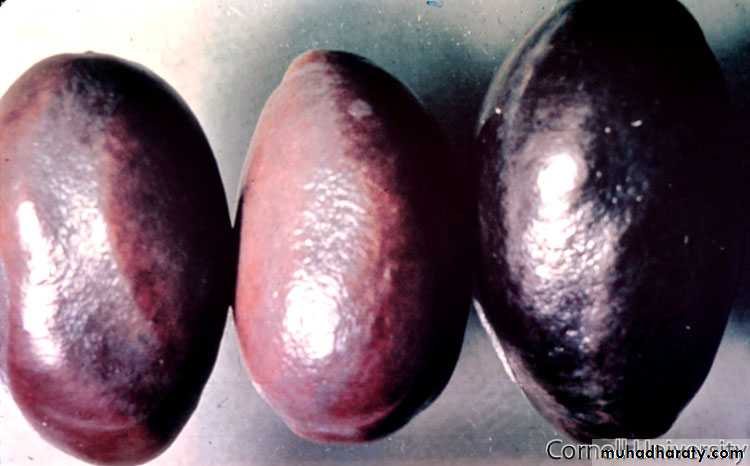
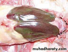
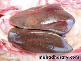
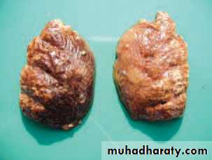
Pic.1organ: liver ( adult)lesion:in the adult bird, liver enlarged and bronze greenish tint.
Acute fowl typhoid
Pic. 2 organ: liver lesion:the enlarged liver, and size of necrosis varies from military to spots with a diameter of 1-2 cm.
Acute fowl typhoid
Pic. 3 organ: spleenlesion:the spleen is (2-3) times bigger, sometime with greyish-white nodules promin of on the surface, representing hyperplastic follicles, (abnormal increase in the number of cell – enlargement of organ or tissue).
Acute fowl typhoid
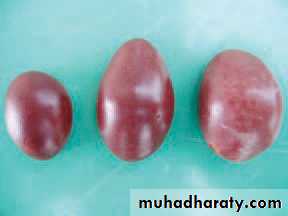
Pic. 4 organ: liver-spleenlesion:jaundice and the liver bronzy and severe congestion of the spleen.
Acute fowl typhoid
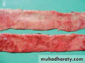
Pic. 5 organ: intestinelesion:enteritis, especially of the anterior part of the small intestine, sometime with ulceration is present.
Acute fowl typhoid
Pic. 6organ: lunglesion:lung brown color, necrosis and followed their organization, sarcoma like nodules.
Acute fowl typhoid
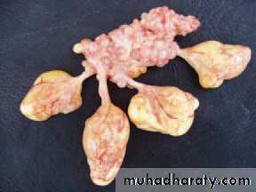
Pic. 7organ: gonadslesion:the ovaries are affected by inflammation and degenerative changes.
Chronic fowl typhoid
Pic. 8organ: ovarian folliclelesion:affected follicle are deformed and appear like thick pendula ting masses (egg should not be used for breeding).
Chronic fowl typhoid
Pic. 9organ: yolk ovarylesion:the going out of yolk from degenerative follicle result in fibrinous adhesive peritonitis.
Chronic fowl typhoid
Pic. 10organ: lunglesion:lung brown discolorations.
Acute fowl typhoidPic. 10organ: lunglesion:greyish-white necrotic foci in chick lung. (similar lesion may be present in the heart and liver).
S. pullorum