Microbiology-p-3 D. Esraa D. T.
Clostridia
Large Gram positive
Straight or slightly curved rods with slightly rounded ends
Anaerobic bacilli
Spore bearing
Saprophytes
Some are commensals of the animal & human gut which invade the blood and
tissue when host die and initiate the decomposition of the corpse (dead body)
Causes diseases such as gas gangrene, tetanus, botulism & pseudo-membranous
colitis by producing toxins which attack the neurons pathways
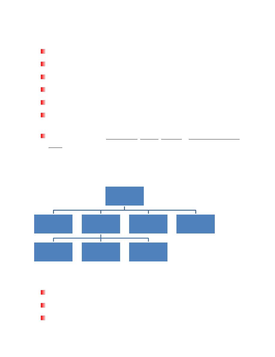
Clostridia of medical importance:
Clostridium Causing Tetanus
Cl. Tetani
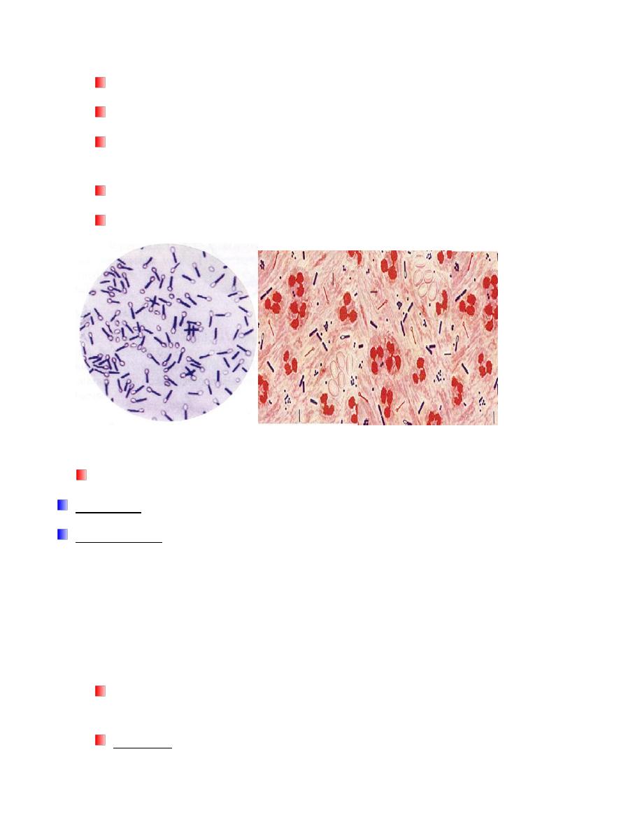
Gram positive, straight, slender rod with rounded ends
All species form endospore (drumstick with a large round end)
Fermentative
Clostridium
Causing
Tetanus
e.g. Cl. tetani
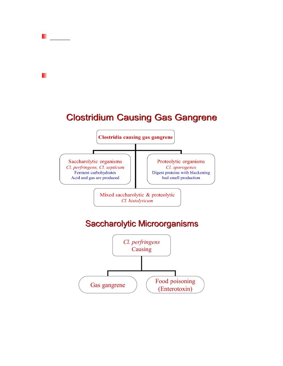
Gas gangrene
Saccharolytic
e.g. Cl. perfringens
&Cl. septicum
Proteolytic
e.g. Cl. sporogenes
Mixed: Cl.
histolyticum
Botulism
e.g. Cl. botulinum
ِAntibiotic
associated diarrhea
e.g. Cl. difficille
Microbiology-p-3 D. Esraa D. T.
Obligate anaerobe
Motile by peritrichous flagella
Grows well in cooked meat broth and produces a thin spreading film when grown
on enriched blood agar
Spores are highly resistant to adverse conditions
Iodine (1%) in water is able to kill the spores within a few hours
Toxins
Cl. tetani produces two types of toxins:
Tetanolysin, which causes lysis of RBCs
Tetanospasmin is neurotoxin and essential pathogenic product
– Tetanospasmin is toxic to humans and various animals when injected parenterally,
but it is not toxic by the oral route
– Tetanospasmin which causes increasing excitability of spinal cord neurons and
muscle spasm
Laboratory Diagnosis of Tetanus
The diagnosis of tetanus depends primarily upon the clinical manifestation of
tetanus including muscle spasm and rigidity.
Specimen: Wound exudates using capillary tube
Microbiology-p-3 D. Esraa D. T.
Culture:
– On blood agar and incubated anaerobically
– Growth appears as a fine spreading film.
Gram stain is a good method for identifying Clostridium
– Cl. tetani is Gram positive rod motile with a round terminal spore giving a
drumstick appearance

Microbiology-p-3 D. Esraa D. T.
Clostridium perfringens
Large Gram-positive bacilli with stubby ends
Capsulated
Non motile (Cl. tetani is motile)
Anaerobic
Grown quickly on selective media
Can be identified by Nagler reaction
Toxins
The toxins of Cl. perfringens
– toxin (phospholipase C, lecithinase) is the most important toxin
Lyses of RBCs, platelets, leucocytes and endothelial cells
Increased vascular permeability with massive hemolysis and bleeding tissue
destruction
Hepatic toxicity and myocardial dysfunction
– -toxin is responsible for necrotic lesions in necrotizing enterocolitis
– Enterotoxin is heat labile toxin produced in colon → food poisoning
Laboratory Diagnosis
Specimen: Histological specimen or wound exudates
Histological specimen transferred aseptically into a sterile screw-capped
bottle & used immediately for microscopical examination & culture
Specimens of exudates should be taken from the deeper areas of the
wound where the infection seems to be most pronounced
Microscopical examination (Gram, Spore stain etc)
Gram-positive bacilli, non motile, capsulated & sporulated
Microbiology-p-3 D. Esraa D. T.
The spore is oval, sub-terminal & non bulging
Spores are rarely observed
Culture: Anaerobically at 37C
On Robertson's cooked meat medium → blackening of meat will observed
with the production of H2S and NH3
On blood agar → β-hemolytic colonies
Biochemical Tests
Cl. perfringnes characterized by:
It ferments many carbohydrates with acid & gas
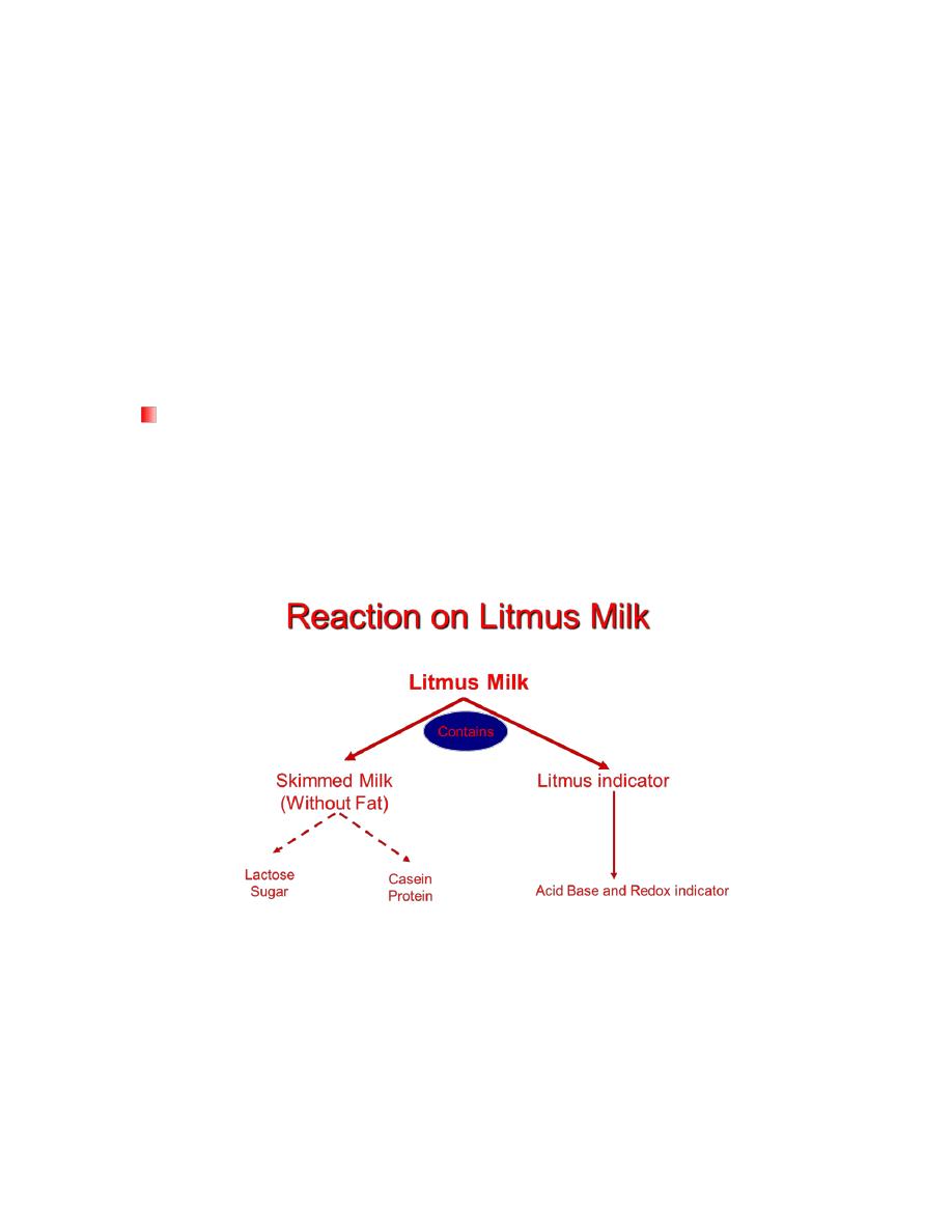
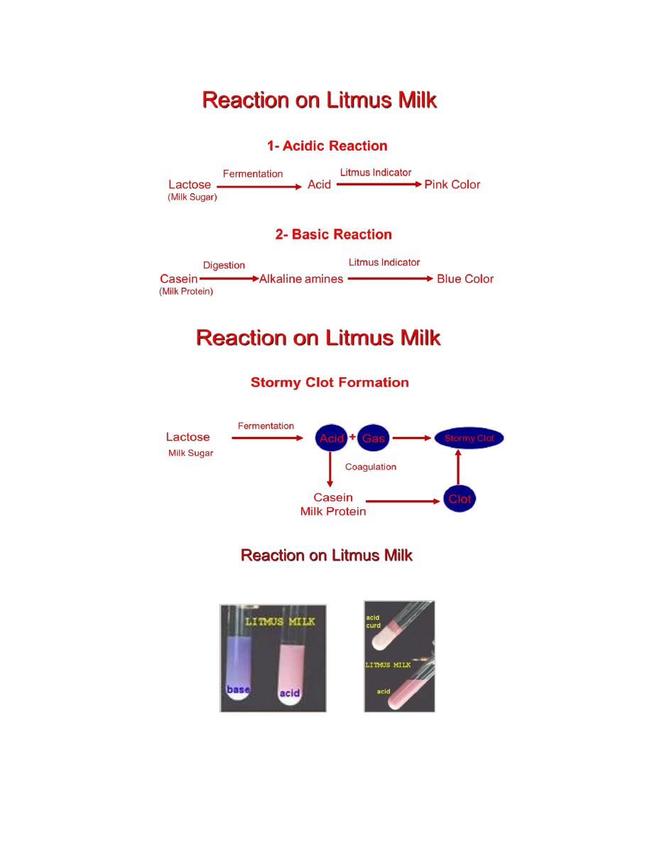
It acidified litmus milk with stormy clot production
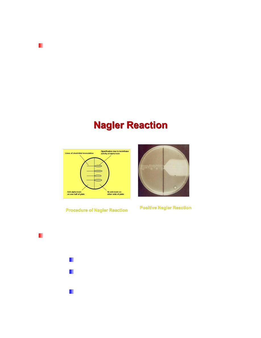
Nagler reaction is positive
Microbiology-p-3 D. Esraa D. T.
Microbiology-p-3 D. Esraa D. T.
Nagler’s Reaction
This test is done to detect the lecithinase activity
– The M.O is inoculated on the medium containing human serum or egg yolk
(contains lecithin)
– The plate is incubated anaerobically at 37 C for 24 h
– Colonies of Cl. perfringens are surrounded by zones of turbidity due to
lecithinase activity and the effect is specifically inhibited if Cl. perfringens
antiserum containing antitoxin is present on the medium
Anaerobic Cultivation
Removal of oxygen & replacing it with inert gas
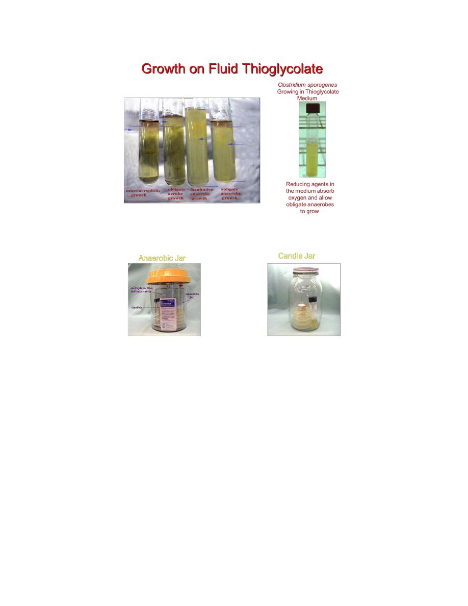
– Anaerobic Jar
It is especially plastic jar with a tightly fitted lid
Hydrogen is introduced from commercially available hydrogen
generators envelop
10 ml of water is added to envelop immediately before placing it in
the jar

Microbiology-p-3 D. Esraa D. T.
Hydrogen and carbon dioxide will release and react with oxygen in
the presence of catalyst to form water droplet
Anaerobic indicator (Methylene blue) is placed in the jar
Methylene blue is blue in oxidized state (Aerobic condition) while
turns colorless in reduced state (Anaerobic condition)
Anaerobic Cultivation
Culture Media containing reducing agent
– Thioglycollate broth
It contains
– Sodium thioglycollate (Reducing agent)
– Rezazurin (redox indicator)
– Low percentage of Agar-Agar to increase viscosity of medium
– Cooked Meat Medium
It contains
– Meat particles (prepared from heart muscles) which contain
hematin & glutathione that act as reducing agent
Microbiology-p-3 D. Esraa D. T.
