
COLONIC DIVERTICULOSIS
A diverticulum is a blind pouch leading off the alimentary tract, lined by mucosa, that
communicates with the lumen of the gut
. Congenital diverticula have all three layers of the
bowel wall (mucosa, submucosa, and most notably the muscularis propria) and are distinctly
uncommon. The prototype is Meckel diverticulum,
Virtually all other diverticula are acquired and either lack or have an attenuated muscularis
propria.
Acquired diverticula may occur anywhere in the alimentary tract, but by far the most
common location is the colon
, giving rise to diverticular disease of the colon, also called
diverticulosis.
It is attributed to the consumption of a refined, low-fiber diet, resulting in reduced stool bulk
with increased difficulty in passage of intestinal contents. Exaggerated spastic contractions of
the colon isolate segments of the colon in which the intraluminal pressure becomes markedly
elevated, with consequent herniation of the bowel wall through the anatomic points of
weakness
MORPHOLOGY
Most colonic diverticula are small flasklike or
spherical outpouchings, usually 0.5 to 1 cm in diameter

TUMORS OF THE SMALL AND LARGE INTESTINES
Epithelial tumors of the intestines are a major cause of morbidity and mortality worldwide. The
colon, including rectum, is host to more primary neoplasms than any organ in the body.
Colorectal cancer ranks second to bronchogenic carcinoma among the cancer killers.
Adenocarcinoma constitute the vast majority of colorectal cancers and represent 70% of all
malignancies arising in the GIT. Curiously, the small intestine is an uncommon for benign or
malignant tumors despite its great length,
Whereas the small bowel represents 75% of the
length of the alimentary tract, its tumors account for only 3% to 6% of gastrointestinal tumors,
with a slight preponderance of benign tumors.
The classification of intestinal tumors is the
same for the
small and large intestine.

Terminology
A
polyp is
a tumorous mass that protrudes into the lumen of the gut; traction on
the mass may create a stalked, or
pedunculated,
polyp. Alternatively, the polyp
may be
sessile
, without a definable stalk. Polyps may be formed as the result of
abnormal mucosal maturation, inflammation, or architecture. These polyps are
non-neoplastic
and do not have malignant potential; an example is the
hyperplastic polyp. Those polyps that arise as the result of epithelial proliferation
and dysplasia are termed
adenomatous polyps or adenomas. They are true
neoplastic lesions (“new growth”) and are precursors of carcinoma.

Non-neoplastic Polyps
hvperplastic polyps
, which are small (less than 5 mm in diameter), nipple-like, hemispherical,
smooth protrusions of the mucosa. They may occur singly but are more often multiple.
Although they may be anywhere in the colon, well over half are found in the rectosigmoid
region. Histologically, they contain abundant crypts lined by well-differentiated goblet or
absorptive epithelial cells, separated by a scant lamina propria. The vast majority of
hyperplastic polyps have no malignant potential.
Juvenile polyps
are essentially hamartomatous proliferations, mainly of the lamina propria,
enclosing widely spaced, dilated cystic glands. They occur most frequently in children
younger than 5 years old but also are found in adults of any age; in the latter group they may
be called
retention polyps
. usually large in children (I to 3 cm in diameter) but smaller in
adults. In general, they occur singly and in the rectum, and being hamartomatous they have no
malignant potential. Juvenile polyps may be the source of rectal bleeding and in some cases
become twisted on their stalks to undergo painful infarction
.

Neoplastic polyp (Adenomas):
Adenomas are neoplastic polyps that range from small, often
pedunculated tumors to large lesions that are usually sessile. Because
the incidence of adenomas in the small intestine is very low, this
discussion focuses on those adenomas that arise in the colon.

adenomatous lesions arise as the result of epithelial proliferation and dysplasia, which may range
from mild to so severe as to represent transformation to carcinoma.
Adenomatous polyps are segregated into three subtypes on the basis of the epithelial architecture:
• Tubular adenomas
: mostly tubular glands.
•
Villous adenomas
: villous projections
•
Tubulovillous adenomas
: a mixture of the above .
Tubular adenomas are by far the most common;
5% to 10% of adenomas are tubulovillous, and only 1% are villous.
The malignant risk with an adenomatous polyp is correlated with three interdependent features:
1. polyp size,
2.
histologic architecture
3. , and severity of epithelial dysplasia
, as follows:
• Cancer is rare in tubular adenomas smaller than 1 cm in diameter.
• The likelihood of cancer is high (approaching 40%) in sessile villous adenomas more than 4 cm
in diameter.
• Severe dysplasia, when present, is often found in villous areas
.

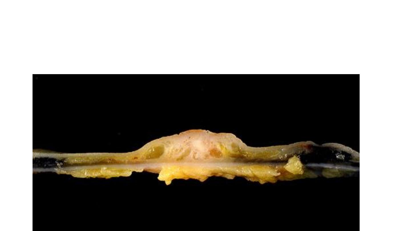
MORPHOLOGY
Tubular adenomas
Histologically the stalk is covered by normal colonic mucosa but the head is composed
of neoplastic epithelium. all degrees of dysplasia may be encountered, ranging up to
cancer confined to the mucosa (intramucosalcarcinoma) or invasive carcinoma-like
masses
.

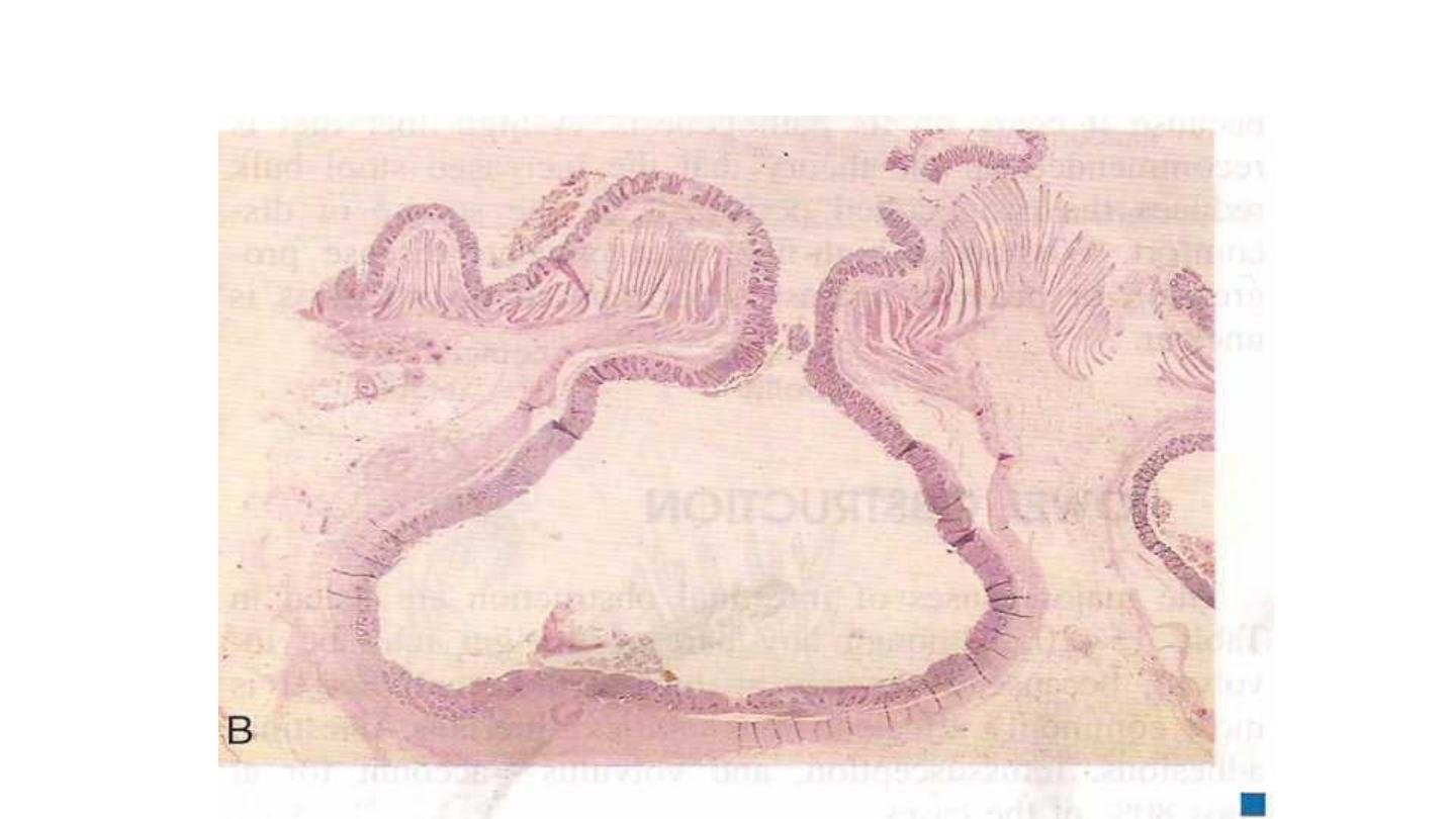
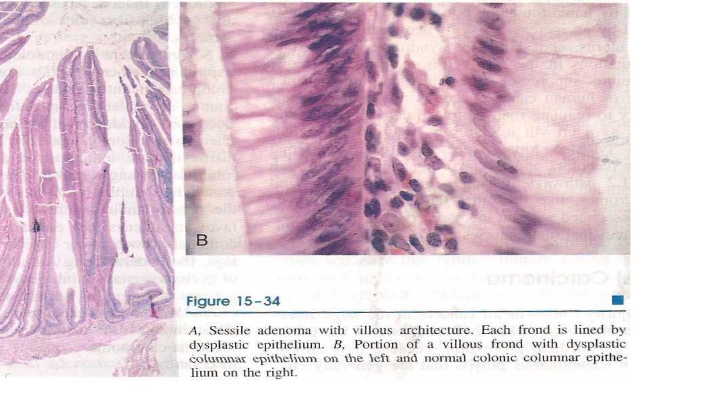
Villous adenomas
are larger and more ominous of the epithelial polyps. They tend to occur
in older persons, most commonly in the rectum and rectosigmoid. but they may be located
elsewhere. They generally are sessile covered by dysplastic, columnar epithelium. All degrees
of dysplasia may be encountered, and invasive carcinoma is found in up to 40% of these
lesions, the frequency being correlated with the size of the polyp.
tubulovillousa denomos
are composed of a broad mix of tubular and villous areas. They
are intermediate between the tubular and the villous lesions in their frequency of having a
stalk or being sessile, their size, the degree of dysplasia, and the risk of harboring
intramucosal or invasive carcinoma
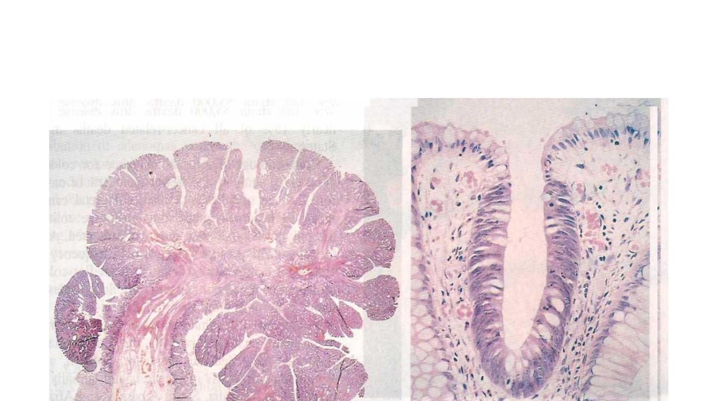
Pedunculatcd adenoma showing a fibrovascular stalk covered by normal colonic mucosa and a head that
contains abundant dysplastic epithelial glands, hence the blue color. B, A small focus of adenomatous
epithelium in an otherwise normal (mucin-secreting. clear) colonic mucosa, showing how the dysplasric
columnar epithelium (deeply stained) can populate a colonic crypt (“tubular’ architecture).

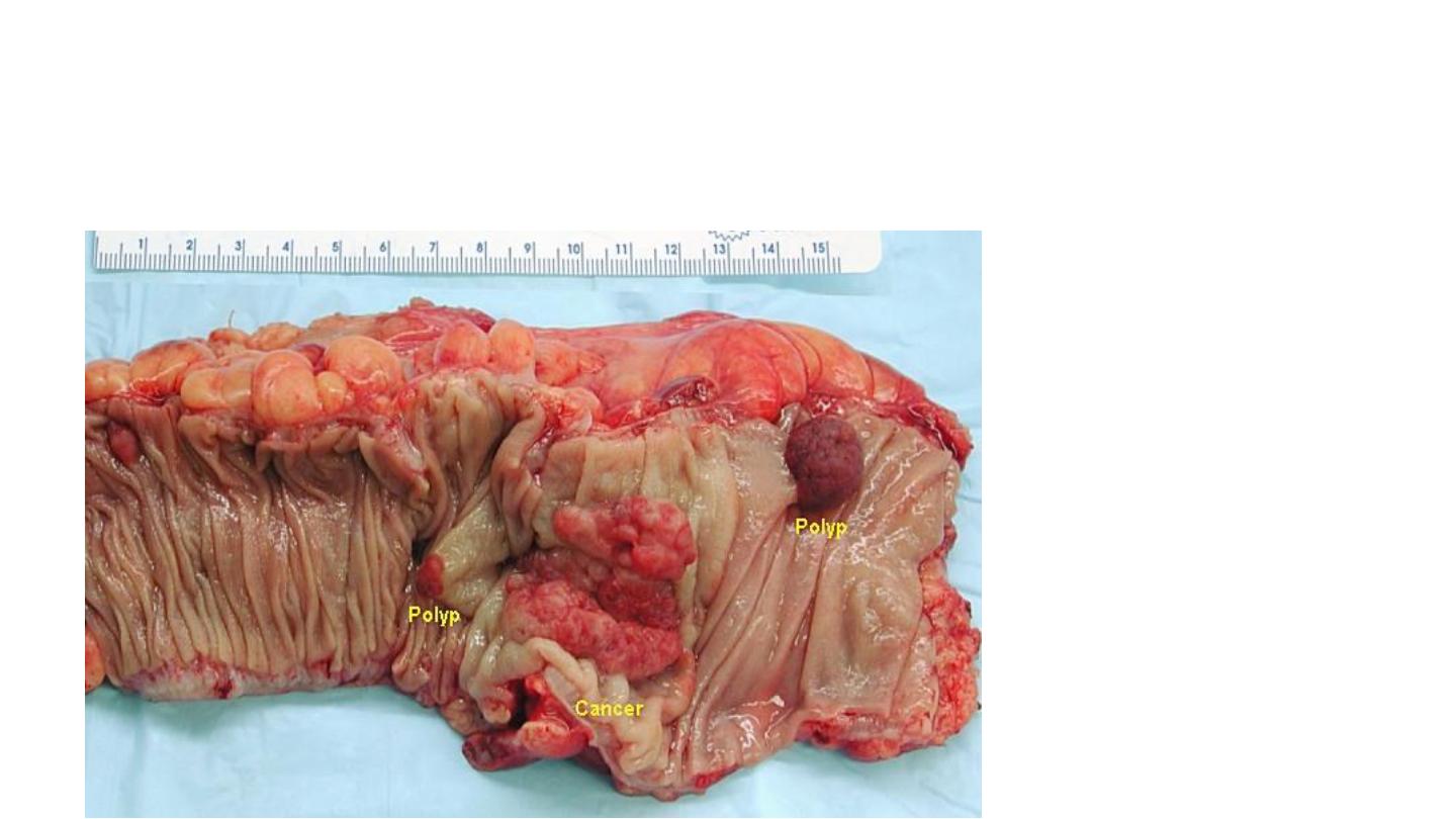
Familial Polyposis Syndromes
Familial polyposis syndromes are uncommon autosmal dominant disorders.
Their importance lies in propensity for malignant transformation.
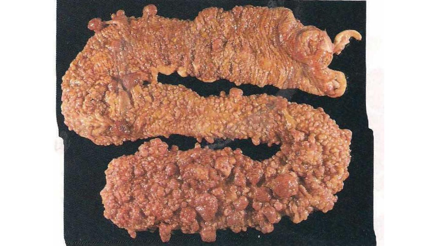
In familial adenomatous polyp (FAP), patients typically develop 500 to 2500
colonic adenomas that carpet the mucosal surface ; a
minimum number of 100
is
required for the diagnosis. Multiple adenomas may also be present elsewhere in
the alimentary tract. Most polyps are
tubular adenomas
; occasional polyps have
villous features. Polyps usually become evident in adolescence or early
adulthood.
The risk of colonic cancer is virtually 100% by midlife, unless a
prophylactic colectomy is performed
. The genetic defect underlying
FAP
has been
localized to the APC gene on chromosome(5q2).

Colorectal Carcinoma
A great majority (98%) of all cancers in the large intestine are adenocarcinomas.
Epidemiology
.
• The peak incidence for colorectal cancer is 60 to 70 years of age.
• When colorectal cancer is found in a young person, preexisting ulcerative colitis or one of
the polyposis syndromes must be questioned.
• Males are affected more often than females.
• Colorectal carcinoma has a worldwide distribution, with the highest incidence rates in the
United States.

Environmental factors
, particularly dietary practices, are implicated in these striking geographic
contrasts.
The dietary factors receiving the most attention are
(1)a low content of unabsorbable vegetable fiber
(2)high content of refined carbohydrates,
(3)a high fat content (as from meat),
(4)decreased intake of protective micronutrients such as vitamins A, C, and B.
It is theorized that reduced fiber content leads to decreased stool bulk, increased fecal retention
in the bowel, and an altered bacterial flora of the intestine. Moreover, high fat intake enhances
the synthesis of cholesterol and bile acids by the liver, which in turn may be converted into
potential carcinogens by intestinal bacteria. Refined diets also contain less of vitamins A, C, and
E, which may act as oxygen radical scavengers.
Several recent epidemiologic studies suggest that use of aspirin and other NSAIDs exerts a
protective effect against colon cancer.

MORPHOLOGY
About 25% of colorectal carcinomas are in the cecum or ascending colon, with a similar
proportion in the rectum and distal sigmoid. An additional 25% are in the descending colon and
proximal sigmoid; the remainder are scattered elsewhere. Hence, a substantial portion of cancers
is undetectable by digital or proctosigmoidoscopic examination.
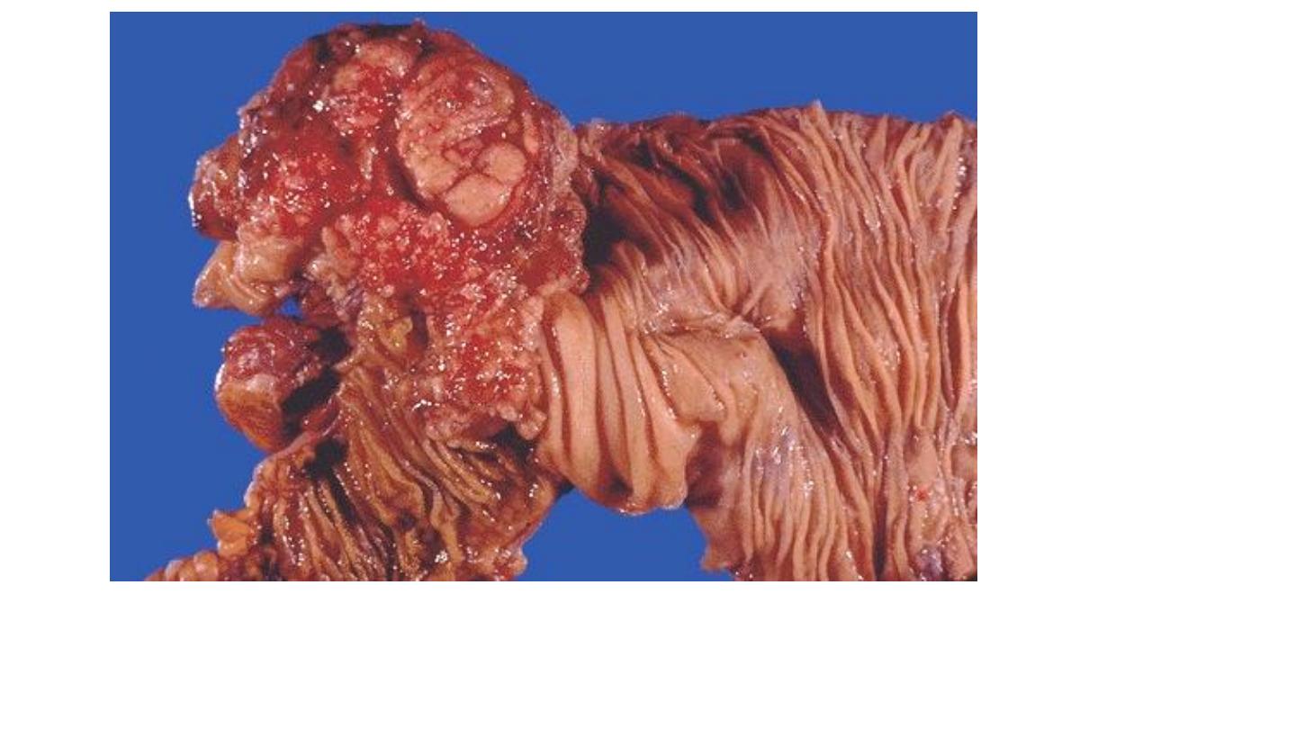
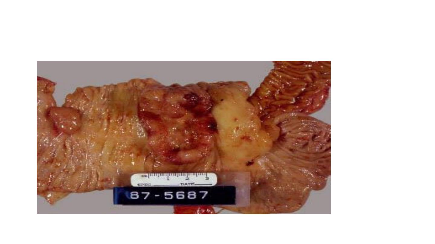
• Tumors in the proximal colon tend to grow as polypoid, exophytic masses. Obstruction is
uncommon.
• When carcinomas in the distal colon are discovered, they tend to be annular, encircling lesions
that produce so-called napkin-ring constrictions of the bowel and narrowing of the lumen.
• all colon carcinomas are microscopically similar. Almost all are adenacarcinamas that range
from well-differentiated to undifferentiated, and anaplastic masses.
• Many tumors produce mucin which is secreted into the gland lumina or into interstitium of the
gut wall. Because these dissect through the gut wall, they facilitate extension of the cancer and
worsen the prognosis.
• Cancers of the anal zone are predominantly squamous cell in origin
.

All colorectal tumors spread by direct extension into adjacent structures and by metastasis
through the lymphatics and blood vessels.
Serum markers for disease, such as elevated blood levels of
carcinoembryonic antigen
, are of
little diagnostic value, because they reach significant levels only after the tumor has achieved
considerable size and has very likely spread. Moreover, “positive carcinoembryonic antigen
levels may be produced by carcinomas of the lung, breast, as well as non-neoplastic disorders .
Because APC mutations occur early in colon cancers, molecular detection of APC
mutations in epithelial cells, isolated from stools, is being evaluated as a diagnostic test
.
The single most important prognostic indicator of colorectal carcinoma is the extent (stage)
of the tumor at time of diagnosis
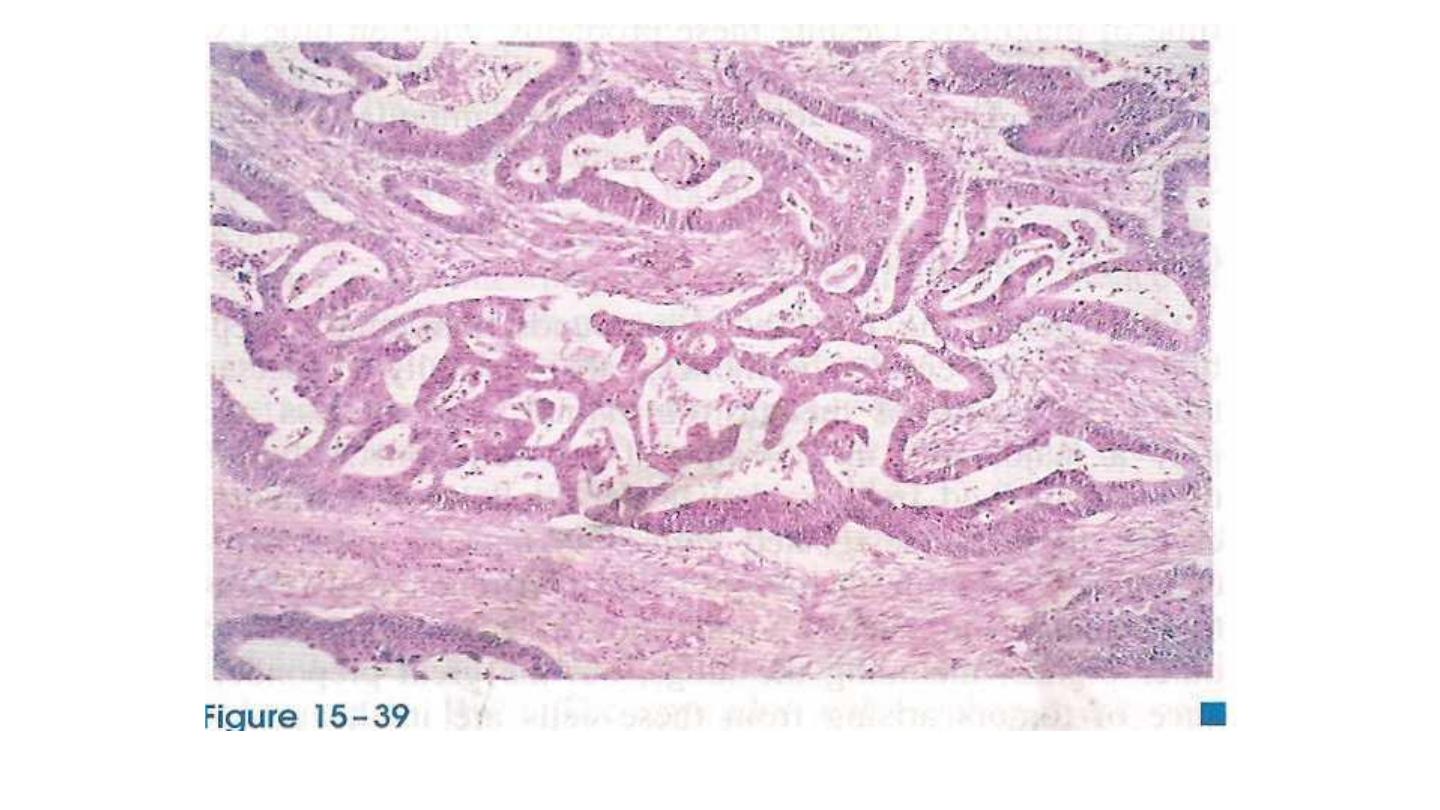
Invasive adenocarcinoma of colon showing malignant glands
infiltrating the muscle wall
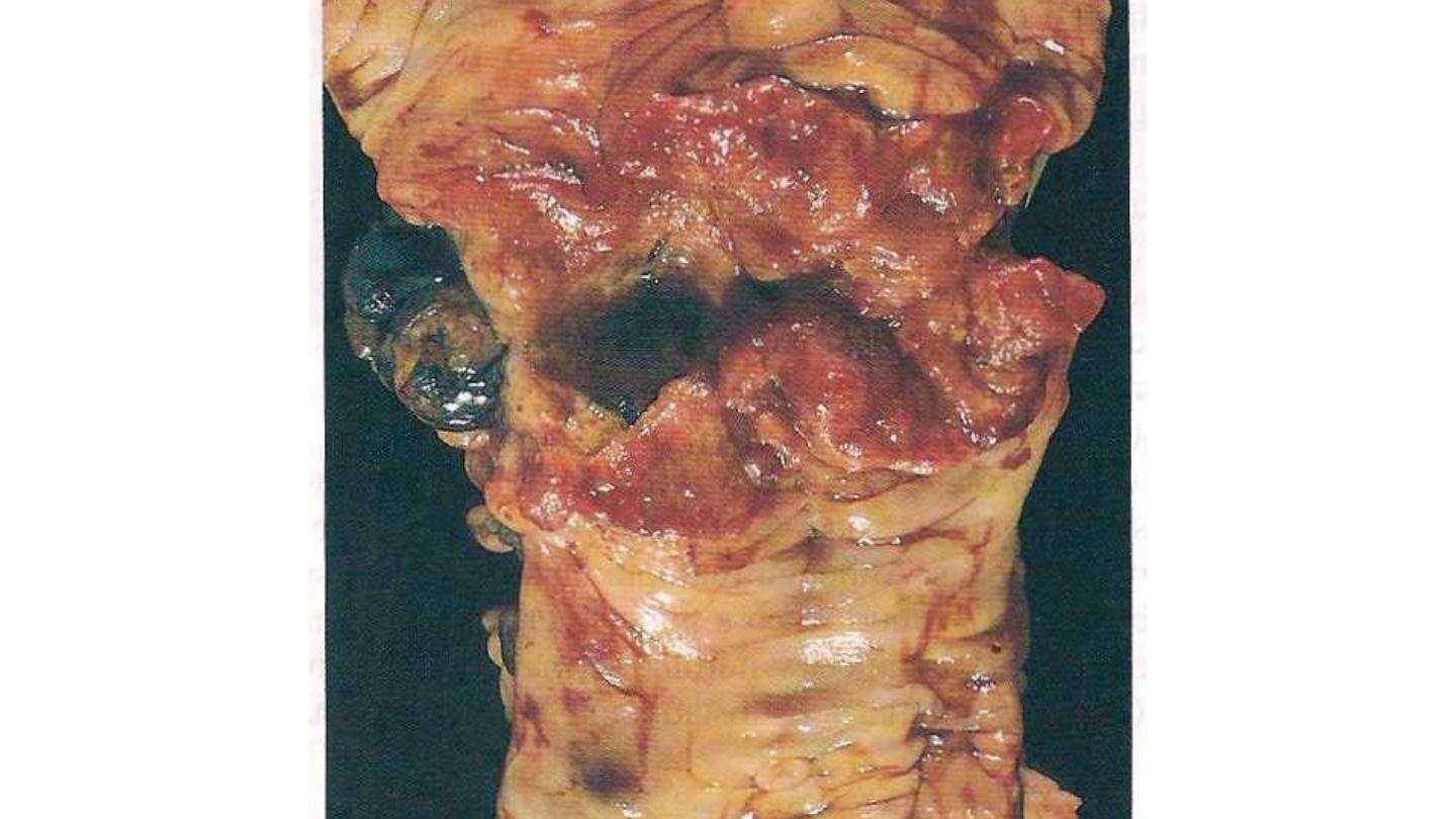
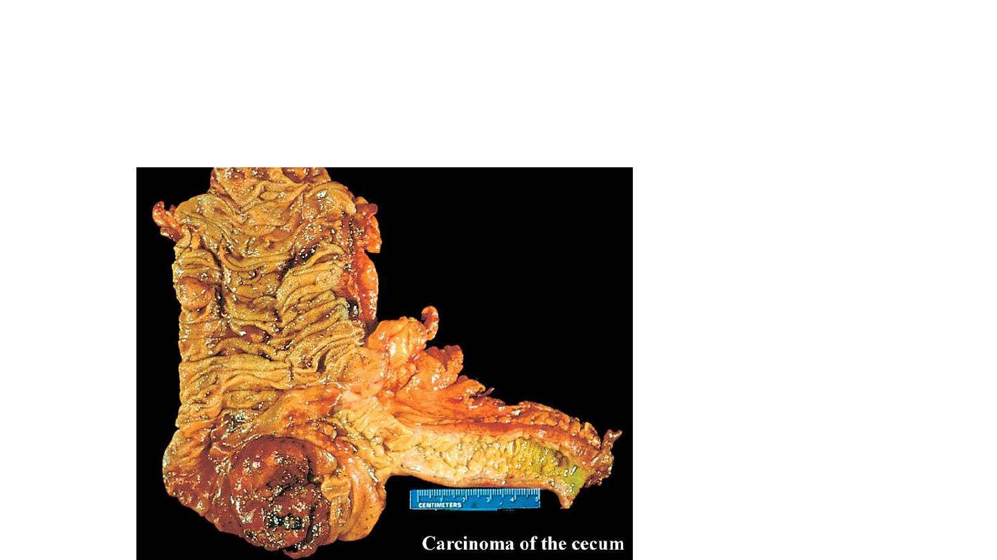
tumors in the proximal colon: polypoid, exophytic masses that extend
along one wall of the cecum and ascending colon
This is an adenocarcinoma of the cecum which demonstrates an exophytic growth pattern, as the bulk of the mass
is within the bowel lumen. The patient had iron deficiency anemia.
Polypoid (protuberant)
Ulcerated, with sharply demarcated margins
)
*
Diffusely infiltrating
Colorectal Cancer and Early Detection
• Colorectal cancer can be prevented through regular screening and the
removal of polyps
• Early diagnosis means a better chance of successful treatment
• Screening should begin at age 50 for all “average risk” individuals or
sooner if you have a family history of colorectal cancer, symptoms, or
a personal history of inflammatory bowel disease