Management of open fractures:-
An open (compound ) fracture should be given immediate attention under aseptic conditions. After 6 to 8 hours the contaminating organisms begin to multiply and infection supervenes. The treatment should includes:1- remove all foreign materials and dead and devitalized tissue leaving healthy well vascularized tissue to combat infection.
2- immobilize the bone segments to prevent further trauma.
3- grossly contaminated wounds with severe damage to soft tissue and contamination of the exposed bone must be cleaned by clipping the hair and cleansing the surrounding area. The area is then draped, inspected, excised and debrided.
The decision about closing the skin depends on 3 factors:
1- time since injury.
2- degree of contamination and infection.
3- extent of the injury.
If the operation is performed within 6-10 hours and the contamination has been slight primary closure is indicated. If more time has passed and severe contamination or infection is present the wound should be left open and covered with sterile gauze and topical antibiotics. The skin may be closed later when the infection has been controlled or the wound may be left open until completely healed.
Clinical tests of bone union:-
1- Absence of mobility between the fragments.
2- Absence of pain when an attempt is made to bend the fracture.
Radiological features of bone union:-
1- Bony trabeculae bridging the fracture and joining fragments.
2- Total or nearly total obliteration of the fracture line.
Factors affecting bone union:-
1- Age of patient. In very young animals a fracture may heal in 2 weeks, in an elderly patient it may take 3-6 months.
2- Location and type of fracture. Diaphyseal fractures heal more slowly than metaphyseal fractures. Multiple fractures usually take longer than the more simple types.
3- History of fracture. Presence of infection, multiple surgical procedures, inadequate fixation, impaired circulation and inadequate reduction.
4- Lapse of time since reduction and fixation.
5- Types of fixation and its application.
Clinical signs of fracture:-
1- Local swelling.
2- Deformity or change in angulation.
3- Abnormal mobility.
4- Loss of function.
5- Localized tenderness.
6- Crepitus.
Radiographic examination of the fracture:-
Radiographs of the injured areas should be obtained. At least 2 views at right angels are essential for accurate diagnosis and selection of best procedure for reduction and immobilization.
Treatment of fracture:
Treatment of fracture performed by the following steps:
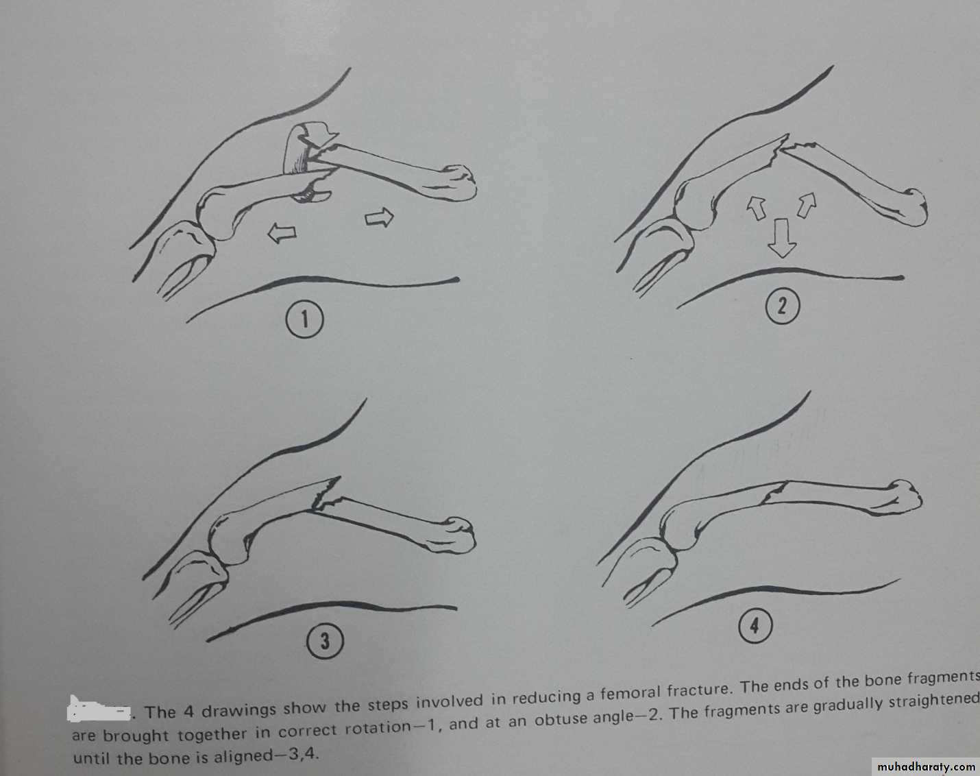
Reduction:-
This involves replacement of bone fragments as nearly as possible to their original position. Good muscle relaxation facilitates reduction and is best achieved by inducing general anesthesia. Methoxyflurane is a particularly useful anesthetic agent for orthopedic surgery.
Reduction can be accomplished by:
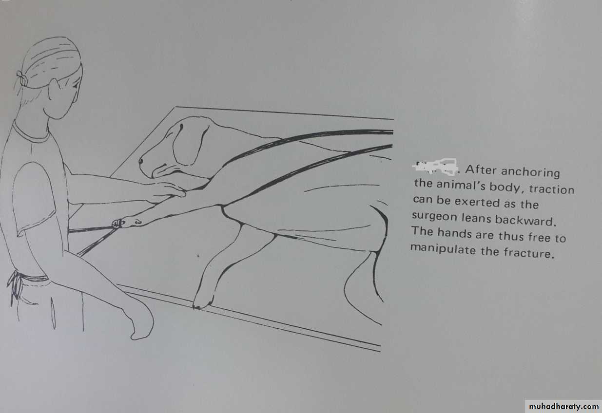
A- Manipulation ( closed reduction) : usually is achieved by manipulation along with application of traction and countertraction. Closed manipulation is the standard method for reduction of simple fractures particularly those with minimum soft tissue covering. The manipulation should be done as gently as possible to prevent injury to the soft tissues.
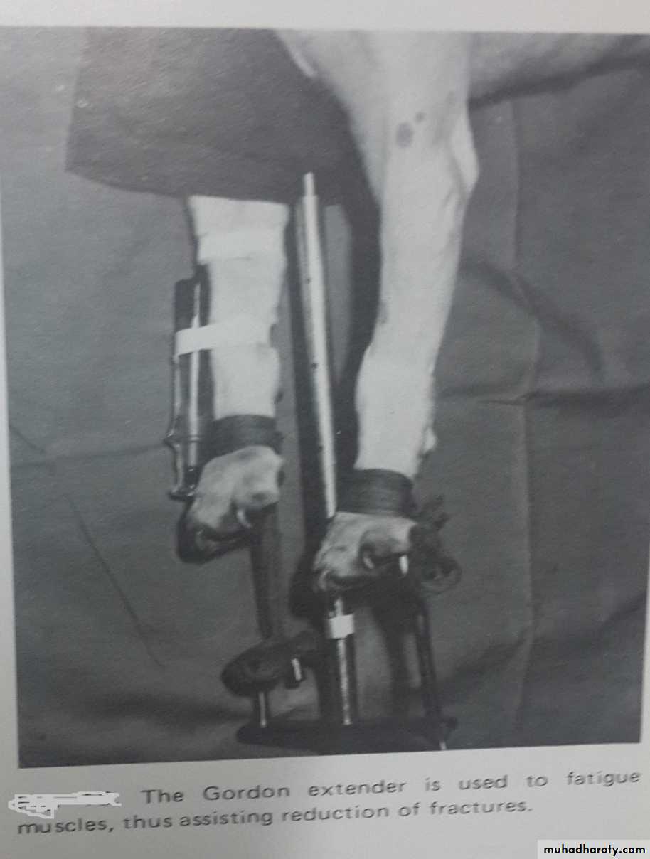
B- Reduction by mechanical traction: by using Gordon extender, force is applied slowly allowing a few minutes to elapse between application of additional traction. From 10-20 minutes are ordinarily required to fatigue muscles and overcome spastic contraction.
C- Open reduction: when other methods fail or in some cases the preferred method, the fracture is reduced following exposure of the bone. Following open reduction an internal fixation device is usually applied. Application of traction and countertraction and manipulation with forceps.