GENERAL FEATURES
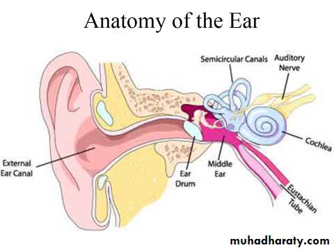
• Ear is a peripheral sense organ concerned with hearing and equilibrium.• It is subdivided into three parts:
the external ear, middle ear or tympanic cavity and the internal ear or labyrinth.
• STRUCTURE
• External Ear• The external ear consists of the auricle that collects sound waves and the external acoustic meatus that conducts these waves to the tympanic membrane (ear drum).
• The auricle and the outer third of external acoustic meatus are made of elastic cartilage covered by thin skin. This skin is provided with hair, sebaceous and ceruminous (modified sweat glands) glands.
• The tympanic membrane separates the external ear from the middle ear. It is a trilaminar structure, lined externally by stratified squamous epithelium and internally by simple cuboidal epithelium and is supported in the middle by fibrous tissue
Middle Ear
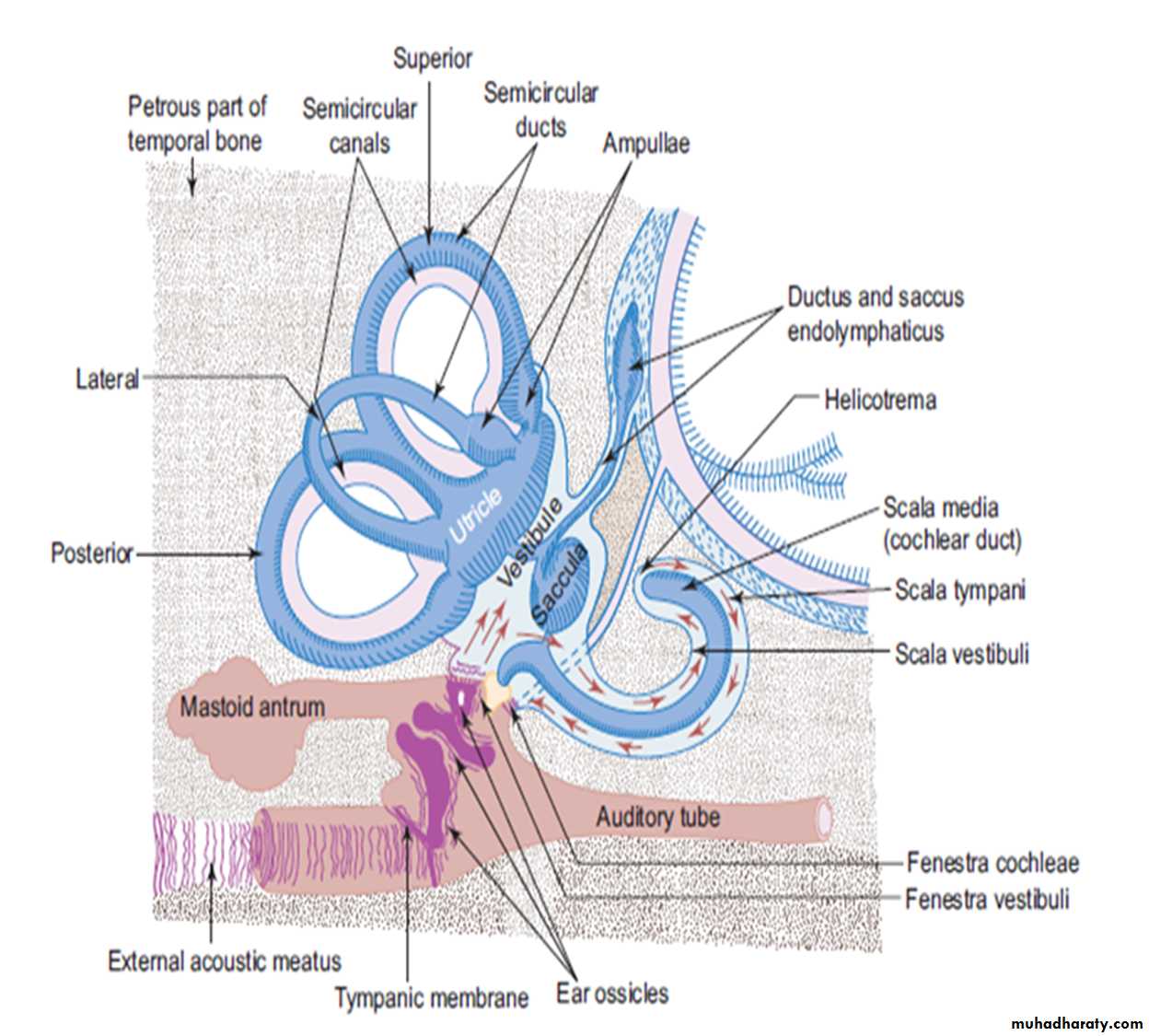
• The middle ear is a narrow air space compressed mediolaterally, occupying the petrous part of temporal bone. It contains a chain of three tiny auditory ossicles (malleus, incus and stapes) whose function is to transmit the vibrations of tympanic membrane to the perilymph of internal ear.
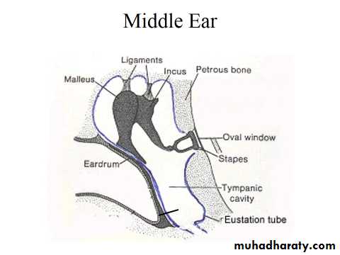
• The middle ear also has two small muscles (tensor tympani and stapedius) attached to the ear ossicles. These muscles reflexly contract to dampen excessive vibration caused by loud noise.
• Middle ear cavity communicates anteromedially with the nasopharynx through auditory tube. This helps to equalize air pressure on both surfaces of tympanic membrane.
Internal Ear
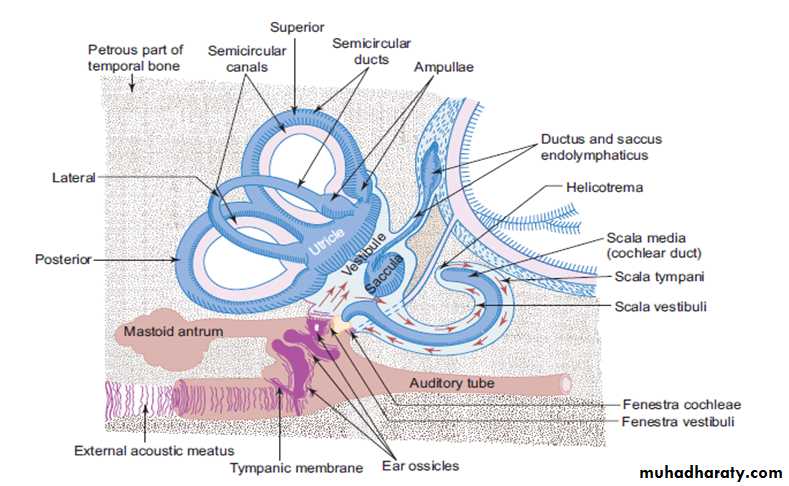
The internal ear consists of a bony labyrinth, comprising a series of cavities within the petrous part of temporal bone and a membranous labyrinth, comprising a series of membranous sacs and ducts present within the bony labyrinth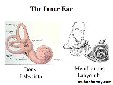
• The bony labyrinth is lined by endosteum and contains a clear fluid, perilymph, in which the membranous labyrinth is suspended.
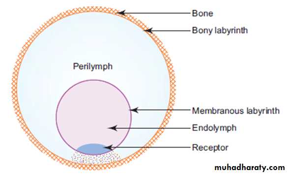
Fig. :General organization of labyrinth .
• The membranous labyrinth is lined by simple squamous epithelium and contains endolymph.
• The squamous epithelial lining of the membranous labyrinth is specialised in certain regions to form receptor organs.
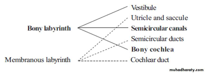
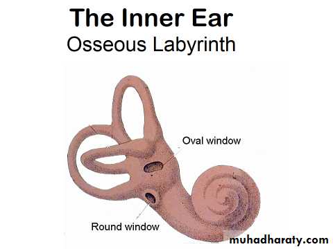
Bony Labyrinth
Bony labyrinth consists of three parts, vestibule, semicircular canals and bony cochlea. These structures are described, as follows:1. Vestibule
It is the centrally situated cavity containing utricle and saccule (the larger and the smaller sac respectively) of membranous labyrinth. It communicates anteriorly with cochlea and posteriorly with three semicircular canalsthrough five orifices (one being common to two of the canals).
In its lateral wall there are two openings:
• The fenestra vestibuli (oval window) closed by the base of stapes and
• Fenestra cochleae (round window) closed by the secondary tympanic membrane.
2. Semicircular canals
• They are three in number (superior, posterior and lateral) and are arranged at right angle to each other, so that all the three planes are represented.• Each canal has a swelling at one end called the ampulla. These canals open into the vestibule through five orifices.
• The semicircular canals contain semicircular ducts.
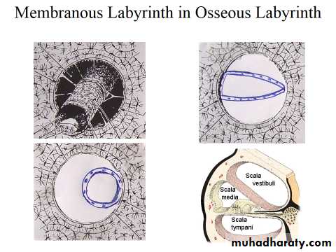
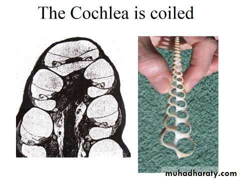
3. Bony cochlea
It consists of a central pillar of bone, the modiolus, around which the cochlear canal makes two and a half spiral turns.Each successive turn is of decreasing radius, so that the whole structure is conical in shape, resembling
the shell of a snail.
Its base is directed towards the internal acoustic meatus and is pierced by the cochlear part of the vestibulocochlear nerve.
• A spiral bony shelf or ledge, the osseous spiral lamina winds round the modiolus like a thread of a screw and projects into the interior of bony canal.
• The endosteum over it is thickened to form limbus spiralis.
• The endosteum on the opposite outer wall is thickened to form spiral ligament which projects into the interior of the canal as a prominence called crista basilaris.
• Extending between the osseous spiral lamina and crista basilaris is the basilar membrane.
• The cochlear duct of the membranous labyrinth, which is triangular in cross section occupies the interior of cochlear canal.
• In cross section, cochlea shows three compartments, the scala vestibuli above, the scala tympani below (these belong to the canal), and scala media in the middle (this belongs to the duct).
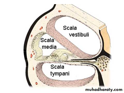
• The scala vestibuli and scala tympani communicate with each other at the apex of cochlea through a narrow space, the helicotrema.
• The perilymph in the scala vestibuli is separated from the tympanic cavity by the base of stapes at the fenestra vestibuli.
• The perilymph in the scala tympani is separated from the tympanic cavity by the secondary tympanic membrane at the fenestra cochlea (Fig. ).
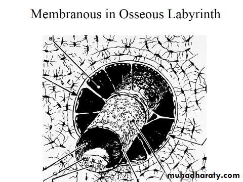
Membranous Labyrinth
• Membranous labyrinth is lodged within the bony labyrinth.
• It consists of two sacs, the utricle and saccule besides semicircular and cochlear ducts. All these structures freely communicate with one another and form a closed system of membranous sacs and ducts.
: schematic diagram of ear , red arrows indicate the direction of vibration of perilymph
The structures are described as follows:1. Utricle and saccule
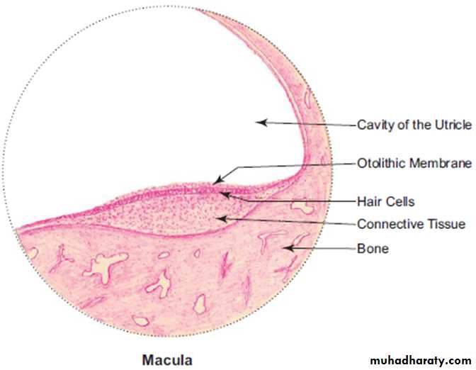
The utricle and saccule are two membranous sacs lodged in the bony vestibule. These two sacs are connected with each other and with the saccus endolymphaticus by means of a Y-shaped utriculosaccular duct. (The saccus endolymphaticus lies beneath the dura mater on the posterior surface of petrous part of temporal bone.)
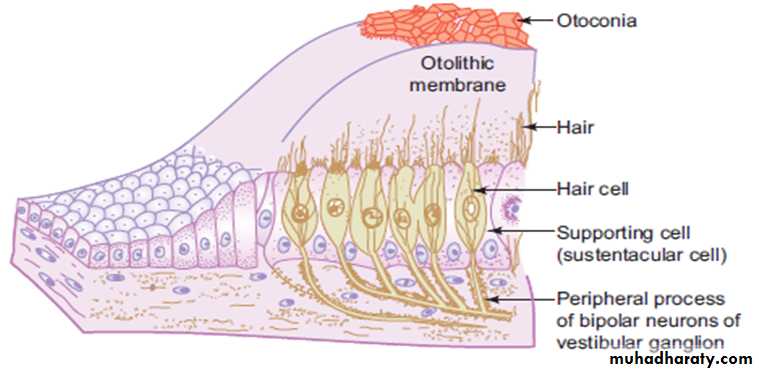
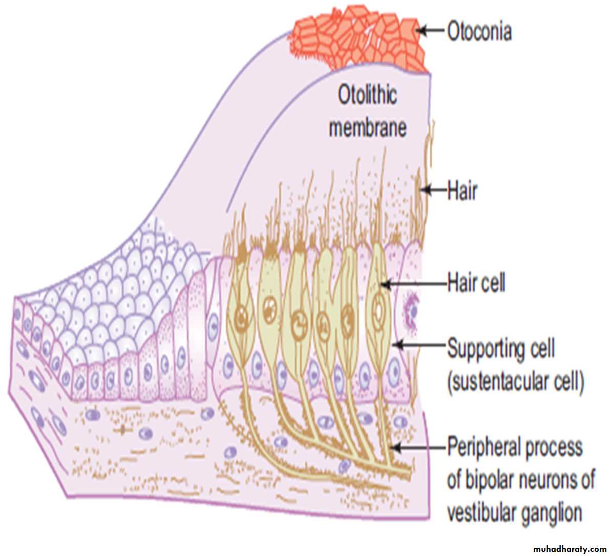
The saccule is connected to the cochlear duct by ductus reuniens (Fig.). The epithelial cells lining the wall are specialized in certain regions to form sensory receptors, the maculae of utricle and saccule.
Maculae are receptors found on the lateral wall of utricle and medial wall of saccule. The macula consists of two types of cells: namely, the sustentacular cells & hair cells.
The sustentacular cells are columnar supporting cells with microvilli on their free surface. Hair cells are fl ask shaped sensory cells lying between sustentacular cells. Hair cells are innervated by vestibular part of the vestibulocochlear nerve.
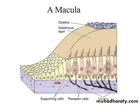
• Each hair cell is provided with long microvilli (stereocilia) of varying length and single nonmotile kinocilium.
• The stereocilia and kinocilia are embedded in a thick gelatinous plaque of glycoprotein called otolithic membrane, secreted by sustentacular cells.
• It contains numerous crystalline bodies called otoliths or otoconia (Fig.). Maculae are sensitive to orientation of the head in relation to gravity or other acceleration forces. It is an organ of static balance or position sense.
Macula
Macula of Utricle.Presence of:
Otolithic membrane and otoconia;
Hair and sustentacular cells.
2. Semicircular ducts
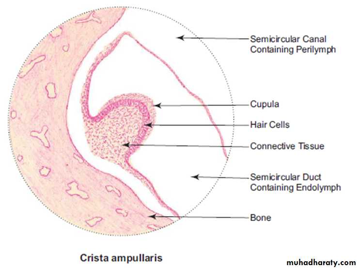
• Semicircular ducts are three in number, small in size and found within the semicircular canals.• These ducts have the same confi guration as the semicircular canals. Sensory receptors are found in the ampullae of semicircular ducts as transverse thickening called crista ampullaris.
• Crista ampullaris is similar in structure to macula, but the glycoprotein layer is very thick and conical in shape in cross section. It is called cupula (Fig.). There are no crystalline bodies in the cupula. Crista is sensitive to angular movement of head (changes in rotational velocity). It is an organ of kinetic balance (movement sense).
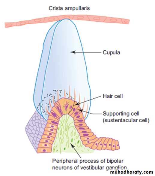
Crista Ampullaris.
Presence of:• Cupula;
• Conical-shaped sense organ comprising hair and sustentacular cells.
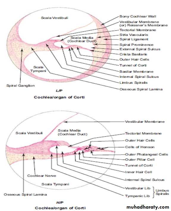
3. Cochlear duct:
Cochlear duct (scala media) lies within the bony cochlear canal and is triangular in cross section. The base (outer wall) is formed by stria vascularis, which is a stratified vascular epithelium lining the spiral ligament. Stria vascularis is responsible for maintaining the correct ionic composition of endolymph. The apex of cochlear duct is at the osseous spiral lamina.• The roof is formed by vestibular (Reissner’s) membrane which separates the scala media from scala vestibuli.
• The floor is formed by basilar membrane which separates the scala media from scala tympani.
• The basilar membrane supports the organ of Corti, which is the receptor sense organ of hearing (Fig.).
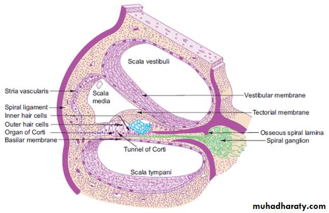
Fig. :cross section of coch lear duct in situe
Organ of Corti
The organ of Corti consists of two groups of cells. These are:
(a) Hair (sensory) cells
• Inner hair cells
• Outer hair cells
(b) Supporting cells
• Inner and outer pillar cells
• Inner and outer phalangeal cells
• Border cells
• Hensen’s cells
• In the center of the organ of Corti there is a canal, the tunnel of Corti, bounded by inner and outer rows of pillar cells. The pillar cells are rod-like cells containing tonofi brils.
• On the inner aspect of the inner row of pillar cells is a single row of columnar cells, the inner phalangeal cells. They support the bases of inner hair cells, arranged in a single row. The inner hair cells are fl ask-shaped cells. They bear stereocilia (hair) which are arranged in the form of letter ‘U’.
• On the outer aspect of the outer row of pillar cells, there are three to fi ve rows of outer phalangeal cells. They support the same number of rows of outer hair cells. Outer hair cells are much taller than the inner hair cells and their hair (stereocilia) are arranged in the form of letter ‘W’.
• Both inner and outer hair cells do not reach the basilar membrane and they are supported by the inner and outer phalangeal cells through their apices. The hair cells have no kinocilium and have only stereocilia (hair). The hair project into a gelatinous layer, the tectorial membrane, which overhangs the organ of Corti from the limbus spiralis. The hair cells are innervated by peripheral processes of bipolar neurons whose cell bodies are situated at the base of the osseous spiral lamina as spiral ganglion. Their central processes pass through the modiolus as cochlear nerve.
• The organ of Corti is limited internally by the border cells. These are columnar cells arranged in a single row on the inner
• aspect of inner phalangeal cells.
• Hensen’s cells limit the outer boundary of the organ of Corti. They are arranged in several rows on the outer aspect of the outer phalangeal cells.
• Stereocilia are agitated due to vibration of basilar membrane caused by conduction of sound from bone to fluid.
Cochlea.
Presence of:
• Organ of Corti;
• Scala vestibuli, scala media, scala tympani;
• Spiral ganglion.