
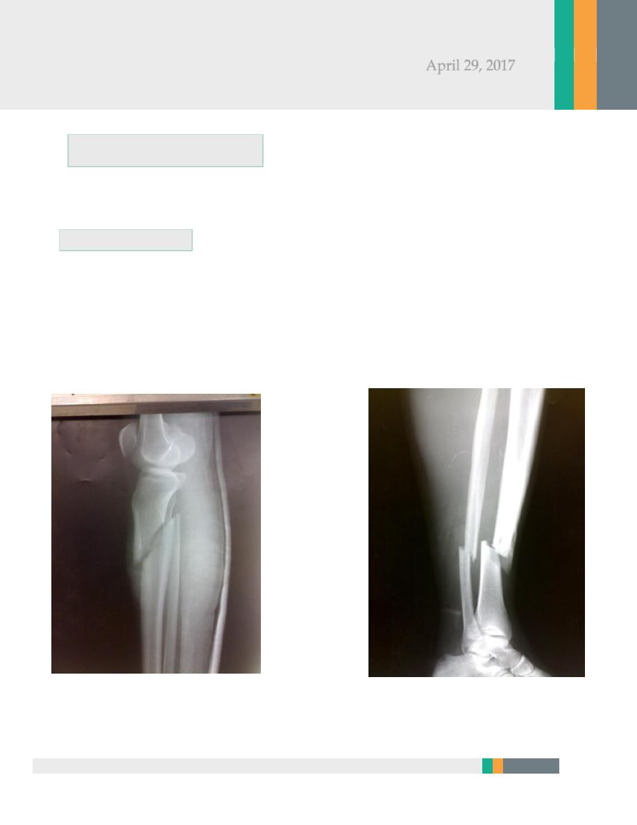
Indirect force: (low energy)
Twisting: spiral fractures of both bones
Angulatory: oblique fractures with butterfly segment.
Direct force:
Transverse (low energy) or comminuted (high energy) fractures usually with
skin and soft tissue damage
.
Surgery
Zakho hospital
Orthopedics Dr. Muthana
(
Fractures of tibia and fibula)
April 29, 2017
2
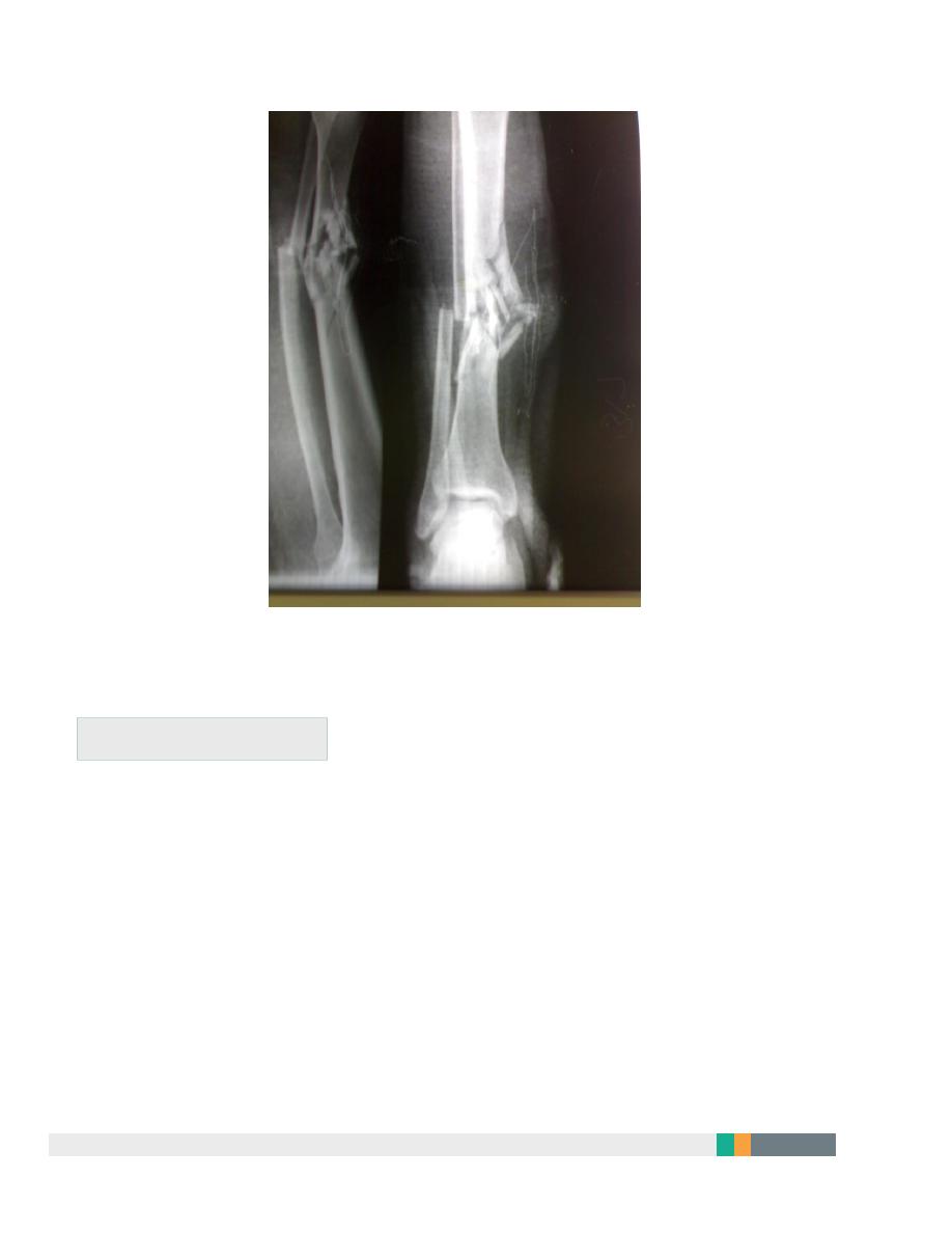
Examine for:
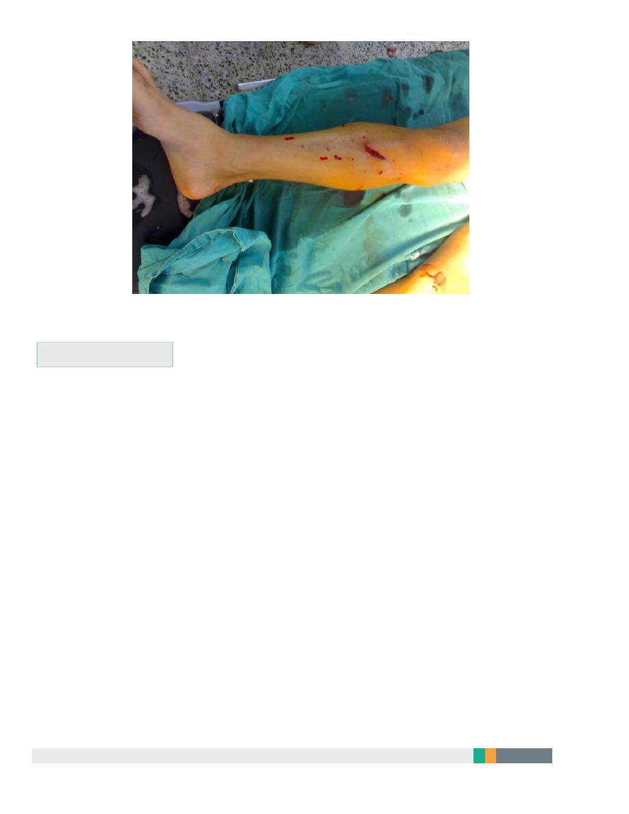
Size of skin and soft tissue damage.
Distal pulses, sensation, movements.
Signs of compartment syndrome.
X-ray must include the knee and ankle.
3
Management
Objectives:
To limit soft tissue damage and preserve skin cover.
To obtain and hold fracture alignment.
To recognize compartment syndrome.
To start early weight bearing to promote healing.
To start joint movement as soon as possible.
4
Treatment of low energy fractures
1. Including closed & Gustilo I & II fractures treated by non-operative
methods; MUA for displaced fractures.
2. Full-length cast from upper thigh to metatarsal necks with the knee in
slight flexion and the foot plantigrade.
3. Elevate the leg and observe for 2-3 days for compartment syndrome.
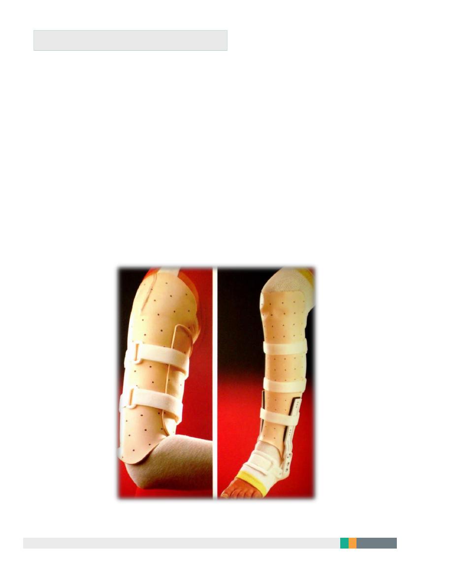
4. After 4-6 weeks change to below knee functional brace and allow weight
bearing and knee movement.
5. Healing is complete in 8 wks in children and in 16 wks in adults.
5
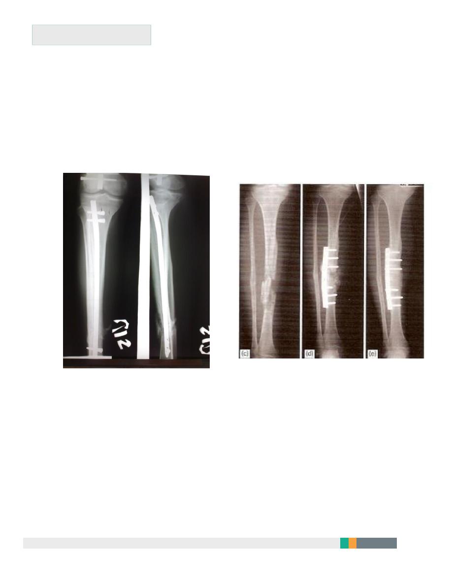
Skeletal fixation
Locked intramedullary fixation is the method of choice for diaphyseal
(shaft) fractures.
Plate and screws for fractures near bone ends (metaphyseal) and for
children with closed unstable fractures.
6
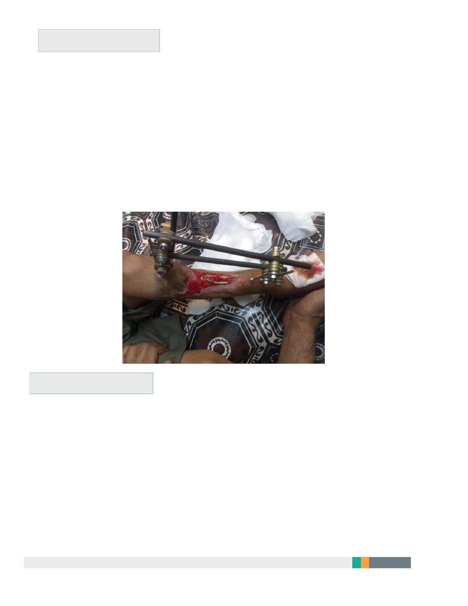
Open fractures:
Antibiotics & anti-tetanus.
Debridement; thorough wash with normal saline, removal of dead
tissues and debris.
Stabilization preferably using external fixation.
Soft tissue cover using flaps or grafts.
Rehabilitation.
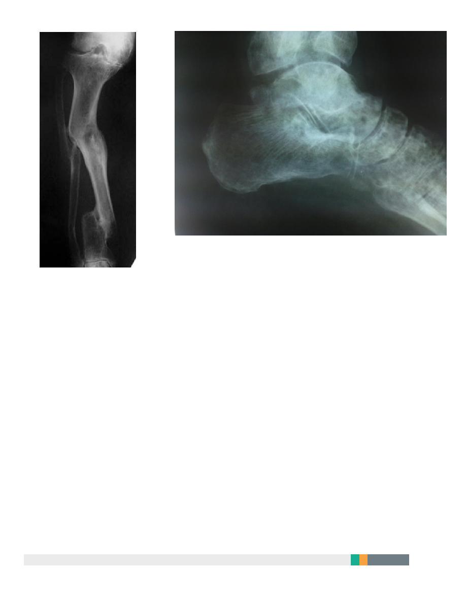
Complications
Infection after open fractures and open surgery.
Malunion: shortening >1.5cm, angulation > 7degrees or rotation should
be corrected.
Delayed union and non-union; bone grafting and good fixation is
required.
Joint stiffness.
Complex regional pain syndrome.
7
A.L.Y
8
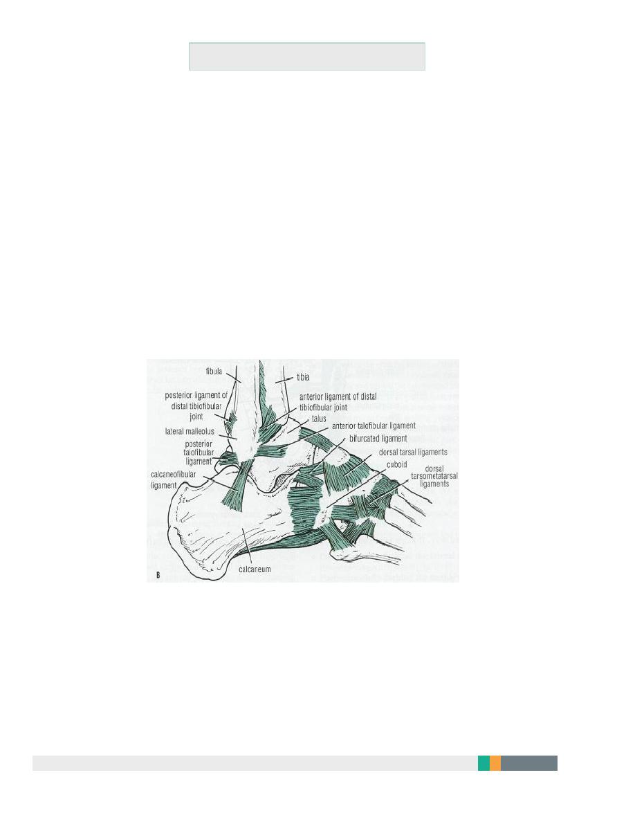
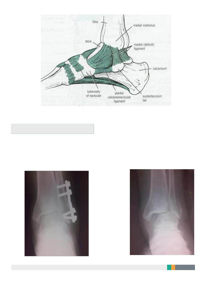
Ankle ligament injuries
Range from:
Stretching of the ligament.
Partial tear: healing restores full function.
Complete tear: joint instability.
Usually involves lateral ankle ligaments (ant. Talofibular lig., talocalcaneal
lig., and post. Talofibular lig.).
Medial calcaneal lig. (deltoid lig.) can result from abduction or eversion
injury
.
9
Clinical features:
Bruising, swelling, tenderness (usually distal and anterior to lateral Maleolus
in anterior talofebular lig. Injury).

10
Treatment
Partial tears:
Elastic bandage and gentle active exercise.
Complete tears:
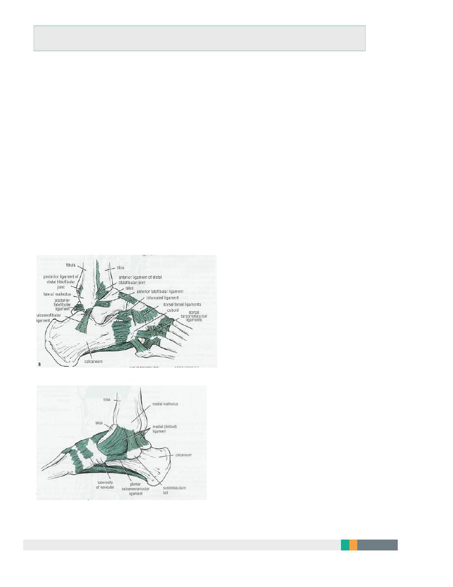
Cast immobilization from below knee to toes for 6 wks then physiotherapy.
If this regime fails; operative repair is done.
Complications:
Recurrent sprains.
Recurrent giving way or instability.
11
FRACTURES AND FRACTURE- DISLOCATIONS OF THE ANKLE
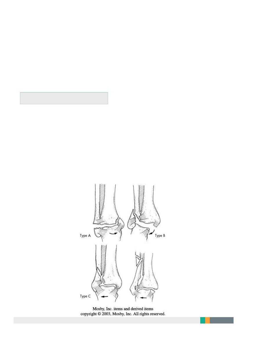
Injury may involve one or more of the following structures:
Lateral maleolus.
Medial maleolus.
Tibiofibular syndesmosis.
Lateral collateral ligament (ant. And posterior talofebular lig., and
calcaneofibular lig.).
Medial collateral (deltoid) lig.
Tibial articular surface (tibial plafond).
Position of the talus
.
ANKLE LIG. LATERAL
ANKLE LIG. MEDIAL
Clinical features
12
History of severe inj. In young athletes or simple inj. In elderly
osteoporotic.
Ankle twist.
Intense pain, swelling, inability to stand and deformity.
Look for points of tenderness espetially over medial or lateral malleolus.
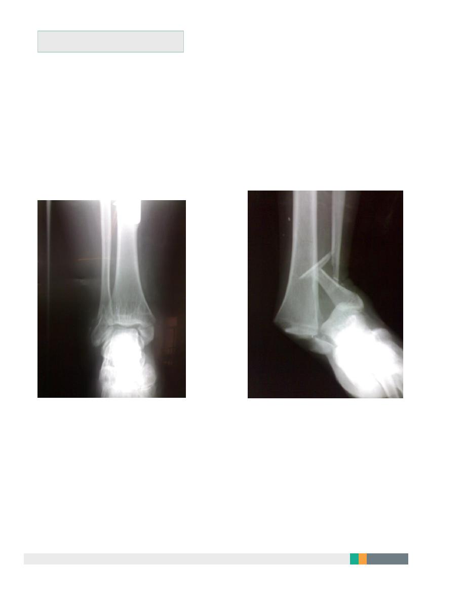
Danis-Weber classification
(based on the level of fibular fracture)
Type A: a fibular fracture below syndesmosis and oblique fracture medial
maleolus.
Type B: fibular fracture at syndesmosis + disruption of ant. Fibers of
tibiofibular lig. + fracture of medial malleolus or rupture of deltiod lig.
Type C: fibular fracture above syndesmosis+ tibiofibular lig. (syndesmosis)
is torn. Unstable fracture-subluxation of ankle mortise.
13
Principals of treatment
Treatment within hours or after several days until swelling subside by
elevation and splintage.
Anatomical reduction is a must to avoid later osteoarthritis.
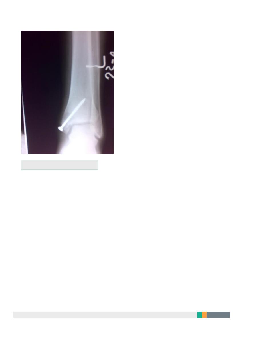
Fibular fracture below tibiofibular lig. Ankle jt. is stable; reduce the
fracture and immobilize in a cast.
Fibular fracture above tibiofibular lig. Ankle jt. is unstable; usually need
internal fixation.
14
Medial maleolus fracture fixation
Postoperative management
Below knee cast for 4 wks in low-level injuries, and 6-8 wks in fractures
treated by surgery.
Complictions
Joint stiffness and prolonged swelling. Treat by compression stocking
and elevation.
Complex regional pain syndrome.
Osteoarthritis
15
Fractures of Talus
Rare and usually due to considerable violence- FFH, car accidents,…
May involve the body, neck, head or dislocation of the talus.
X-ray: difficult to diagnose. May need to repeat several days later to see
the fracture.
C-T in difficult cases.
Treatment
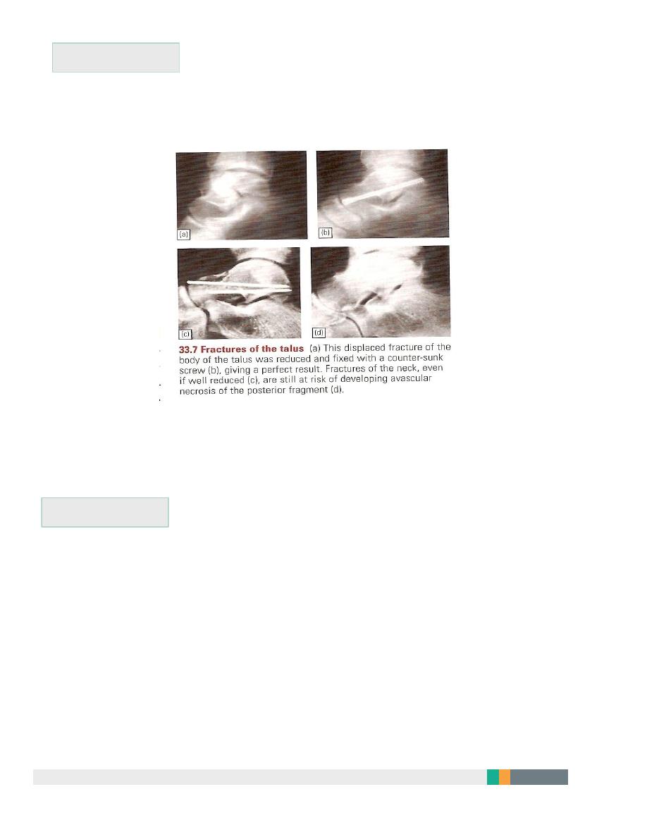
Trivial displacement: Below knee plaster with knee plantigrade for 6-8
wks
Displaced fractures or fracture dilocations: urgent reduction by closed
manipulation; if fails, ORIF
16
Complications
Non-union.
Avascular necrosis of body after fracture of neck.
Oseoarthritis.
Calcaneal fractures
Usually after FFH.
Associated injuries: spine, pelvis, hip or base of skull.
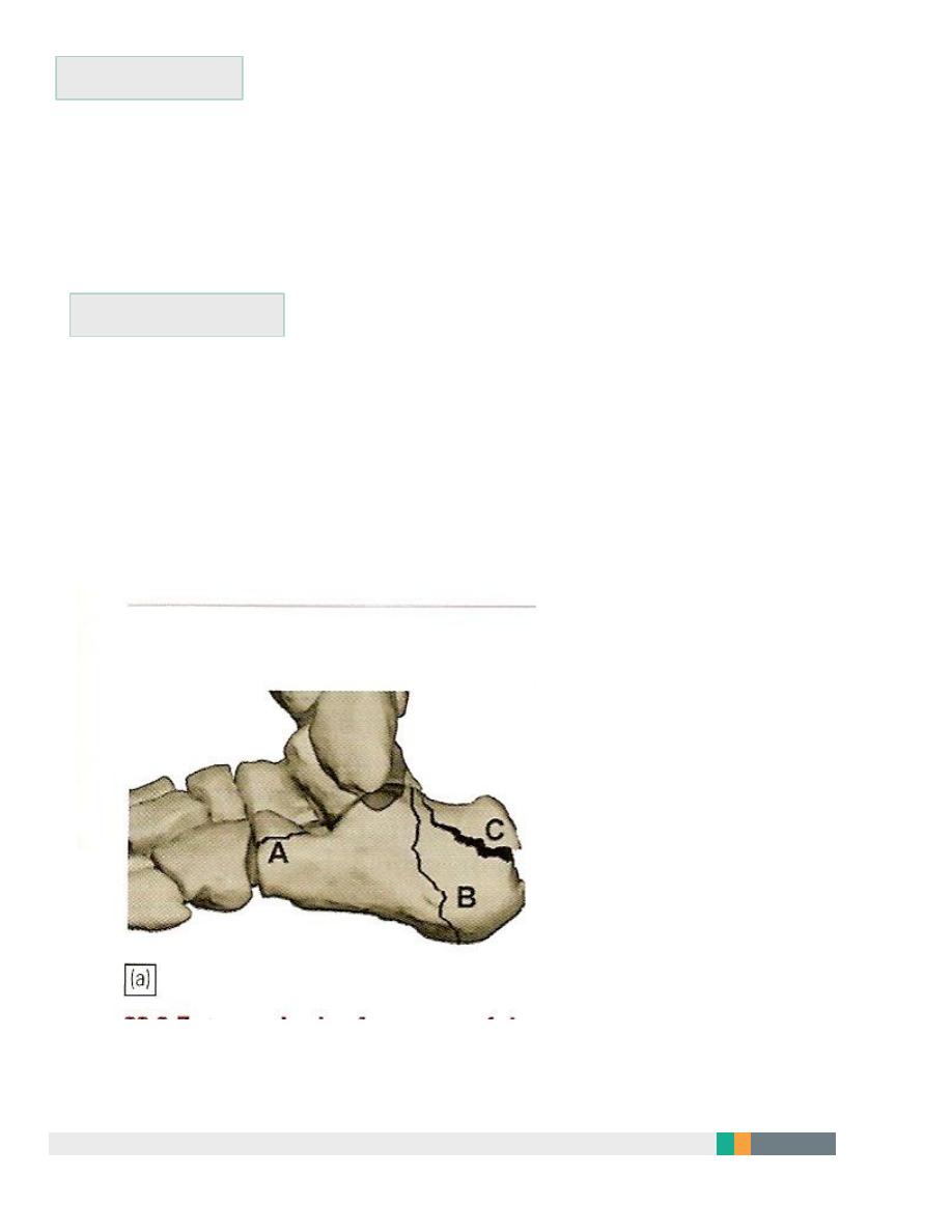
Extra-articular fractures: need closed treatment. Have good prognosis.
Intra-articular fractures: involve superior articular surface
May be comminuted.
17
Special features
Foot painful, swollen, bruised and the heel look broad. Movement is
painful.
Signs of compartment syndrome: intense pain and diminished
sensation.
Necessary to X-ray the knees, spine, and pelvis.
Treatment
Admit to hospital, elevate the leg and apply ice-packs until swelling
subside.
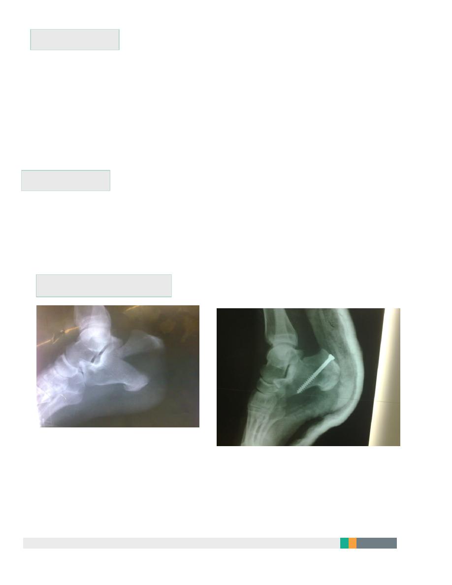
Undisplaced fractures: closed treatment.
Displaced fractures: ORIF with screws.
Calcaneal tuberosity fracture
Complications
Broadening of the heel
Talocalcaneal stiffness and OA

18
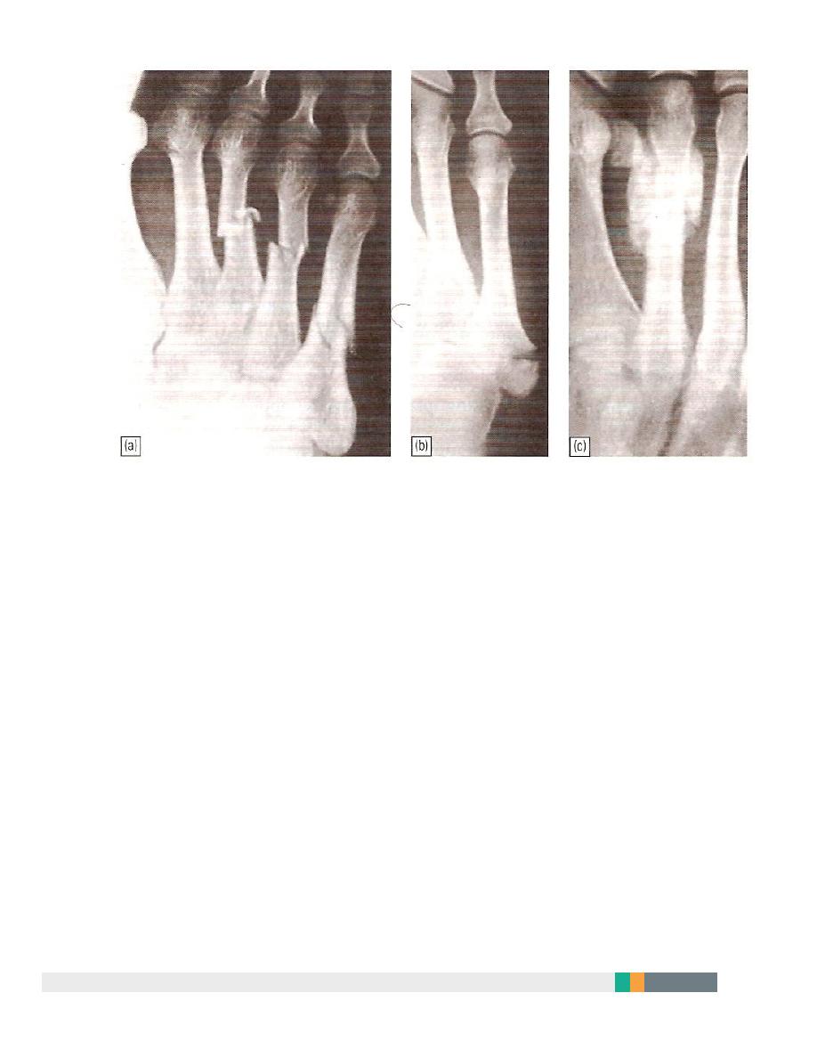
Metatarsal fractures
Mechanism of injury:
Direct blow.
Twisting.
Repetitive stress.
Treatment:
Walking plaster for 3 wks.
Displaced fractures; Kirschner wire fixation.
Stress injury (march fracture)
Young adult (often recruit or a nurse)
Painful foot after overuse.
Tender lump in shaft of metatarsal (usually 2
nd
).
X-ray: at first normal but later show hairline fracture and callus.
Radioisotope shows early increased activity.
Avulsion of base of fifth metatarsal
Result from forefoot inversion injury.
Base of 5
th
MT avulsed by peroneus brevis tendon.
Treatment: Below knee walking cast for 3wks.
19
A.L.Y