1
2

3
Lec:1
Surgery
History, Physical exam and Specialized
methods of urological diagnosis
History:
Pain: Location (CVA, genitals, suprapubic) Onset, Quality ( colicky, burning ) Severity,
Radiation .
Associated symptoms: Fever, Chills, Weight loss, Nausea, Vomiting .
Irritative symptoms: Frequency, Nocturia, Dysuria, Urgency .
Obstructive Symptoms: Hesitancy, Straining, Intermittency, decreased force or caliber
of stream, Prolonged voiding, post-void dribble, Incomplete emptying .
Incontinence: Stress incontinence, Urge inco., Overflow inco., history of neurological
problems, past pregnancies and method of delivery, past abdominal-pelvic operations
Urine: Hematuria, Pneumaturia, foul smell, Colour (cloudy, white, orange)
.
Urethral discharge: ( colour, amount, smell ), Sexual history, UTIs, external skin
lesions, lymphadenopathy .
Others: Renal calculi, Infertility, Erectile dysfunction, Congenital disorders, family
history of urological disease.
Physical Exam:
Inspection
Abdomen : masses, Scars from previous operations, Suprapubic distention, hair
distribution .
Penis : Circumcision, Phymosis/Paraphymosis, Epispadias, Hypospadias, Urethral
discharge, Superficial ulcers or vesicles, Venereal warts, Meatal stenosis, Balanitis .
Scrotum : Testicular atrophy, Testicular asymmetry, Dilated veins (varicocele ) on
Standing, Scrotal erythema/edema/cysts/hemangiomas .
Palpation
Kidneys, Bladder, Penis, Testes, Vas deference, Epididymis, Prostate .
Abdomen : Masses, CVA tenderness, Suprapubic distention/ tenderness (examine for
dullness on percussion), lymphadenopathy.
Penis : Peyronie’s plaques, Penile masses, Penile tenderness .
Scrotum : Scrotal tenderness/masses (size, consistency, location, mobility, shape),
hernia, hydrocele, spermatocele.
Spermatic cord : ( varicocele, fusiform enlargement, thickening of the cord ), absence
of vas deference .
Epididymal : size/induration/tenderness .
Prostate : on DRE ( Digital Rectal Exam )
4
Size, Consistency ( rubbery, hard, boggy, indurated ), Nodularity ( size, location ),
Tenderness, Warmth .
SPECIALISED METHODS OF UROLOGICAL DIAGNOSIS
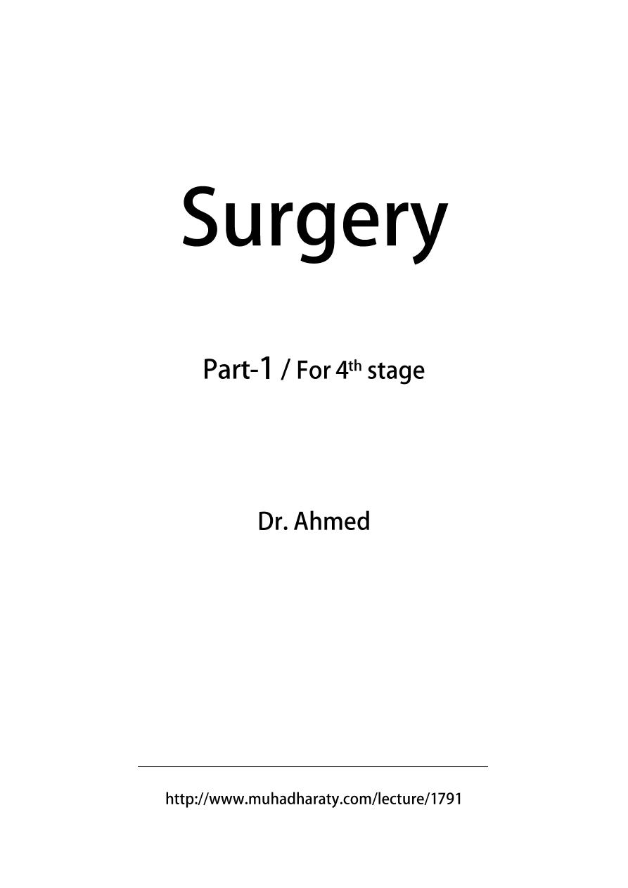
Clinical examination
Fig. bimanual palpation of the kidney
Laboratory investigation
Urine analysis
This is best performed on a mid-stream specimen of urine. After cleansing the
external urethral meatus, the first 20 ml or so of urine (containing bacteria and cells
from urethra) are discharged before collecting the next part of the voided urine in a
sterile container.
Chemical tests
"Dipstick" = a strip coated with chemicals for measuring the urine pH and for
detecting the presence of glucose, protein or blood; bilirubin, urobilinogen, ketones
and nitrites can also be detected.
The urine pH
Varies between 4.5 and 8.0 Persistently alkaline urine (pH > 8.0) suggest infection
with urea-splitting organism such as Proteus mirabilis
Protein
The amount of protein in the urine is normally less than 100 mg/24 h. Dipstick will
only detect levels greater than 300 mg/l Transient proteinuria (e.g. UTI) or persistent
(glomerulopathia)
Glycosuria
Usually diabetes mellitus, rarely renal glycosuria
Microscopy
Microscopical examination of urine directly of the urinary sediment studied after
centrifugation
Red blood cells
White blood cells
Epithelial cells
Casts - from glomerular disorders (hyaline or cellular)
Crystals - related to stone disease
Bacteria - Gram stain should be performed; if tuberculosis is suspected, the urinary
sediment should be stained using the Ziehl-Nielsen methods
Ova - schistosomiasis
Culture

5
The specimen should be plated out promptly or refrigerated until processing to
prevent multiplication of bacteria after voiding.
Significant infection is present if there are more than 100 000 (= 10 5 ) organism/ml ,
whilst counts less than 10 000 (=10 3 )/ml suggest contamination.
Antibiotic sensitiveness is determined using culture plates with antibiotic discs that
inhibit the growth of susceptible organisms.
If tuberculosis is suspected, three early morning samples of urine (EMU) are taken
and cultured on Lowenstein-Jensen medium .
Blood tests
Renal function studies
the plasma urea (normal range 2,3-6,9 mmol/l) and creatinine (normal range 50-120
µmol/l)
creatinine clearance (normally 100-140 ml/min) - closely approximates to the
glomerular filtration rate (GFR)
Haematology
Anaemia - tumours, renal impairment
White blood cell count - may raised in infections
ESR - elevated in certain disorders, tumors, retroperitoneal fibrosis
Other tests
PSA - see BPH and prostate cancer
Diagnostic imaging
Plain abdominal X-ray (KUB)
The KUB (a plain X-ray to include the kidneys, ureters and bladder) is useful to detect:
Radio-opaque urinary calculi (90% of calculi) unless they overlie areas of the bony
skeleton
Soft tissue masses in the renal areas and pelvis
Gallstones (10%)
Pelvic phleboliths
Calcified lymph nodes
Sclerotic deposits in prostate cancer (osteoplastic metastases
for other tumours are more typical osteolythic metastases)
Intravenous urography (IVU)
After a plain film, iodine-containing contrast medium is injected intravenously and
serial films are taken to follow its excretion by the kidneys
The nephrogram phase - on the initial film 1-3 minutes after injection, contrast
medium is in the glomeruli and proximal tubules so that a clear image of the renal
outline is obtained
The pyelogram phase - subsequent excretion of contrast medium outlines the
collecting systems, renal pelvis, ureter and bladder, showing any structural
abnormalities or filling defects

6
The procedure may be complicated by allergic reaction to the contrast medium,
ranging in severity from a mild urticarial rash to anaphylactic shock .
Ultrasound
The most frequently used radiological techniques in urological disorders
Almost all urological out-patient department are able to perform ultrasound
immediately after physical examination
Colour-flow Doppler techniques - measuring blood flow
CT scanning (computed tomography)
Multidetector Spiral CT
It enables reconstructions in different planes and biphasic CT angiography
PET/CT
Combination of positive emission tomography and CT.
It allows precise localisation of tumours.
MRI (magnetic resonance imaging)
Arteriography
Renal arteriography is used in diagnosis of renal vascular disorders, renal tumours and
renal trauma; therapeutic embolisation of the renal artery can be performed at the
same time to control bleeding from the kidney.
Iliac arteriography is useful for assessing the pelvic tumours or trauma, and
therapeutic embolisation of the internal iliac artery is occasionally used for
uncontrollable bladder haemorrhage, pelvic trauma, and priapism.
Other radiological techniques
Antegrade pyelogram - contrast medium is injected via a small-bore needle passed into the
collecting system under local anaesthetic or via a percutaneous nephrostomy
Ascending ureterogram - using a catheter inserted into the ureteric orifice at cystoscopy
Urethrography (ascending and descending) - contrast medium is instilled directly into
urethra (ascending urethrography), contrast medium is passed out and is performed with
micturition
cystogram (it can demonstrate vesicourethral reflux) and descending urethrography.
Urethrography is useful for diagnosis of urethral stricture mainly.
Lymphography - following injection of contrast medium into a lymphatic in the foot is used
to demonstrate the iliac and para-aortic nodes in pelvic malignancy; nowadays, it has been
replaced by CT scanning
Radionuclide studies
Renal scintigraphy
It is useful mainly for dynamic diagnosis - upper urinary tract obstruction, assessing of renal
function of both kidneys.
Bones scintigraphy
It is most widely used in the detection of bony metastases from prostatic, bladder and renal
carcinoma.

7
Lec:2
Surgery
Specialized methods of Urological Treatment
CATHETERISATION
Catheters are used mainly therapeutically to relieve urinary retention.
Types and sizes of catheters
Material :
- soft silicon coated latex (silicon is highly resistant to incrustation) , it can be introduced up
to 4 weeks
- 100% silicon - it can be introduced up to 8 weeks (it is better, but more expensive)
Types of catheters :
1. One way catheter : for dilatation of urethral stricture, to discover residual urine (better it
is performed by ultrasound), to introduce contrast medium into the bladder
2. Two way catheter : self-retaining balloon catheter = Foley catheter
3. Three way catheter : for irrigation (lavage) of bladder (by bleeding to bladder after
prostatectomy, due to bladder tumour)
Division by a tip of catheter :
Nelaton - straight round tip
Tiemann - curved pointed tip
Size of catheters :
French scale = Charrie scale = circumference in mm diameter is size in F (Ch) divided by
( = 3.14) e.g. catheter 18 F has diameter 6 mm
ENDOSCOPY
Two types of endoscopes:
1. rigid .
2. flexible .
Examination is more difficult, an endoscope is more expensive, through this
endoscope can be passed only flexible instruments, but for patient is this
flexible endoscopy more pleasant
Panendoscopy : Uretroscopy and cystoscopy = endoscopy of urethra and urinary
bladder
Ureteroscopy
: Endoscopy of ureter
Ureterorenoscopy : Endoscopy of ureter and renal pelvis too
8
Endoscopic operations
Lithotripsy : disintegration of stone in bladder and ureter
Transurethral resection of prostate (TURP = TUPE) and bladder tumour ( TURT )
Nephroscopy
It is used mainly for treatment of stones = nephrolitholapaxy
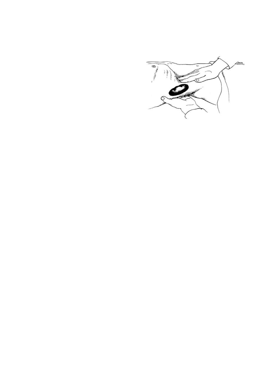
Nephrostomy
To the dilated renal pelvis is introduced under ultrasound and/or radiological control
catheter (= percutaneous nephrostomy).
Percutaneous nephrostomy step by step:
Needle puncture of dilated renal pelvis under X-ray or/and ultrasonography control
Guide wire passed down needle Dilatation of channel over guide wire
"pig-tail" catheter inserted over guide wire
BIOPSY
Prostate biopsy
Indication: suspicion on prostate cancer It is performed transrectal (or transperineal)
under mainly ultrasound control
Biopsy of kidney
It is done under ultrasonography or CT control. Mainly used in nephrology, biopsy for
kidney tumour is indicated rarely.

9
Lec:3
Surgery
Symptoms of urological diseases
Changes in urine volume
Anuria - diuresis < 100 ml/24h
Oliguria -diuresis < 500 ml/24h
Polyuria- diuresis > 1500 ml/24h
Chages in urine appearance
Haematuria-blood presence in urine
Pyuria : pus presence in urinae
Bacteriuria – bacteria in urine
Leukocyturia- leucocytes in urine
Proteinuria- proteins in urine
Pneumaturia – passage of gass
Stercoruria-stool presence in urine
Voiding disorders
Polakisuria or frequency – going to void too often
Urine retention- inability to void – subvesical obstruction
Overflow incontinence – bladder overdistension
Nocturia- frequet voiding on the night
Stranguria – painful voiding
Urinary incontinence :
o Urgent- overactive detrussor
o Stress – due to raised abdominal pressure
o Reflex
o Mixed
o Overflow incontinece
o Ureteric – ectopic ureteric orifice under the level of sphincter
o Enuresis nocturnal
Dysuria- difficult voiding
o Voiding initiation delay
o Interrupted voiding
o Weak urine stream
o Necessity of abdominal pressure during voiding
Renal pain
Nefralgia
o constant ache in lumbar area caused by distention of renal capsul
o inflamation, tumor, hydronephrosis

11
Renal colic
o colicky pain caused by spam and hyperperistalsis of smooth muscle organ.
o radiats from costovertebral angle, toward the lower anterior abdominal quadrant,
along the corse of the urether into the scrotum or vulva
o passage of blood clot or stone.
Vesical pain
Cystalgia:
o persistant pain localised behinde pubic
Painful contraction
o spasmodic bladder contraction
Urethralgia
o Several pain along the urethra
o Inflammation, tumor, foreign body
Prostatic pain - prostatodynia
o sharp pain promoting to the perineum
Orchalgia
o pain localised it the testes, often propagating along the spermatic cord
o inflammation, tumor, torsion
Haematuria
-
Presence of blood in the urine
-
Danger signal, that cannot be ignored
Macrohematuria:
Total
Initial- prostate, urethra
Terminal- urinary bladder
Microhemturia:
Noticeable just with microscope
4 erytrocytes/fields of view
Dismorphic erytrocytes detected by phase-contrast indicative of glomerular disease
Diferential diagnosis
Trauma
Urolithiasis
Tumor
Inflammation
Glomerulonephritis
BPH
Malformation
Sports
Coagulophaty

11
Examination
Chemical and microscopic examination
Bacterial culturs
PCR for TBC
Urine cytology
Ultrasonography
Pyelolithiasis
Trauma
Dilatation of KPS
Renal tumor, cysts
Tumor of urinary bladder
Anomalies
Cystolithiasis
BHP
CT
Trauma
Tumor of urinary tract
Urolithiasis
Abcess
Cystocopy
Tumor of urinary bladder
Varix in urinary bladder
Cystolithiasis
Fisstula

12

13
Lec:4
Surgery
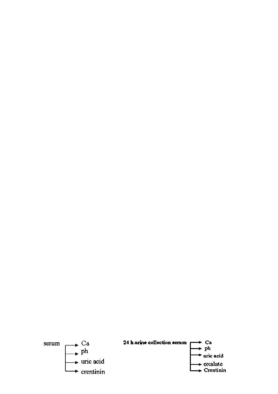
Urinary tract stone (calculi)
urinary calculi: composition ,frequency, and characteristics
Type of stone frequency (%) effect of ph on solubility radiographic density (bone=1.0)
Calcium stones 80
▪
▫ Oxalate 35 little effect 0.50
(monohydrate
and dihydrate)
▫ Phosphate 10 increased at ph <5.5 0.1
▫ Oxalate and 35 variable variable
phosphate
Struvite 10 increased at ph <5.5 0.20
▪
Uric acid 8 increased at ph >6.8 0.05
▪
Cystine 1
increased at ph >7.5 0.15
▪
Other types 1
▪
▫ triamterene
▫ xanthine increased at ph >6.8 0.05
▫ Matrix
(noncrystalline)
Factors associated with urolithiasis
Factor conditions of increased incidence
Genetics /heredity cystinuria – autosomal recessive
renal tubular acidosis type I
medullary sponge kindney
Geography high temperature/humidity
Diet increased intake of calcium or oxalate
Occupation sedentary
Type of stone eticologic factos
▪ Calcium oxalate supersaturation of urine with calcium
▪ Calcium phosphate from (1) renal leak ,(2) intestinal absorption,
▪ Calcium carbonate (3) bone resorption ; hyperoxalueia
▪ Uric acid hyperuricosuria , low uriea ph
▪ Cystine cystinuria
▪ Magnesium ammonium alkaline urine produced by urea-splitting
phosphate (struvite) organisms
▪ Matrix alkaling urine produced by urea-splitting
organisms

14
Epidemiology of stones
:
o USA---- 400.000 hospitalization annually
o Men: Women 3:1
Etiology and Pathogenesis :
o Multifactorial process
o Supersaturation
o Nucleation and aggregation of crystals
- exmp. : Uric acid , cystine , xanthine
o Inhibitors Pyrophosphete , Citrrate , mngnesium , zine
o Matrix : non crystalline mucoprotein – proteus-infection
o Exogenenous sutstances
o Indinavir – antiviral
o Triamterene } rudolucent stones
o Stone of the upper urinary tract
clinical presentation :
o obstration : pain – Hematuria
o nausea , vomiting -- urine infection
o silent stones
Diagnosis :
o urine A.
o KUB , ULS
o IVP
o Axinlor spirel CT.
Treatment :
o Conservative Treatment – Hydation
Low grade obstruction
Stone 4mm --- 90 % passage
Stone 6mm --- 20 % passage
intervention :
▫ infection
▫ high grad obstruction
15
o Expectant treatment
narrowing sides
PUJ ( ureteropelvic Junction)
Pelvic brim
Ureterovesical Junction
o Stone extraction
Nepheroscopy ( PCNL
(
Uretoroscopy
o Shock wave hithotripsy
o ureterolithotomy .
Bladder Stones
o outlet obstruction
o Foreign Bodies
o Passed Ureter stones
Clinical Presentation
:
o Pain – Hypogastrim
o Referred to penis
o Intermittent Stream
o Dysuria , Hematuria
Treatment
o Lithotrities (cystolitholapaxy)
o (Mechanical crushing devices)
o Electrolaydraulic Lithotripsy
o uslithotripsy
o pneumatic lithotripsy
o cystolithotomy
o Recurrent stone Disease
o predisposing factors can be identified in 80%

16
Hypercalciuria
o Resorptive Hypercalciuria
o Hyperparathyroidism >> 50% - Ca-oxlaete
o Metastatic bone
o Multiple myeloma
o Immobilization ( Spinal cord injury
(
o Cushings disease
o Hyperthyroidism
o Absorptive Hypercalciuria >> 50% Ca – Stones
o Exaggerated intestinal response to Dvit
o Renal Hypercalciuria >> 10% of Hypercalciuria
o Treatment : Hydrochlorothiazide 50 mg 1x2 potassium supplementation !
Treatment
o Hydration Fluid intake > 3 L .
o Alkalinization - sodium Bicarbonate
o Potassium citrate
o Reduction of uric acid load
o 90 gr. Protien
o Allopurinol ( 200 – 600mg )
Hyperoxalueria
o primary Hyperoxaluria → Rare autosomnl recessive disorder
Treatment : 100 – 400 mg Phyricoxine
o Enteric Hyperoxaluria
inflammatory bowel disease
Small bowel bypass surgery
Fatty acid → bind calcim
o Exogenous Hyperoxaluria
ascprbic acid → 5 gr/ day .
Struvite stones
o Triple – Phosphete stones
Composed of Mg- ammonium – Pho
sphat – Carbonate a patite
PH markedly elerated
Ammomin + bicarbonate in urine – Due to urea splitting organisms .
Protius species 75%
Klebsialla , pseudomonal , providencia Staphylococeus , and ureaplasma
urealyticum

17
Lec:5
Surgery
UTI in adults and children
Urinary Tract Infection in Adults
Incidence
UTI (urinary tract infection) - common, affecting all ages and both sexes.
The most common but one infections (the first - breath infections) .
Clinical syndromes associated with UTI:
Septicaemia (urosepsis)
Renal infection
o Pyelonephritis
o Pyonephrosis
o Renal abscess
o Peri- ET paranephric abscess
o Cystitis - bacterial, abacterial
o Prostatitis
o Urethritis
o Epididymitis, epididymo-orchitis
Methods of introducing UTI
Ascending infection - via urethra to bladder, reflux of infected urine up to ureter and/or
spread of organisms along peri-ureteric lymphatics
Infection via a fistula (e.g. vesico-colic)
heamatogenous infection (via renal artery)
Aetiology and pathogenesis
The urinary tract is normally sterile above the distal urethra
The chiefly defence mechanisms:
Hydrokinetic = the dilution of bacteria by the flow of urine

18
Mucosal = mainly secretion of immunoglobulin A (Ig A) and phagocytic capability of
the urothelium itself
Factors predisposing to infection
UTI - commoner in women:
1. Due to shorter urethra
2. Opening of urethra at the vaginal vestibule, which is readily contaminated with
faecal organism
3. In many young women, infection are precipitated by sexual intercourse, bacteria-
laden secretion from the perineum entering the urethra during sexual activity (so
called honey- moon cystitis)
In either sex UTI may develop:
1. Incomplete bladder emptying (residual urine) due to outflow obstruction (BPH,
urethral stricture …)
2. Bladder diverticula
3. Neuropathic bladder
4. Upper urinary tract stasis due to obstruction of ureter, mega ureter, stones
Vesico-ureteric reflux interferes with both ureteric and bladder emptying and is
commonly accompanied by infection
Calculi, bladder tumors and foreign bodies (e. g. catheters) are predispose to infection,
as may instrumentation of the urinary tract
Factors that suppress the immune response (diabetes mellitus, cytotoxic or
immunosuppressive agents)
Common urinary pathogens
1- Ascending infection Bacteria
Gram-negative - Escherichia coli - klebsiella spp, proteus spp, pseudomonas spp.
Gram-positive cocci - streptococcus faecalis - staphylococcus aureus
Chlamydia trachomatis
L-organism - ureaplasma urealyticum, mycoplasma hominis
Fungi - candida spp
2- Haematogenous infection
Bacteria - mycobacterium tuberculosis
Fungi
Parasites - schistosoma spp
Viruses - cytomegalovirus, adenovirus type 11

19
Clinical manifestation
Symptoms
Lower UTI
o Voiding symptoms - frequency, urgency, micturition with discomfort, burning
sensation (= dysuria)
o Occasionally hematuria
Upper UTI
o Loin pain
o Systemic disturbance - fever, sweating, rigors
o Some patients have lower UTI as well (often upper UTI follow lower UTI)
Physical signs
Fever and tachycardia
Tenderness in the loin and in the suprapubic region
Diagnosis
The presence of pus cells on microscopy
The presence of significant number (over 10 5 per ml) of organism in a mid-stream
specimen of urine (MSU)
Microbiology laboratories determines antibiotic sensitivities
Specialized microbiological techniques may be required in certain circumstances (e. g.
Tuberculosis, fungal infection, viral infection)
Further investigation
Cystitis in young sexually active women investigation is not required for the first attack
unless it is accompanied by haematuria or loin pain
investigation is indicated in this group of women for recurrent infections, in older
women, pregnant women, children, men, diabetes mellitus, neuropathy, known urinary
stones or urinary tract anomaly - urinary tract ultrasound, if indicated IVU, blood count,
the serum urea and creatinine
Treatment
Antibiotics commonly used to treat UTI:
o Nitrofurantoin
o Co-trimoxazol (sulfamethoxazol + trimethoprim) and trimethoprim alone

21
o Ampicillin, amoxycillin, co-amoxycillin (clavulic acid + amoxicillin)
o Gentamicin
o Quinolones (norfloxacin, ciprofloxcin)
o Cefalosporins
High fluid intake and regular emptying of the bladder to promote hydrostatic clearance
of bacteria
Attention to personal hygiene for women with recurrent infection
In patients with collections of infected urine or pus (e.g. pyonephrosis, perinephric
abscess) drainage is usually required
Upper Urinary Tract Infection
Acute renal infection
Most result from ascending infection (75% of patients have preceding lower-tract
symptoms)
Some they are result of haematogenous spread
There is important to distinguish between infection alone and infection combined with
upper-tract obstruction; the latter combination may lead to rapid obstruction of renal
tissue unless prompt drainage of the obstructed kidney is established
Acute pyelonephritis
Acute inflammation of the pelvic epithelium, with bacteria entering the collecting duct and
fornices to produce inflammation of the renal parenchyma
Renal carbuncle
An abscess in the renal parenchyma and is usually due to haematogenous spread of organisms
(typically staph. aureus from foil, infected infusion site, contaminated needles in drug addicts)
Pyonephrosis
Infection within an obstructed kidney rapid destruction of kidney
Perinephric abscess
It result from any of the above infective processes

21
Initially the infection is confined by Gerota’s fascia (= perinephric abscess), but may
rupture through this (= paranephric abscess) and to reach the skin (in Petit’s lumber
triangle) , the psoas muscle or the bowel; it may even rupture through the diaphragm to
reach the pleura and lungs
Clinical symptoms
Loin pain, fever, tachycardia, scoliosis in severe cases
Mass may be palpable in the loin
Septicaemia and shock
Investigation
Urine should be examined for pus cells and bacteria (urine culture), blood culture (all
patients with pyrexia or clinical suspicious of septicaemia)
Ultrasound (urinary tract, liver, spleen)
Plain abdominal X-ray, chest X-ray, IVU[
Management
Septicaemic patient
Rapid intravenous fluid replacement
Intravenous hydrocortisone or methylprednisolone
Parenteral bactericid antibiotics
Subsequent management depend on the pattern of infection, basic treatment is are
antibiotics.
Acute pyelonephritis
Antibiotics for 7-14 days, guided by the result of urine culture and sensitivity
Renal carbuncle
Drainage
By aspiration of the abscess under ultrasound or CT control
By open surgery

22
Pyonephrosis
Drainage by percutaneous nephrostomy or with a ureteric catheter passed retrogradely
from the bladder at cystoscopy
After improvement ascendant pyelography or descendent pyelography (nephrostogram)
identification of obstruction
Renal scintigraphy determines remaining renal function
Treatment of obstruction (e. g. ureteroscopy for ureterolithiasis, nephrectomy if kidney
function is by scintigraphy under 10-15 %.
Perinephric abscess
Surgical drainage or nephrectomy, if function in the affected kidney is very poor
Chronic pyelonephritis
Combination of renal scarring and urinary infection
It may follow vesico-ureteric reflux and infection
Repeated episodes of acute pyelonephritis
Differential diagnosis of other types of interstitial nephritis or hypoplasia of kidney is
difficult
Treatment
Eradication of infection to prevent further renal damage. Nephrectomy, if:
Renal function is under 10 (15) %.
Severe secondary hypertension.
Xantogranulomatous pyelonephritis
The result of granulomatous reaction within kidney to chronic infection
Treatment
Nephrectomy
Lower Urinary Tract Infection
Acute bacterial cystitis
Usually result of ascending bacterial infection from the perineum
Particularly common in women (due to short urethra)

23
Clinical features:
Frequency and urgency of micturition with dysuria
There may be suprapubic pain, urine often has a fishy smell or may be blood stained =
haemorrhagic cystitis)
Association of loin pain and fever suggest spread of infection to the kidney (acute
pyelonephritis)
Management
MSU (including urine culture) before treatment to confirm the diagnosis
Antibiotics for a 5 days period this can be changed, if necessary, on the basis of
antibiotics sensitivity tests
Analgetics and spasmolytics (the best in combination e.g. Algifen®)
Resolution of symptoms = MSU to repeat at 2 weeks and at 3 months to ensure
eradication of infection
Chronic and recurrent bacterial cystitis
Clinical symptoms
Similar to acute cystitis
Histologically cystic changes (cystitis cystica) and squamous metaplasia
Treatment
In women self-help advice
Increase fluid intake
Pass urine every 2 hours
Regular washing of the vulva and vaginal introitus
Wipe from front to back after bowel actions
Empty the bladder after sexual intercourse (if the symptoms are precipitated by sex)
Infection (antibiotics)
Long-term low dose antibiotics (6-12 months), e. g. furantoin 100 mg daily,
trimethoprim 100 mg twice daily, co-trimoxazol one tablet (480 mg) one or twice daily
Immunotherapy (e.g. Uro-Vaxom®)
In women, whose infections are precipitated by sexual intercourse, voiding and single
dose of antibiotics after intercourse may be prevent infection developing

24
Abacterial cystitis
Trauma, toxic drugs (e. g. severe haemorrhagic cystitis is caused by cyclofosfamid),
chemicals, irradiation, viruses and related organism such as chlamydia trachomatis
Interstitial cystitis
Special type of chronic abacterial cystitis. Well recognise syndrome of unknown
aetiology.
Diagnosis and treatment are very complicated.
Asymptomatic Bacteriuria
1-2 % schoolgirls, 3-5% of adult women, 0.5% schoolboy, 0.5% of adult male.
Management
Exclude some abnormalities of the urinary tract
Active treatment – pregnant woman due to 30% risk of developing acute pyelonephritis
Other treatment is doubtful
Urinary Tract Infection in Children
Two special problems:
1- Symptoms of urinary infection in small children may be non-specific
2- Collection of urine, particularly in small girls, may be difficult
By coincidence UTI anomalies of urinary tract - 3 groups of children with UTI:
1- Anomalies, which can be lead to rapid deterioration in renal function - reflux,
obstruction
2- Relatively harmless anomalies - duplication of upper tract, bladder anomalies
3- Normal urinary tract