The Skeletal System
Dr.Haythem Ali AlsayighDepartment of Human Anatomy and Histology
UNIVERSITY OF BABYLON
COLLEGE OF MEDICINE
The Skeletal System summary
1-The skeletal system develops from mesenchyme, which is derived from the mesodermal germ layer and from neural crest.2-Some bones, such as the flat bones of the skull, undergo membranous ossification; that is, mesenchyme cells are directly transformed into osteoblasts
3-In most bones, such as the long bones of the limbs, mesenchyme condenses and forms hyaline cartilage models of bones.
4-Ossification centers appear in these cartilage models, and the bone gradually ossifies by endochondral ossification
The skull consists of the neurocranium and viscerocranium (face).
The neurocranium includesa membranous portion, which forms the cranial vault,
and a cartilaginous portion,the chondrocranium, which forms the base of the skull.
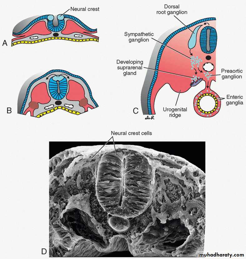
Neural crest cells form the face, most of the cranial vault,
and the prechordal part of the chondrocranium (the part that lies rostral to the notochord).
Paraxial mesoderm forms the remainder of the skull.
Limb
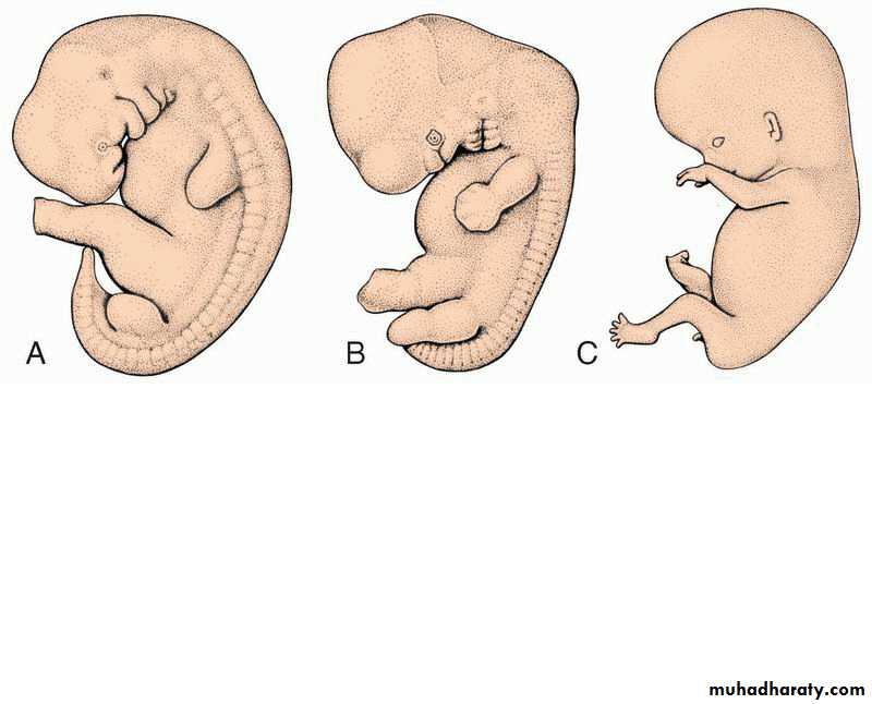
Limbs form as buds along the body wall that appear in the fourth week.
Lateral plate mesoderm forms the bones and connective tissue, while muscle cells migrate to the limbs from the somites.
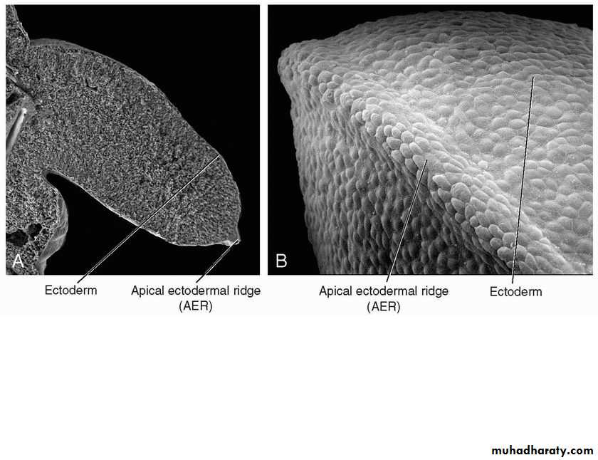
The AER regulates limb outgrowth, and the ZPA controls anteroposterior patterning.
Many of the genes that regulate limb growth and patterning have been defined (Fig. 9.17).
vertebra
The vertebral column and ribs develop from the sclerotome compartments of the somites,and the sternum is derived from mesoderm in the ventral body wall.
A definitive vertebra is formed by condensation of the caudal half of one sclerotome and fusion with the cranial half of the subjacent sclerotome .
The many abnormalities of the skeletal system include vertebral (spina bifida), cranial (cranioschisis and craniosynostosis), and facial (cleft palate) defects. Major malformations of the limbs are rare, but defects of the radius and digits are often associated with other abnormalities (syndromes).
Musculo system
Most muscles arise from the mesoderm.Skeletal muscles are derived from paraxial mesoderm,
including (1) somites, which give rise to muscles of the axial skeleton, body
wall, and limbs, and
Musculo system
(2) somitomeres, which give rise to muscles of the head.Progenitor cells for muscle tissues are derived from the ventrolateral (VLL) and dorsomedial (DML) edges (lips) of the prospective dermomyotome.
Musculo system
Cells from both regions contribute to formation of the myotome.
Some cells from the VLL also migrate across the lateral somitic frontier into the parietal layer of the lateral plate mesoderm. This frontier or border separates two mesodermal domains in the embryo: (1) the primaxial domain that surrounds the neural tube and contains only somitederived cells (paraxial mesoderm) and (2) the abaxial domain that consists of the parietal layer of lateral plate mesoderm in combination with somite-derived cells that migrate across the frontier into this region. Abaxial muscle precursor cells differentiate into infrahyoid, abdominal wall (rectus abdominus, external and
internal obliques, transversus abdominus), and limb muscles. Primaxial muscle precursor cells form muscles of the back, some muscles of the shoulder girdle, and intercostal muscles
(Table 10. Muscles of the back (epaxial muscles) are innervated by dorsal primary rami;
muscles of the limbs and body wall (hypaxial muscles) are innervated by ventral primary rami. Molecular signals for muscle cell induction arise from tissues adjacent to prospective muscle cells. Thus, signals from lateral plate mesoderm (BMPs) and overlying ectoderm (WNTs) induce VLL cells; while signals from the neural tube and notochord (SHH and WNTs) induce DML cells. Connective tissue derived from somites, parietal mesoderm, and neural crest (head region) provides a template for establishment of muscle patterns.
Most smooth muscles and cardiac muscle fibers are derived from splanchnic mesoderm.
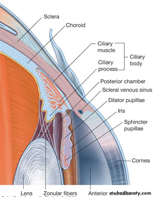
Smooth muscles of the pupil, mammary gland, and sweat glands differentiate from ectoderm.
skeletal system develops from:
1. Paraxial mesoderm.2. Lateral plate mesoderm; somatic layer :
Shoulder bones
Pelvic girdle
Bones of limbs
Sternum
3. Neural crest;
Bones of face
and skull
The skeletal system develops from:
1. The paraxial mesoderm.2. The somatic layer of lateral plate of mesoderm (as the bones of the shoulder and pelvic girdle, bones of the limbs).
3. The neural crest cells; in the head region they differentiate into mesenchyme and participate in formation of bones of the face and skull
The paraxial mesoderm:
At 3rd weekParaxial develops into:
Somitomeres: head region
Somites: from occiput to caudal end of trunk.
first pairs of somites ;occipital region at 20th day
proceeds cephalocaudally forming 3 somitic pairs each day
End of 5th week: 42-44 somitic pair as :
4=occipital 8=cervical.
12=thoracic. 5=lumber.
5=sacral. 8-10=coccygeal.
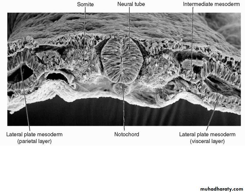
During the 3rd week, the mesoderm on the side of the midline i.e. Paraxial, develops into segmented tissue blocks on the sides of the neural tube, these are called the somitomeres , they develop in the head region and form the head mesenchyme. These blocks develop more to form the somites in the regions from the occiput to the caudal end of the trunk. The pairs of somites occurs first in the occipital region at the 20th day, and then proceeds cephalocaudally forming 3 somitic pairs each day. Finally, 42-44 somitic pair develops at the end of the 5th week as following:
4=occipital, 8=cervical. 12=thoracic. 5=lumber.
5=sacral. 8-10=coccygeal.
The first occipital and the last 5-7 coccygeal disappear. The other somites form the axial skeleton.
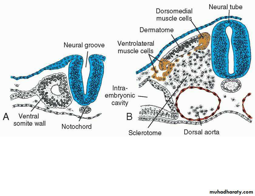
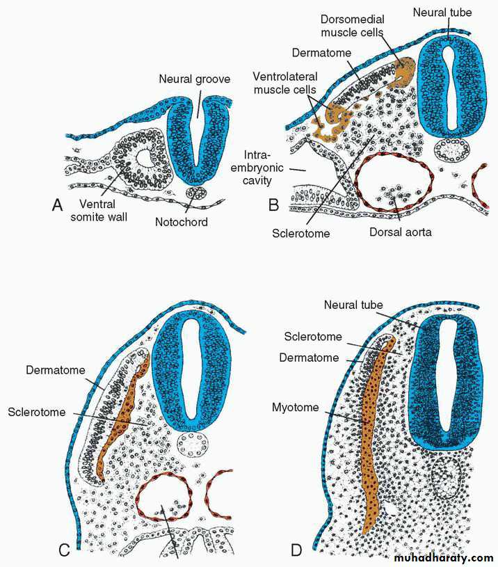
During the 4th week, each of the somites has three parts:
The paraxial mesoderm:
Parts of Somites
4th weekVentromedial part:
(sclerotome) : differentiate to loosely C.T.
Surrounding notochord and spinal cord.
This mesenchymal tissue differentiates later to:
• Fibroblast
• Chondroblast
• Osteoblast
vertebral column
Parts of Somites
Ventrolateral part;Form muscle of body wall and limbs.
Dorsomedial part;
form back muscles
Cells between these two groups ;
form dermatome which forms dermis and subcutaneous tissues
of the skin having segmental nerve supply.
Cells from both muscle precursor groups become mesenchymal and migrate beneath the dermatome to create the dermomyotome.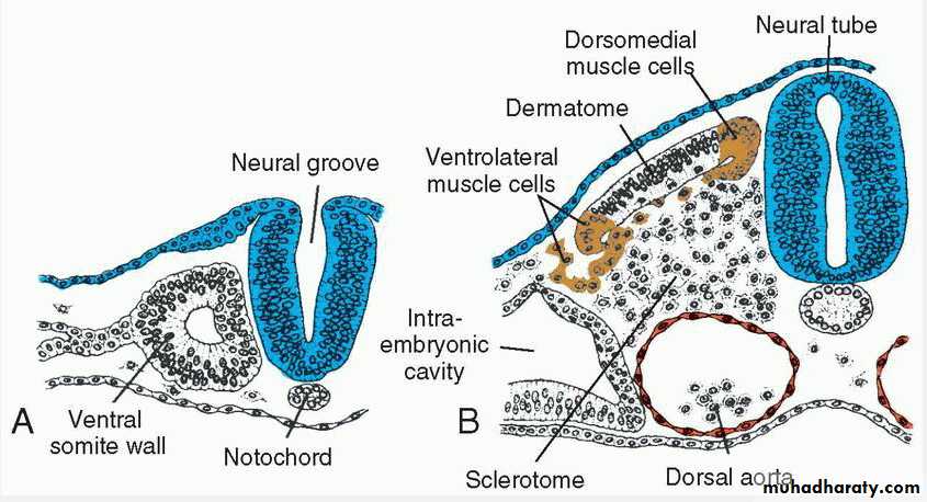
Skull
The skull can be divided into two parts: the neurocranium, which forms a protective case around the brain, and the viscerocranium, which forms the skeleton of the face.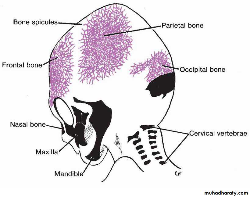
Development of the skull
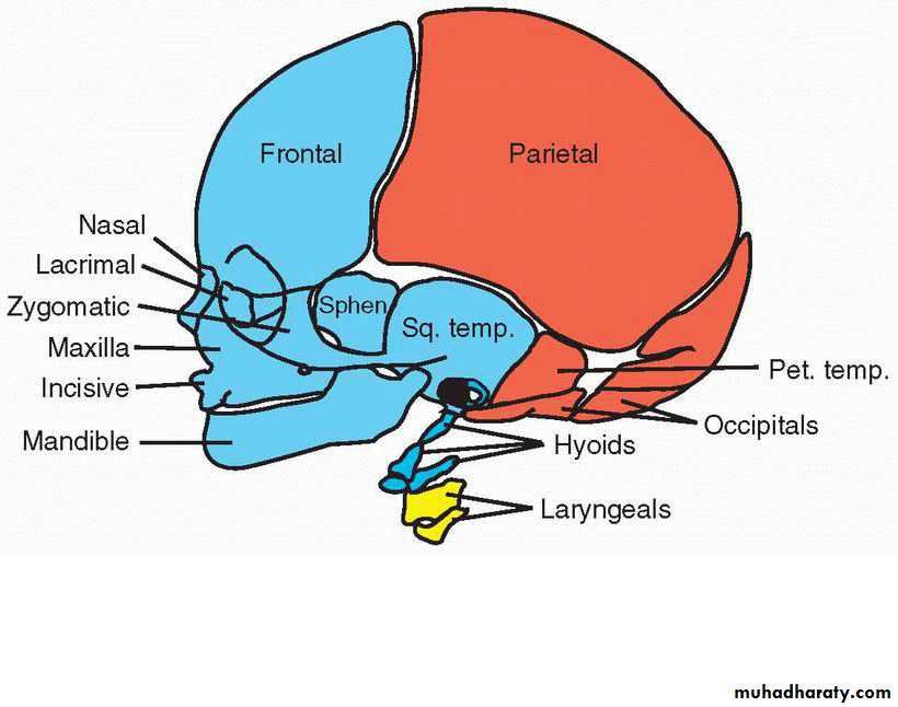
• Neurocranium: from 2 portions:• Membranous portion:
• flat bones of brain vault
• Derived from neural crest and occipital paraxial mesoderm
• Bony specules
• Enlarged after birth by osteoblastic activity at outer and osteoclastic activity absorbing inner surface
Development of the skull
• Neurocranium: from 2 portions:• Membranous portion:
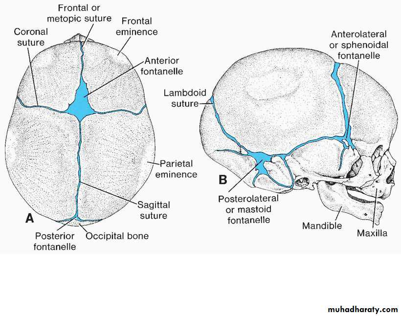
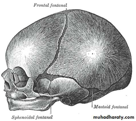
• At birth; sutures (CT seams)
• Derived from NC cells and PM
• Fontanelles
• Allow for molding
Closure of Fontanelles
In humans, the sequence of fontanelle closure is as follows:
Posterior fontanelle closes 1-3 months after birth
Sphenoidal fontanelle close around 6 months
Mastoid fontanelle closes from 6 to 18 months
Anterior fontanelle is the last to close between 1-2 0r 3 years.
Development of the skull
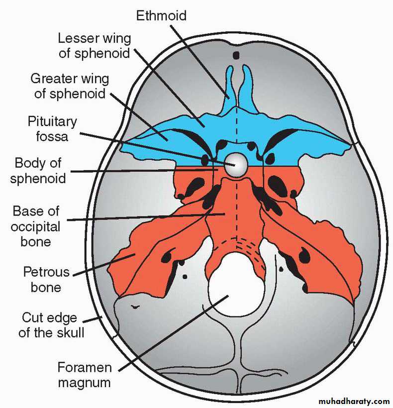
• Neurocranium: from 2 portions:• Cartilagenous portion: (chondrocranium)
• Forms bones of base of skull& floor of cranial cavity.
• consists of a number of separate cartilages
• prechordal chondrocranium; from NC cells
• chordal chondrocranium; from occipital sclerotomes
Development of the skull
• Viscerocranium:
• Maxillary process:
• Mandibular process:
• with dorsal tip of mandibular process gives rise to incus, the malleus, and the stapes
Development of the limb bone
4th weeklimb buds project from ventrolateral body wall
extensions from somatic layer of LPM covered by ectoderm.
6th week; flat plates of hand and foot develop distally.
Later, constrictions appear between parts of the limb
Development of the limb bone
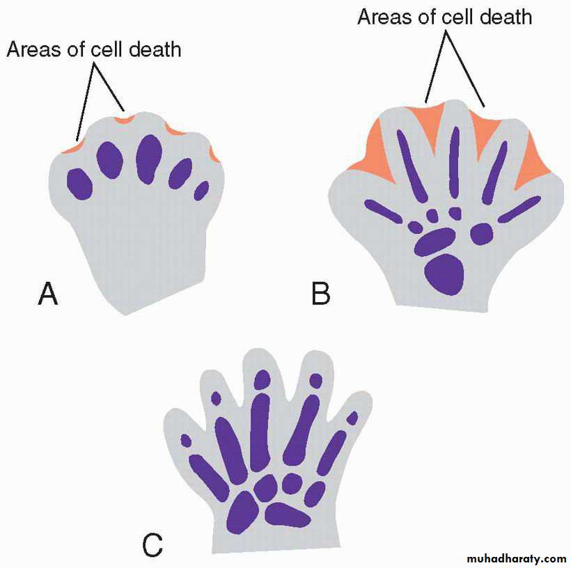
AER appearance induces elongation and formation of the limb buds.Segmental death in AER will form digits
Development of UL precedes LL by about 1-2 days.
Development of the limb bone
7th week, UL rotate 90 degrees laterally, and LL rotates 90 degrees medially.Hyaline cartilaginous model of long bones forms at 6th week
Ossification occur at 12th week by presence of primary ossification centers which will form the diaphysis
completely ossification of diaphysis occur at birth
epiphysis are still cartilagenous
Development of the vertebral column
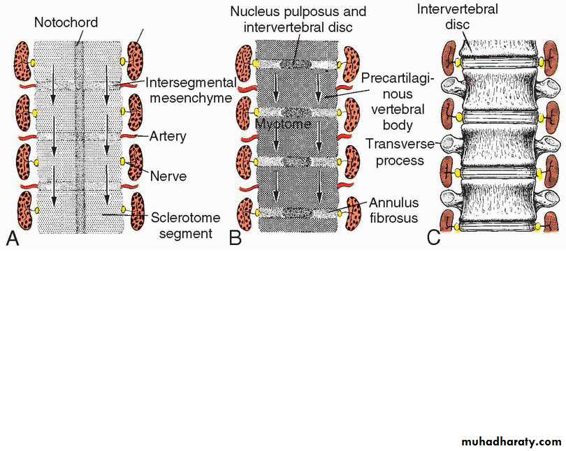
Vertebrae: derived from the sclerotomes of somitesMigrate to surround notochord and spinal cord during 4th week.
sclerotome of each somite undergoes a process called resegmentation
Resegmentation occurs when the caudal half of each sclerotome grows into and fuses with the cephalic half of each subjacent sclerotome
The mid-region form intervertebral disc:
- nucleus pulposus from notochord
- peripheral annulus fibrosis from medial regions of sclerotomes
Development of the vertebral column
Development of the ribs and sternum
Ribs :Bony portion from the costal processes of thoracic vertebrae (thoracic sclerotomes)
Costal cartilages from sclerotome cells that migrate across the lateral somitic frontier into the adjacent lateral plate mesoderm
sternum :
Two sternal bands formed in parietal LPM on either side of the midline
Fuse to form cartilaginous models of manubrium, sternebrae, and xiphoid process
Abnormality of the skeletal system
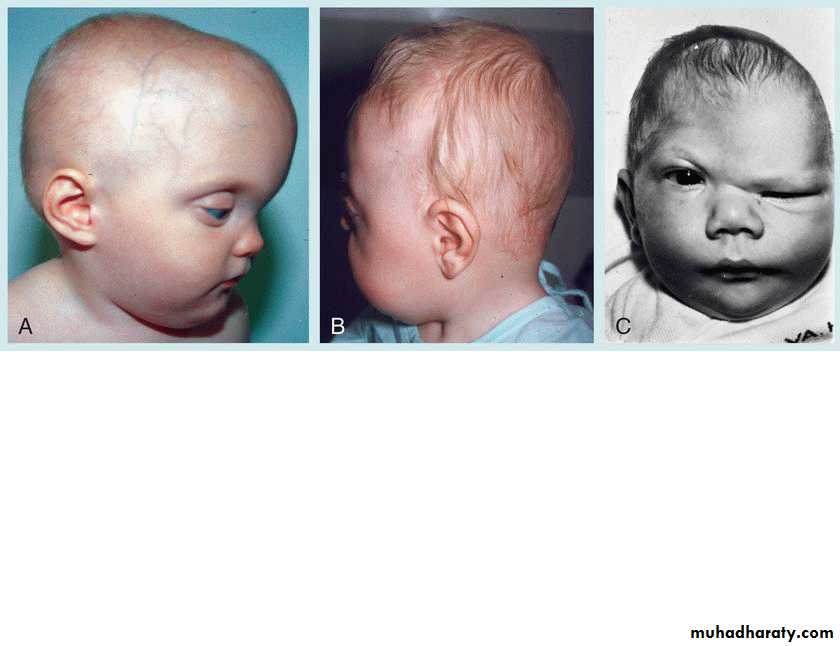
Skull Anomalies :• Cranioschisis
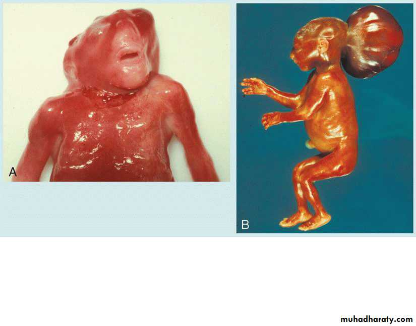
Anencephaly
Meningocele
Meningoencephalocele
Abnormality of the skeletal system
Skull Anomalies :• Craniosynostosis
• Microcephaly
•
Abnormality of the skeletal system
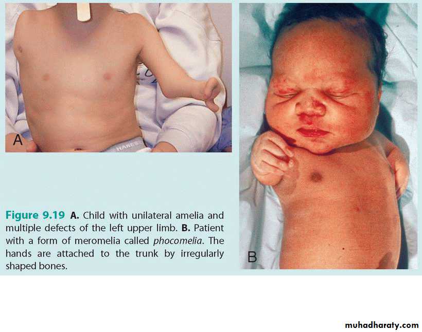
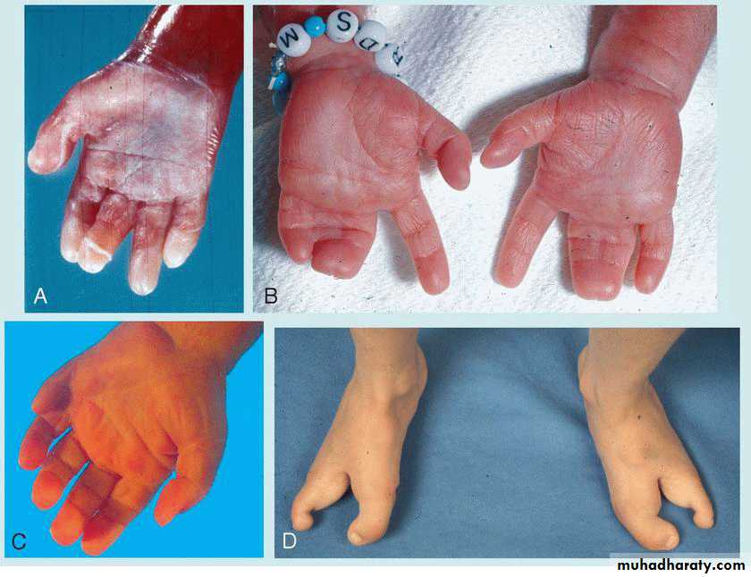
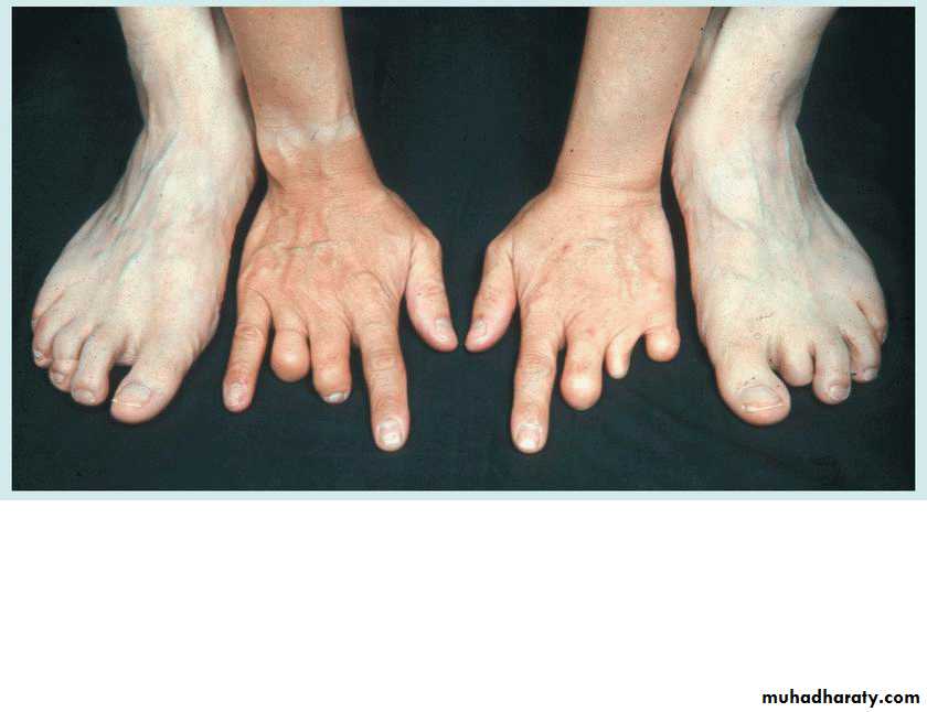
Limb Anomalies :• Absence of limbs: -meromelia , -Amelia
• Micromelia
• Polydactyly
• Ectrodactyly
• Syndactyly
•
Abnormality of the skeletal system
Limb Anomalies :• Cleft of hand or foot; lobester claw
• Club foot
• Congenital absence or deficiency of radius
• Amputation by amniotic bands.
• CDH.
•
Abnormality of the skeletal system
• Vertebral anomalies:• Fusion .
• Scoliosis .
• Abnormal number
• Malunion of vertebral arches: Cleft vertebra and spina bifida
The muscular system:
Development of smooth muscles
from visceral layer of LPMSM of the GIT wall and the vascular walls.
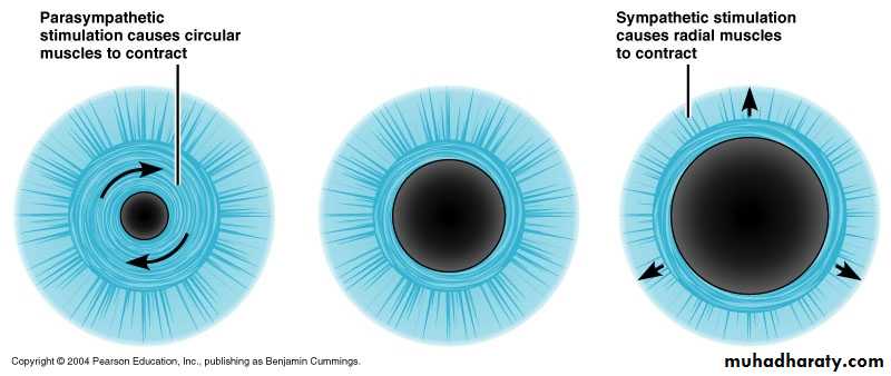
Ectoderm: pupillary, breast muscles and sweat glands muscles
Development of cardiac muscles
From the visceral layer of LPMCardiac myoblasts adhere to one another by the intercalated discs.
Myoblast Not fuse together.
Later special bundle of muscles form the Conductive system
Development of skeletal muscles
From somitesHead region: from paraxial mesoderm (somitomeres & somites) and mesoderm of pharyngeal arches.(iris from optic cup ectoderm)
The somites form skeletal muscles in two regions:
• Dorsomedial region of somites--- Epiaxial M
• Ventrolateral region of somites--- Hypaxial M
Development of skeletal muscles
• Dorsomedial region of somites:
5th week myotomes form epimeres
dorsal extensor muscles of vertebral coulumn.
supplied segmentally by dorsal rami
Development of skeletal muscles
• Ventrolateral region of somites:Development of skeletal muscles
7th week, limb muscles differentiateregulated by connective tissues and bones of the limbs
derived from somites, somatic LPM and NCS
upper limb: opposite lower 5 cervical and upper 2 thoracic somites
lower limb: opposite lower 4 lumber and upper 2 sacral
Nerves: supply muscles , promote development and provide sensory innervation for the dermatomes.
Regardless of their domain, each myotome receives its innervation from spinal nerves derived from the same segment as the muscle cells originate.