background image

background image

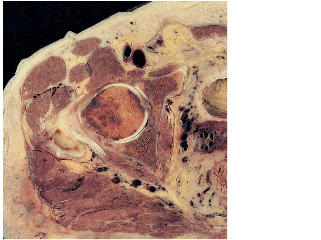
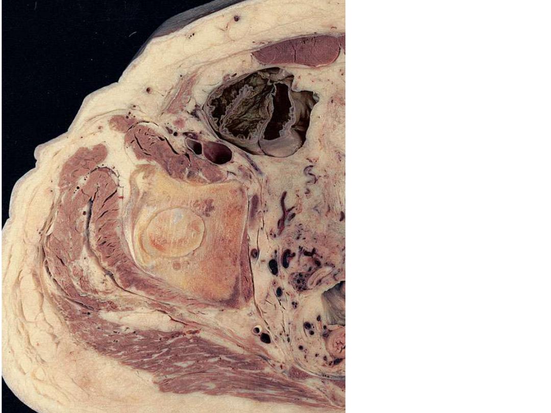
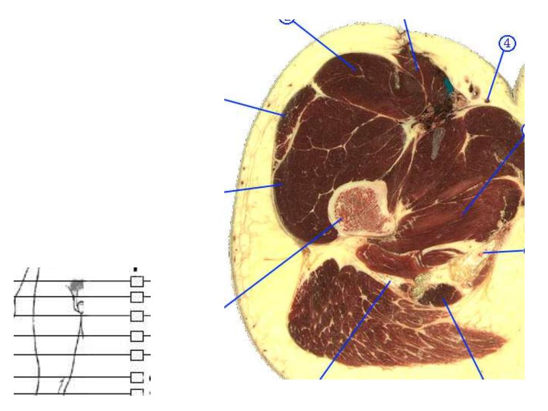
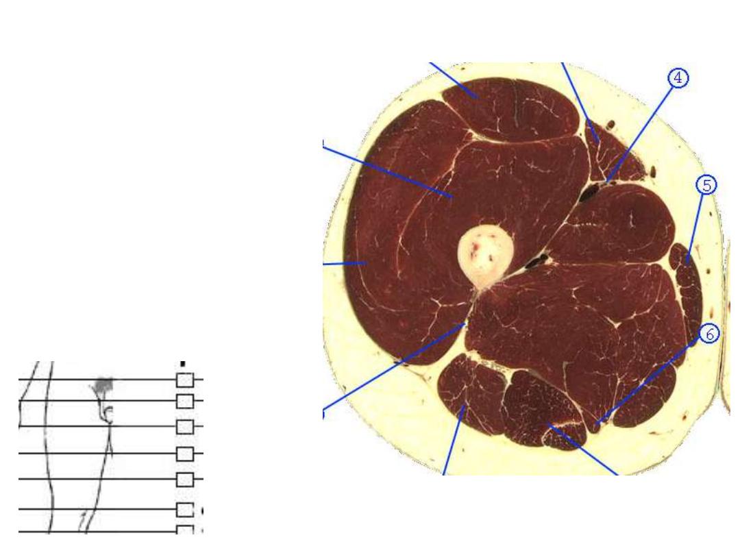
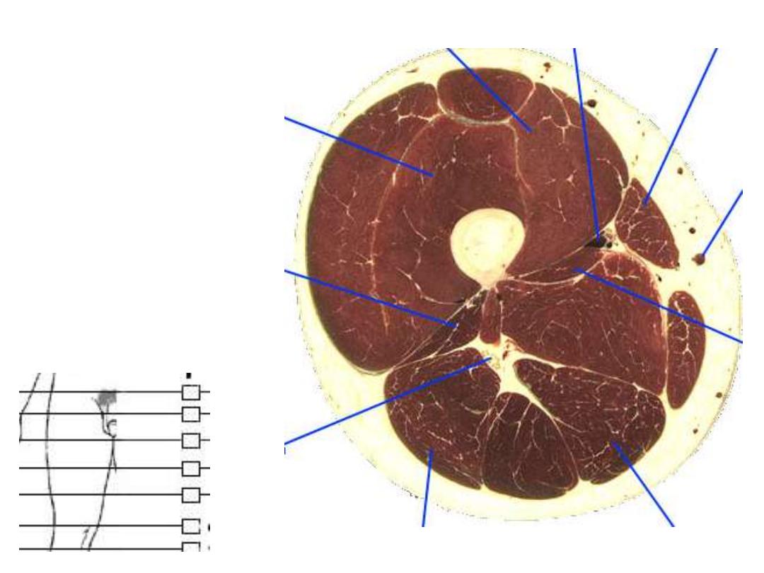
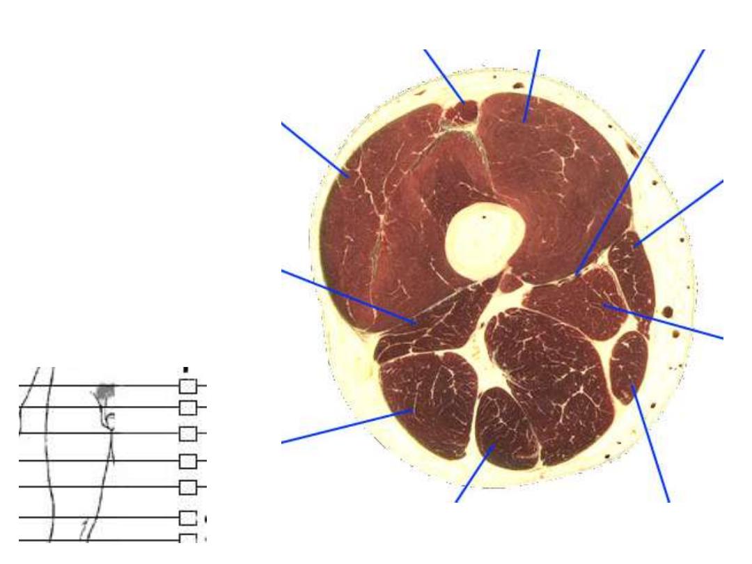
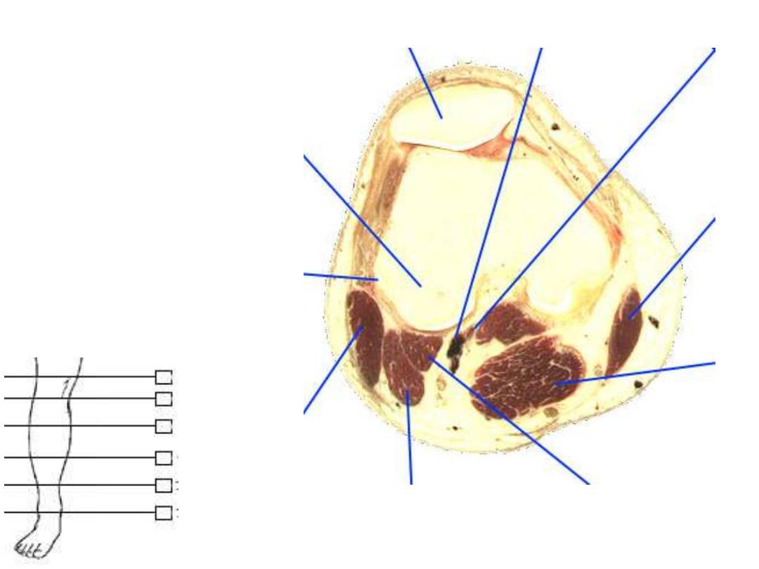
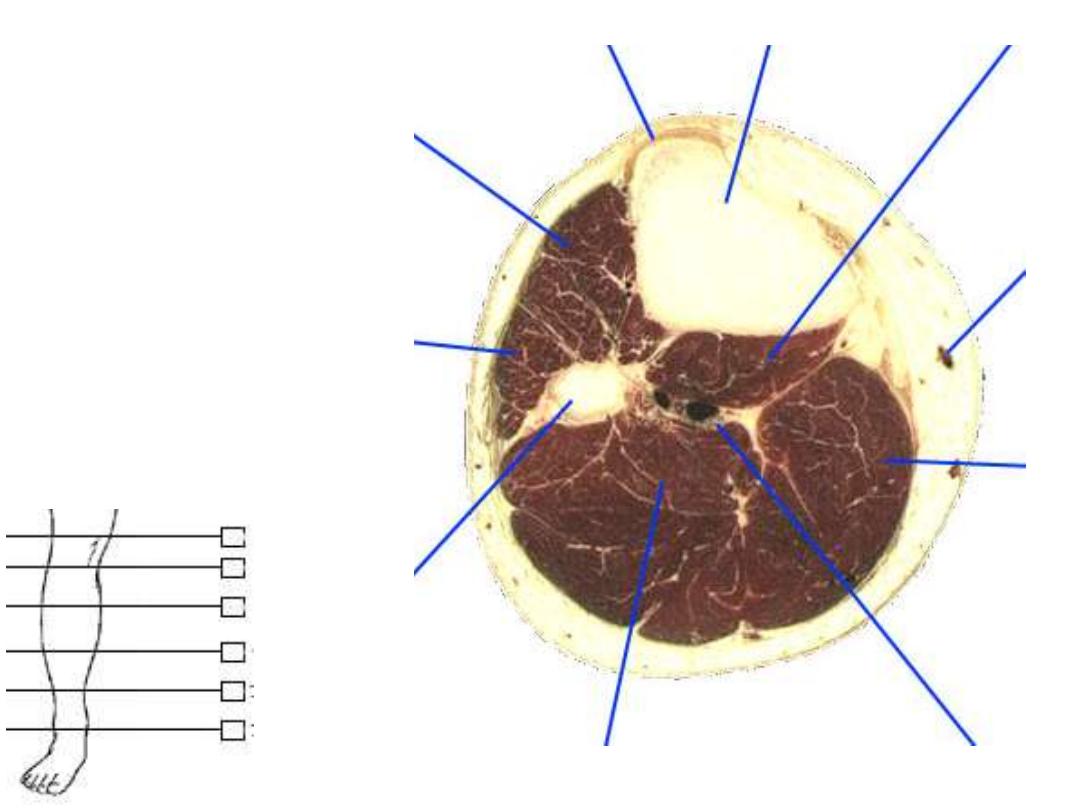
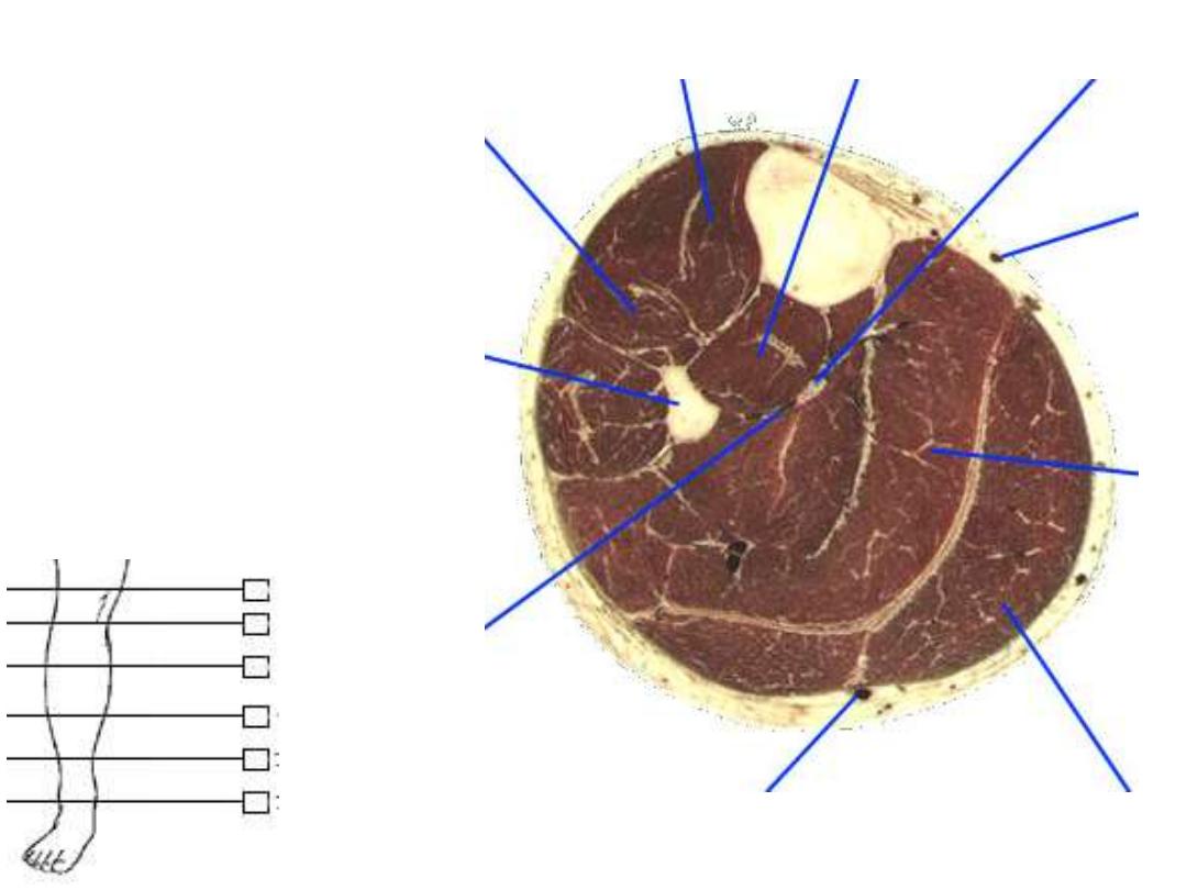
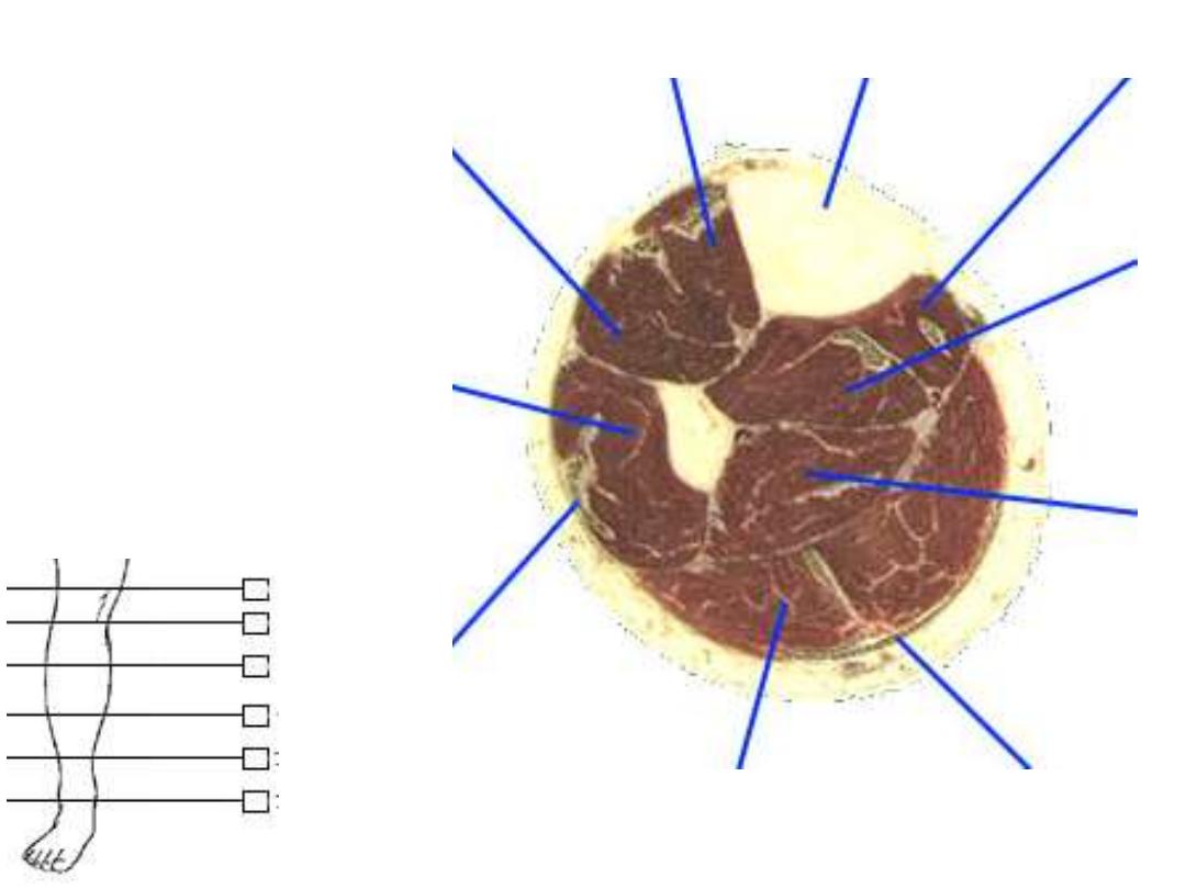
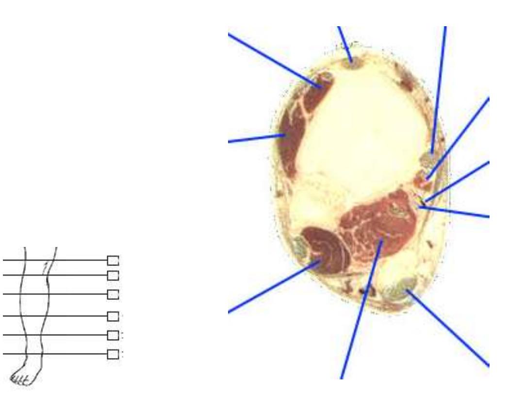
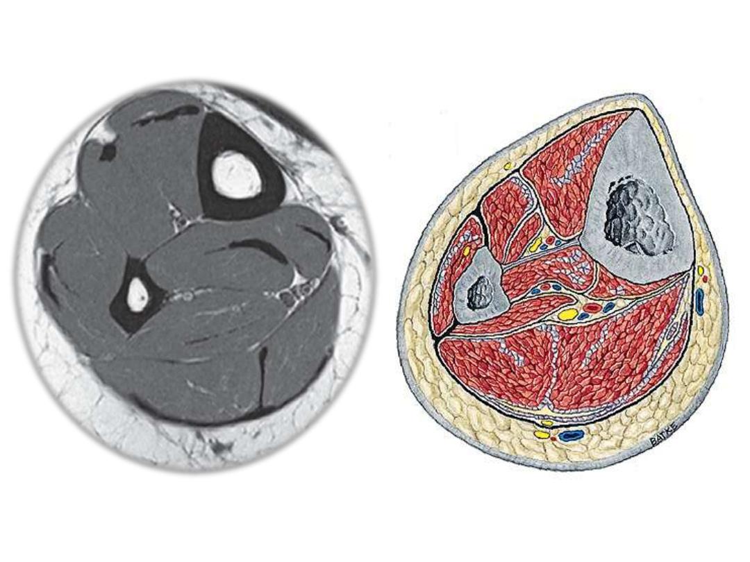
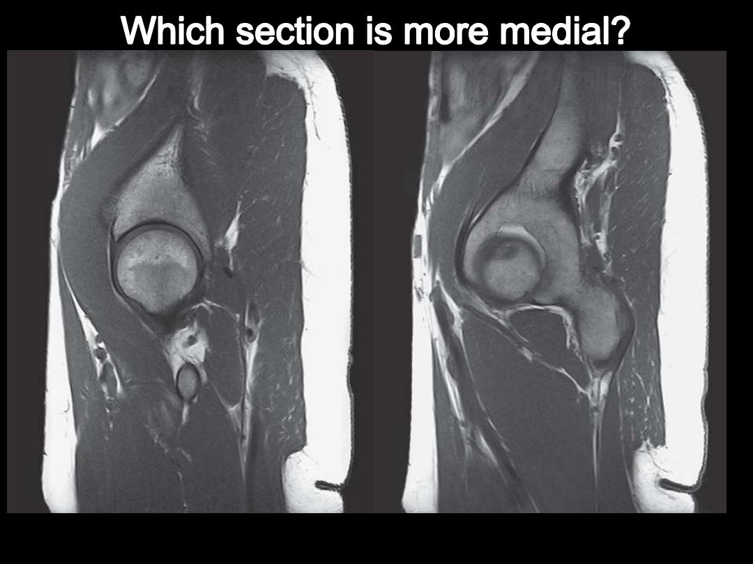
To view different sections for lower limb 

To define the 3D anatomy of lower limb 

background image

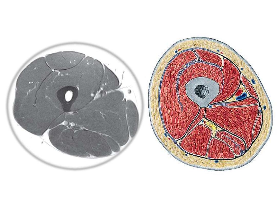
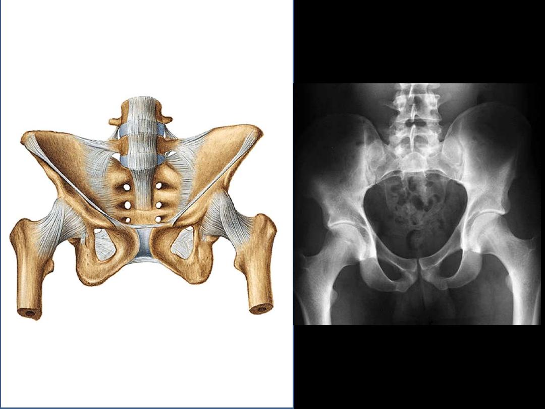
Basic radiological points … 

Bones, ligaments, cartilages, 

muscles, joints, air … 

background image

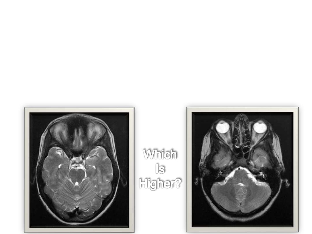
What will happen when successive sections are viewed? 

-

New structures appear 

-

Some structures disappear 

-

Change in position 

-

Change in dimensions 

-

Change in shape 

-

Change in relations 

background image

background image

background image

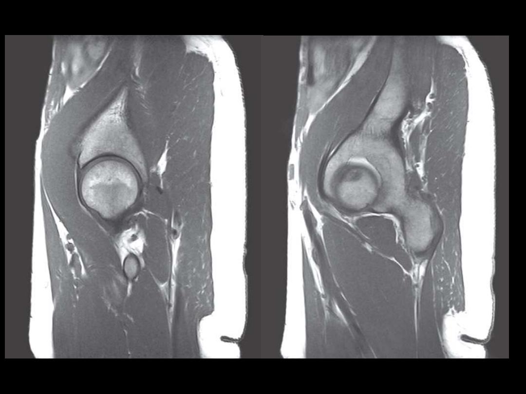
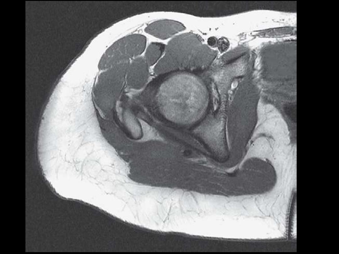
Tis section is a bit higher 

than the previous one.. 

Why? 

background image

1 

2 

3 

4 

5 

6 

7 

8 

9 

10 

X 


background image

1 

2 

3 

4 

5 

6 

7 

8 

9 

10 

X 


background image

1 

2 

3 

4 

5 

6 

7 

8 

9 

10 

X 


background image

1 

2 

3 

4 

5 

6 

7 

8 

9 

10 

X 


background image

1 

2 

3 

4 

5 

6 

7 

8 

9 

10 

X 


background image

1 

2 

3 

4 

5 

6 

7 

8 

9 

10 

X 


background image

1 

2 

3 

4 

5 

6 

7 

8 

9 

10 

X 


background image

1 

2 

3 

4 

5 

6 

7 

8 

9 

10 

X 


background image

1 

2 

3 

4 

5 

6 

7 

8 

9 

10 

X 


background image

1 

2 

3 

4 

5 

6 

7 

8 

9 

10 

X 


background image

background image

background image

background image

background image

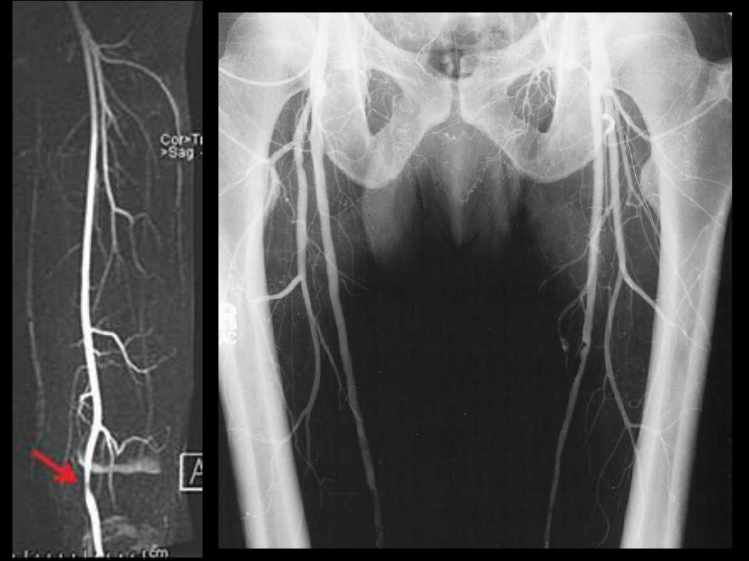
Femoral angiogram 


background image

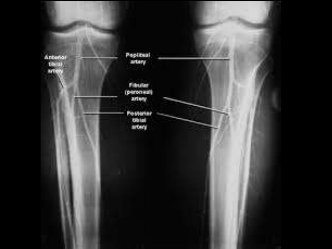
Popliteal angiogram 


background image

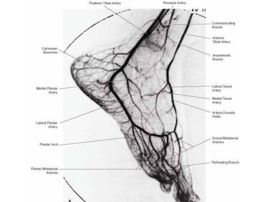
Foot angiogram  


رفعت المحاضرة من قبل: Abbas Ahmed
المشاهدات: لقد قام 9 أعضاء و 275 زائراً بقراءة هذه المحاضرة


تسجيل دخول

أو
عبر الحساب الاعتيادي
الرجاء كتابة البريد الالكتروني بشكل صحيح
الرجاء كتابة كلمة المرور
لست عضواً في موقع محاضراتي؟
اضغط هنا للتسجيل