The Six Kingdoms:
Plants, Animals, Protists, Fungi, Archaebacteria, Eubacteria.How are organism placed into their kingdoms?
Cell type, complex or simple.
Their ability to make food.
The number of cells in their body.
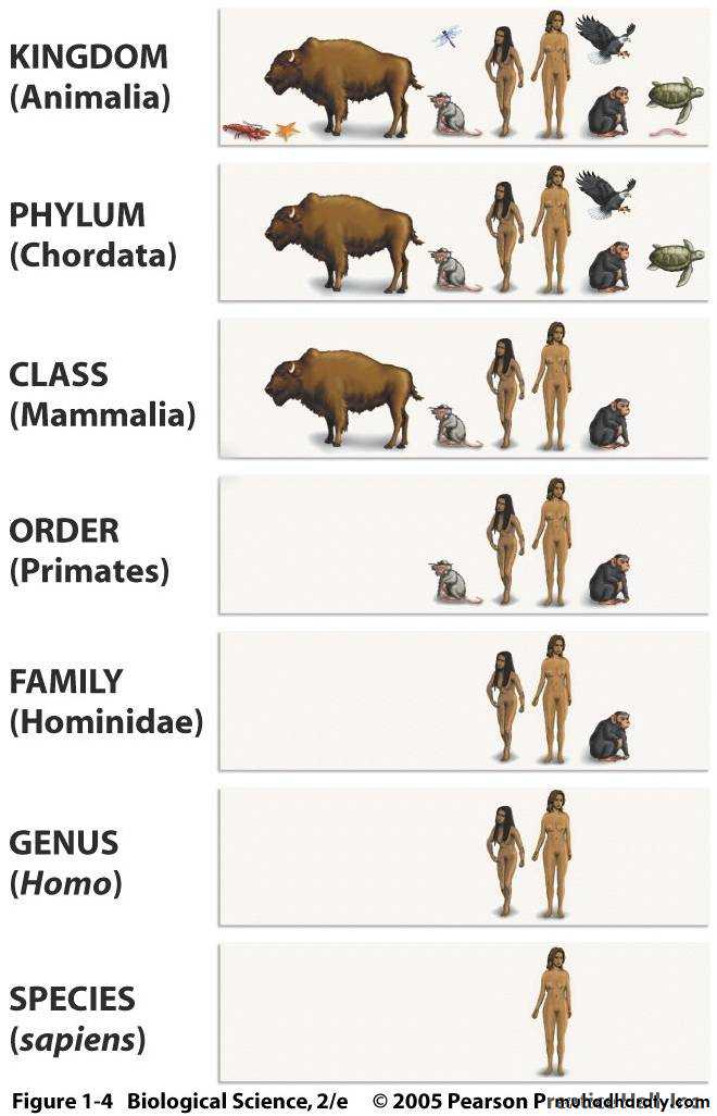
Humans are members of a large group of animals known as mammals (Class Mammalia).
Mammal featuresAll mammals (including humans) have the same distinctive features. These include:
fur or hair growing from the skin.
mammary glands that, in females, produce milk for feeding the young.
three bones (the malleus, incus and stapes) in the middle ear for transmitting sound to the inner ear.
a single bone (the dentary) on each side of the lower jaw.
Humans possess many unique characteristics but we also share a number of similarities with other animals. These similarities and differences are revealed through our genetic make-up, the ways our bodies are constructed and our behavior.
Mammals digestive system
In mammalian, the alimentary canal is the location of all digestion. The alimentary canal is most easily described as a long tube which runs from the mouth, through the body to the anus. In an adult human, it can be up to 6 meters long, and is obviously coiled to preserve space. This canal, in addition to organs which secrete various substances into it make up the digestive system.
Mechanical digestion is the process of chewing breaking larger pieces of food into smaller ones, providing a larger surface area for the digestive enzymes to act on during chemical digestion .
The alimentary canal has a wall with four main layers - the mucosa, submucosa, muscularis externa and serosa.
The mucosa is the layer nearest the lumen and on its inner surface is a thin epithelium, which contains goblet cells. These secrete mucus to :
Lubrication.
Protect the cells from abrasion by food.
It also stops damage by digestive enzymes.
Beneath the epithelium is a layer of connective tissue, and beneath that is a layer of smooth muscle called the muscularis mucosa. It is smooth muscle which allows it to contract slowly and rhythmically for long periods without tiring.
The submucosa consists of connective tissue lying within blood vessels and nerves. It contains a high proportion of collagen and elastin (fibrous proteins).
The musclurais externa, smooth muscle, is made up of two bands - longitudinal muscle and circular muscle. Longitudinal lies lengthwise along the wall of the canal, whereas circular muscle lies around the wall. Their combined contraction and relaxation moves food through the alimentary canal via peristalsis.
The serosa is a thin layer of connective tissue that makes up the outer layer of the wall
Mouth/esophagus
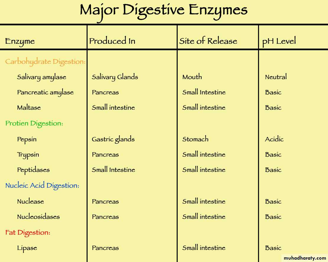
Food is ingested and then masticated (chewed) using molars/premolars which breaks solid food into smaller pieces. Saliva is secreted into the mouth - being mostly water it helps dissolve any soluble components, and the enzyme amylase begins the breakdown of starch to maltose. Swallowing takes the food into the top of the esophagus, and peristalsis takes it to the stomach.
Stomach
The stomach is a small sac with muscles at either end (sphincters), which control the entry and exit of food. When a bolus arrives the stomach, the cardiac sphincter relaxes to allow food to enter, whilst the pyloric sphincter remains contracted so that food can remain there for several hours to form chyme. It then allows the partially digested food to pass into the duodenum.
The mucosa in the stomach is very folded, forming gastric pits, which secrete gastric juice. The epithelium is made up of columnar cells. It also contains chief cells and oxyntic cells, the former of which produce pepsinogen and the latter of which produce hydrochloric acid. Oxyntic cells have many mitochondria and deep invaginations on their surface.
Digestion
Gastric juice is mostly water plus hydrochloric acid from oxyntic cells. This hydrochloric acid gives gastric juice a pH of <1.0, which kills a high proportion of bacteria that is present in food. Chief cells secrete pepsinogen, which does not function as an enzyme and is converted to its active form (pepsin) by the removal of a short length of amino acids.
Gastric juice also contains lipases to break down fats. The walls of the stomach lining is protected by alkaline mucus, coating the entire stomach wall. Only a small, lipid-soluble molecules( alcohol, and aspirin) are absorbed.
Liver and Pancreas
The mix of enzymes, hydrochloric acid with chyme passes into the small intestine when the pyloric sphincter relaxes, and this coincides with the liver and the pancreas releasing juices into the small intestine. One of the liver's functions is to produce bile, which is stored in the gall bladder and then carried along the bile duct into the dueodenum. Bile contains bile salts which help emulsify fats, and are then reabsorbed (mostly) and go back to the liver, where they are then resecreted. Bile also contains hydrogen carbonate ions which help neutralise the acidic mixture of the duodenum.
The pancreas is both an endocrine and exocrine gland - its endocrine function is to secrete insulin and glucagon (produced by the islets of Langerhans) is the secretion of pancreatic juice into the pancreatic duct.
Pancreatic juice consists of enzymes and enzyme precursors. Trypsin and chymotrypsin are secreted in an inactive form, trypsoinogen and chymotrypsinogen. The former gradually changes to trypsin, and is speeded up by an enzyme enterokinase. Trypsin can then act on trypsoinogen and chymotripsinogen to activate them. Other enzymes include carboxypeptidase , lipase and amylase. It also contains hydrogen carbonate ions to neutralise the acidic mixture of food and gastric juice in the duodenum.
Small Intestine
The small intestine consists of three regions - the duodenum, jejunum and the ileum. The structure of the ileum has many numerous tiny folds in the walls, known as villi, made from the mucosa layer, and each villus is around 1mm tall. These villi all have their own microvilli which combined have a great surface area available for absorption. The smooth muscle in the musclaris mucosa can make them contract and sway around for great contact with the food in the lumen, and the villus also have a network of blood capillaries for absorption and transport of food.
Beneath the villi are glands called the crypts of Lieberkuhn, which contain goblet cells to secrete mucus.
The products of digestion, amino acids, fatty acids, glycerol and monosaccharides, can all cross the plasma membranes of the epithelial cells on the villi, passing through these cells to enter either the blood or lymphatic capillaries. Monosaccharides and amino acids go straight into the blood stream, whilst fatty acids and glycerol go into the lymphatic system.
Monosaccharides and aminoacides are absorbed by sodium ions continually pumped out of the base of the epithelial cells into the surrounding tissue fluid, resulting in sodium ions travelling down this created concentration gradient, carrying glucose and amino acids with them as a co-transport.
fatty acids and monoglyceride are transported into the endoplasmic reticulum, where they are used to synthesize triglyceride. Beginning in the endoplasmic reticulum and continuing in the Golgi, triglyceride is packaged with cholesterol, lipoproteins and other lipids into particles called chylomicrons. Water, inorganic ions and vitamins are also absorbed in the ileum, via osmosis, active transport, facilitated diffusion.
Colon
The colon, caecum, appendix and rectum make up the large intestine, and the colon's function is to absorb inorganic ions and water. The epithelium does not contain villi but it does have columnar cells with microvilli, providing a relatively large surface area for absorption. The material that reaches the rectum is mostly indigestible material - cellulose and lignin as well as mucus and cells that have been shed form the canal - it is passed out as faces.
Hormones of gut:
Gastrin is synthesized in G cells, which are located in gastric pits, primarily in the antrum region of the stomach and binds receptors found predominantly on parietal and enterochromaffin-like cells.
Secretin, a digestive hormone secreted by the wall of the upper part of the small intestine (the HYPERLINK "https://www.britannica.com/science/duodenum" duodenum). secretin is released into the bloodstream and stimulates the acinar cells of the pancreas to secrete water and bicarbonate into the pancreatic ducts that drain into the duodenum. By this mechanism, hydrochloric acid secreted by the stomach, which can be damaging to the intestinal lining, is promptly diluted and neutralized. Secretin also inhibits the secretion of gastrin, which triggers the initial release of hydrochloric acid into the stomach, and delays gastric emptying.
Cholecystokinin (CCK) : it is secreted from mucosal epithelial cells in the first segment of the small intestine (duodenum), and stimulates delivery into the small intestine of digestive enzymes from the pancreas and bile from the gallbladder. Cholecystokinin is also produced by neurons in the enteric nervous system, and is widely and abundantly distributed in the brain. Cholecystokinin stimulates the gallbladder to contract and release stored bile into the intestine. It also stimulates the secretion of pancreatic juice and may induce satiety.
Nervous signals of gut
The taste, sight and smell of food all feed into the nervous system, and the medulla oblongata carries impulses from it to the salivary glands as part of the autonomic nervous system. Gastric juice secretion is also stimulated by nerve impulses sent from the brain to the stomach wall
Other mammalian types
Herbivores
Herbivores are animals whose primary food source is plant-based. These animals have evolved digestive systems capable of digesting large amounts of plant material. The plants are high in fiber and starch, which provide the main energy source in their diet. Since some parts of plant materials, such as cellulose, are hard to digest, the digestive tract of herbivores is adapted so that food may be digested properly. Many large herbivores have symbiotic bacteria within their guts to assist with the breakdown of cellulose. They have long and complex digestive tracts to allow enough space and time for microbial fermentation to occur.
Carnivores
Carnivores are meat eating animals and thus have very different dentistry and digestion. They has long pointed teeth known as canines at the front of its mouth, which allow to stab the body of its prey when bites it, killing it. Behind the canines are premolars and molars with extremely sharp edges, and are known as carnassial teeth because they slice past each other with the jaw is closed in a scissor like action that cracks, crushes bones and cuts meat into smaller pieces.
The incisors are used in scraping meat from the surface of the bones. Since meat does not contain starch or cellulose cell walls, amylase is not required. However, compensating for the lack of chemical digestion in the mouth is a very concentrated acid in the carnivore stomach, allowing them to eat 'dangerous' food that is rotten without harm.