
1
Fifth stage
Pediatric
Lec. 4
.د
اوس
22/3/2017
Genetics
Genetic disorders
common, with 2% of live-born babies having a significant congenital malformation and
about 5% a genetic disorder
burdensome to the affected individual, family and society, as many are associated with
severe and permanent disability.
Genetically determined diseases
1. Single gene mutations,((Mendelian disorders)
2. Chromosomal disorders
3. Multifactorially inherited conditions
4. Disorders that show an unusual pattern of inheritance
5. Teratogenically caused conditions
Mendelian inheritance
Disorders with these patterns of inheritance, described by Mendel in 1865, are rare
individually, but collectively numerous, with over 15 000 single gene traits or disorders
described
There are three classic forms of genetic inheritance: autosomal dominant, autosomal
recessive, and X-linked
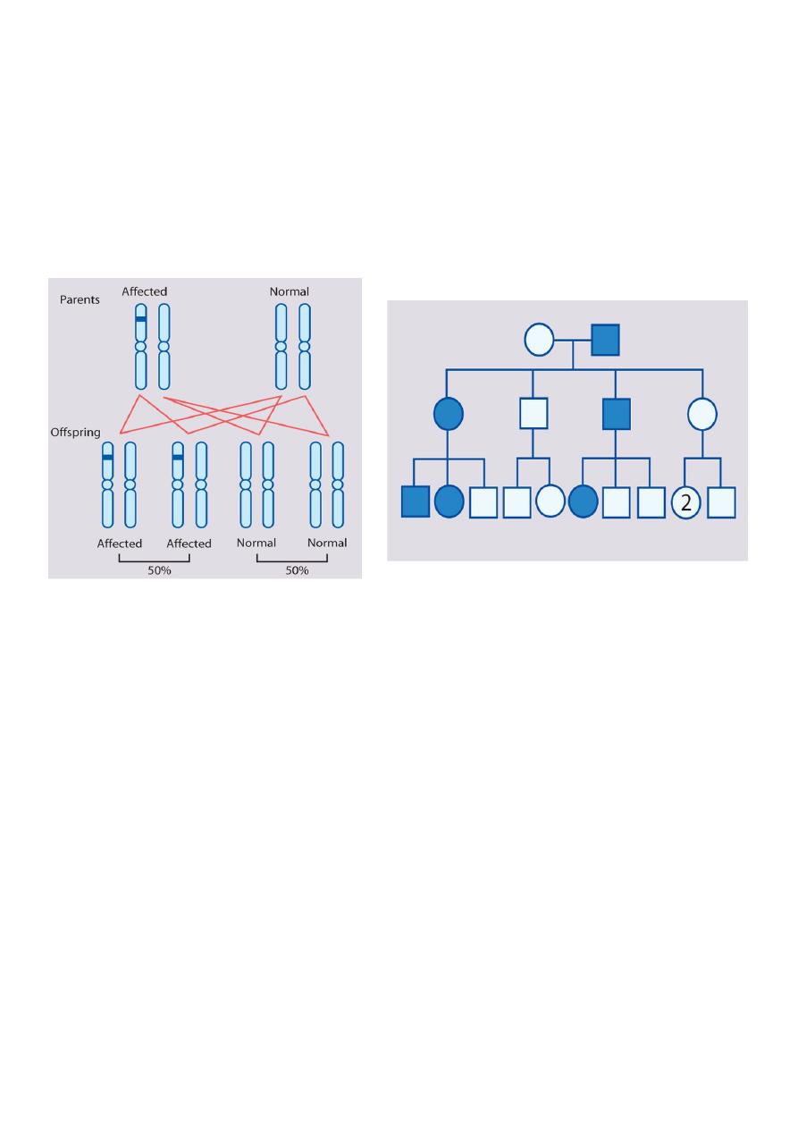
Autosomal dominant inheritance
This is the most common mode of Mendelian inheritance
Autosomal dominant inheritance is determined by the presence of one abnormal gene on
one of the autosomes (chromosomes 1–22).
Male and female offspring each have a 1 in 2 (50%) chance of inheriting the abnormal gene
from an affected parent
2
Rules of Autosomal Dominant Inheritance
Trait appears in every generation
Each child of an affected parent has a 1 in 2 chance of being affected
Males and females are equally affected
Male-to-male transmission occurs
•
Tuberous sclerosis
•
Marfan syndrome
•
Neurofibromatosis
•
Huntington's disease
•
Retinoblastoma
•
Waardenburg syndrome
•
Myotonic dystrophy
•
Familial hypercholestrolemia (LDL receptor defect Type IIa)
•
Adult polycystic kidney disease
•
von Hippel Lindau
•
Familial adenomatous polyposis and Peutz Jeghers Syndrome
3
•
Hereditory spherocytosis
•
Achondroplasia
•
Ehlor's Danlos (vascular type)
•
Acute intermittent porphyria
•
Hypertrophic Obstructive Cardiomyopathy (HOCM)
•
Von Willebrand Disease
•
Polydactyly
•
Osteogenesis Imperfecta (Except Type VII)
•
Hereditary hemorrhagic telengiactasia (Osler-weber-rendu syndrome)
•
Osteopetrosis Type II (Adult type)
•
Hypokalemic Periodic Paralysis
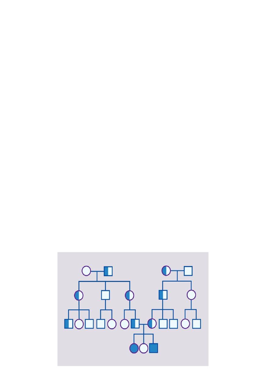
Autosomal recessive inheritance
Many hundred disorders resulting from this type of inheritance are known
An affected individual is homozygous for the abnormal gene, having inherited an abnormal
allele from each parent, both of whom are unaffected heterozygous carriers
4
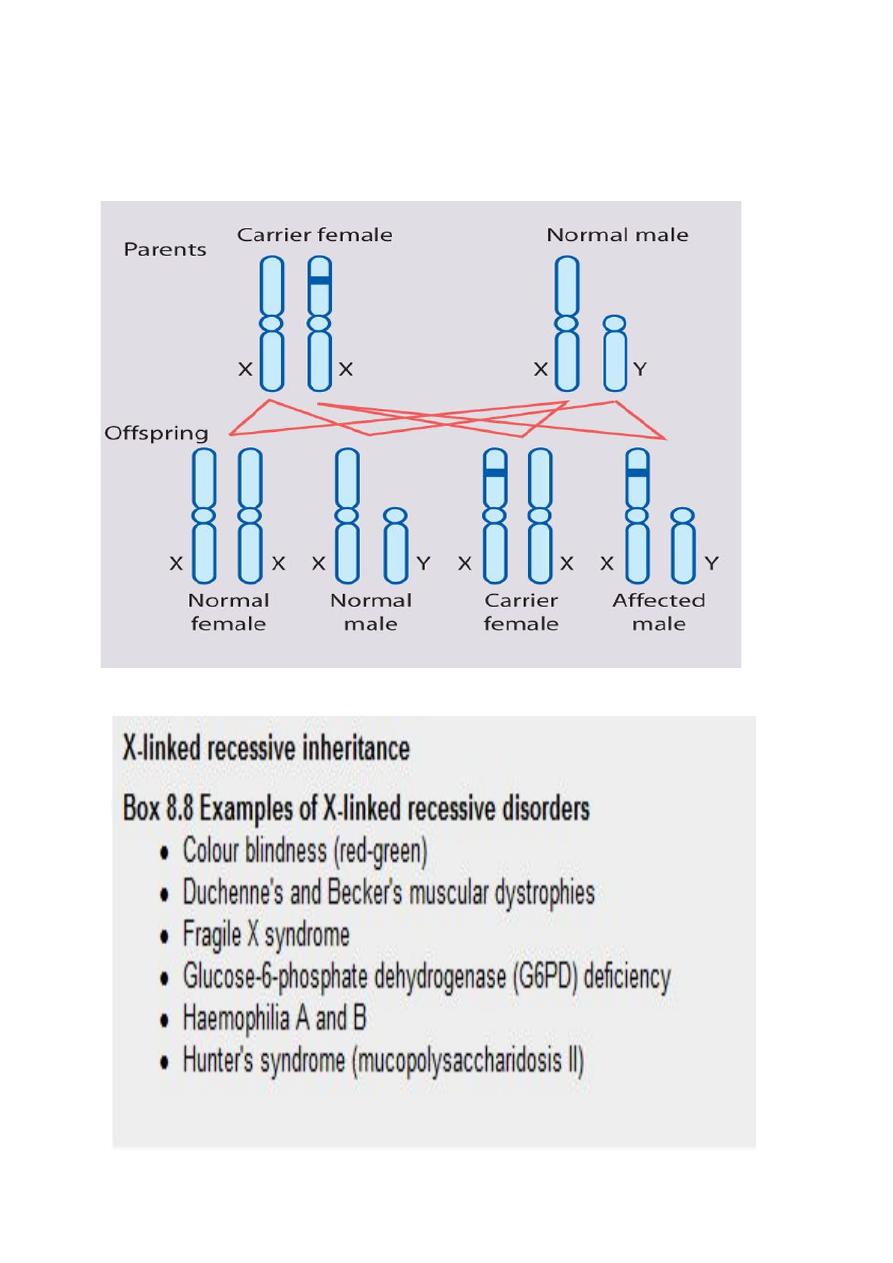
X-linked recessive inheritance
Over 400 disorders have been described in which an abnormal recessive gene is carried on
the X chromosome
1. males are affected
2. occasionally a female carrier shows mild signs of the disease (manifesting carrier)
3. each son of a female carrier has a 1 in 2 (50%) risk of being affected
4. each daughter of a female carrier has a 1 in 2 (50%) risk of being a carrier
5
5. daughters of affected males will all be carriers
6. sons of affected males will not be affected, since a man passes a Y chromosome to his
son
6
Chromosomal abnormalities
Chromosomal abnormalities are either numerical or structural. They occur in approximately
10% of spermatozoa and 25% of mature oocytes and are a very common cause of early
spontaneous miscarriage
Cytogenetics
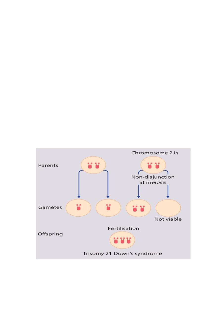
The extra chromosome 21 may result from non-disjunction, translocation or mosaicism
Non-disjunction (94%)
most cases result from an error at meiosis
the pair of chromosome 21s fails to separate, so that one gamete has two chromosome 21s
and one has none
fertilisation of the gamete with two chromosome 21s gives rise to a zygote with trisomy 21
parental chromosomes do not need to be examined.

7
Clinical features
Down's syndrome is usually suspected at birth because of the baby's facial appearance
Typical craniofacial appearance
1. Round face and flat nasal bridge
2. Upslanted palpebral fissures
3. Epicanthic folds (a fold of skin running across the inner edge of the palpebral fissure)
4. Brushfield spots in iris (pigmented spots)
5. Small mouth and protruding tongue ,Small ears
6. Flat occiput and third fontanelle
Other anomalies
1. Short neck
2. Single palmar creases, incurved fifth finger and wide 'sandal' gap between toes
3. Hypotonia
4. Congenital heart defects (40%)
5. Duodenal atresia
6. Hirschsprung's disease
Later medical problems
1. Delayed motor milestones
2. Moderate to severe learning difficulties
3. Small stature
4. Increased susceptibility to infections
5. Hearing impairment from secretory otitis media
6. Visual impairment from cataracts, squints, myopia
7. Increased risk of leukaemia and solid tumours
8. Risk of atlantoaxial instability
9. Hypothyroidism and coeliac disease
10. Epilepsy
11. Alzheimer's disease

8
Edwards' syndrome (trisomy 18) and Patau's syndrome (trisomy 13)
Although rarer than Down's syndrome (1 in 8000 and 1 in 14 000 live births, respectively),
particular constellations of severe multiple abnormalities suggest the diagnosis at birth and
most affected babies die in infancy
The diagnosis is confirmed by chromosome analysis
Clinical features of Edwards' syndrome (trisomy 18)
Low birthweight
Prominent occiput
Small mouth and chin
Short sternum
Flexed, overlapping fingers
Rocker-bottom feet
Cardiac and renal malformations
Clinical features of Patau's syndrome (trisomy 13)
Structural defect of brain
Scalp defects
Small eyes (microphthalmia) and other eye defects
Cleft lip and palate
Polydactyly
Cardiac and renal malformations
Later medical problems
1. Delayed motor milestones
2. Moderate to severe learning difficulties
Small stature
3. Increased susceptibility to infections
4. Hearing impairment from secretory otitis media

9
5. Visual impairment from cataracts, squints, myopia
6. Increased risk of leukaemia and solid tumours
7. Risk of atlantoaxial instability
8. Hypothyroidism and coeliac disease
9. Epilepsy
10. Alzheimer's disease