Structure And Function Of The Skin د.وسام اللامي
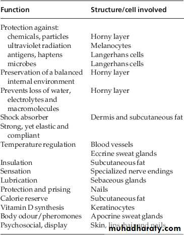
The skin – the interface between humans and their environment – is the largest organ in the body. It weighs an average of 4 kg and covers an area of 2m2. It acts as a barrier, protecting the body from harsh external conditions and preventing the loss of important body constituents, especially water. The skin has three layers. The outer one is the epidermis, which is firmly attached to, and supported by connective tissue in the underlying dermis. Beneath the dermis is loose connective tissue, the subcutis/hypodermis, which usually contains abundant fat.
Epidermis
The epidermis consists of many layers of closely packed cells, the most superficial of which are flattened and filled with keratins; it is therefore a stratified squamous epithelium. It adheres to the dermis partly by the interlocking of its downward projections (epidermal ridges) with upward projections of the dermis (dermal papillae) .The epidermis contains no blood vessels. It varies in thickness from less than 0.1 mm on the eyelids to nearly 1 mm on the palms and soles. As dead surface squames are shed (accounting for some of the dust in our houses), the thickness is kept constant by cells dividing in the deepest (basal or germinative) layer. A generated cell moves, or is pushed by underlying mitotic activity, to the surface, passing through the prickle and granular cell layers before dying in the horny layer. The journey from the basal layer to the surface (epidermal turnover or transit time) takes 30 to 60 days. Stem cells reside amongst these interfollicular basal cells and also amongst the cells of the external root sheath at the bulge in the hair follicle at the level of attachment of the arrector pili muscle.The basal layer, the deepest layer, rests on a basement membrane, which attaches it to the dermis. It is a single layer of columnar cells, whose basal surfaces sprout many fine processes and hemidesmosomes, anchoring them to the lamina densa of the basement membrane.
The spinous or prickle cell layer is composed of keratinocytes. These differentiating cells, which synthesize keratins, are larger than basal cells. Keratinocytes are firmly attached to each other by small interlocking cytoplasmic processes, by abundant desmosomes and by other cadherins . Under the light microscope, the desmosomes look like ‘prickles’. They contain desmoplakins,desmogleins and desmocollins. Autoantibodies to these proteins are found in pemphigus, when they are responsible for the detachment of keratinocytes from one another and so for intra-epidermal blister formation. Tonofilaments are small fibres running from the cytoplasm to the desmosomes. They are more numerous in cells of the spinous layer than of the basal layer, and are packed into bundles called tonofibrils. Many lamellar granules ( Odland bodies or keratinosomes ), derived from the Golgi apparatus, appear in the superficial keratinocytes of this layer. Their contents are discharged into the intercellular space of the granular cell layer to become precursors of the lipids in the intercellular space of the horny layer.
The granular layer, normally consists of two or three layers of cells that are flatter than those in the spinous layer, and have more tonofibrils. These cells contain large irregular basophilic granules of keratohyalin, which merge with tonofibrils. These keratohyalin granules contain proteins, including involucrin, loricrin and pro-filaggrin, which is cleaved into filaggrin by specific phosphatases as the granular cells move into the horny layer.As keratinocytes migrate out through the outer-most layers, their keratohyalin granules break up and their contents are dispersed throughout the cytoplasm. Filaggrin peptides aggregate the keratincytoskeleton, collapsing it, and thus converting the granular cells to flattened squames. These make up the thick and tough peripheral protein coating of the horny envelope.
The horny layer (stratum corneum) is made of piled-up layers of flattened dead cells (corneocytes)– the bricks – separated by lipids – the mortar – in the intercellular space. Together these provide an effective barrier to water loss and to invasion by infectious agents and toxic chemicals. The corneocyte cytoplasm is packed with keratin filaments, embedded in a matrix and enclosed by an envelope derived from the keratohyalin granules. This envelope, along with the aggregated keratins that it encloses, gives the corneocyte its toughness, allowing the skin to withstand all sorts of chemical and mechanical insults. Horny cells normally have no nuclei or intracytoplasmic organelles, these having been destroyed by hydrolytic and degrading enzymes found in lamellar granules and the lysosomes of granular cells.
Keratinization
All cells have an internal skeleton made up of microfilaments (7 nm diameter; actin), microtubules (20 –35 nm diameter; tubulin) and intermediate filaments (10 nm diameter). Keratins ( meaning ‘horn’) are the main intermediate filaments in epithelial cells and are comparable to vimentin in mesenchymal cells, neurofilaments in neurones and desmin in muscle cells. During differentiation, the keratin fibrils in the cells of the horny layer align and aggregate, under the influence of filaggrin. Cysteine, found in keratins of the horny layer, allows cross-linking of fibrils to give the epidermis strength to withstand injury. The cytoskeleton of tonofibrils also maintains the cell shape rigidly. The typical ‘basket weave’ appearance of the horny layer in routine histological sections is artefactual and deceptive. In fact, cells deep in the horny layer stick tightly together and only those at the surface flake off; this is in part caused by the activity of cholesterol sulphatase. Desquamation is normally responsible for the removal of harmful exogenous substances from the skin surface.Other cells in the epidermis
Keratinocytes make up about 85% of cells in the epidermis, but three other types of cell are also found there: melanocytes, Langerhans cells and Merkel cells.
Melanocytes
Melanocytes are the only cells that can synthesize melanin. They migrate from the neural crest into the basal layer of the ectoderm where, in human embryos. They are also found in hair bulbs, the retina and pia arachnoid. Each dendritic melanocyte associates with a number of keratinocytes, forming an ‘epidermal melaninunit’ . The dendritic processes of melanocytes wind between the epidermal cells and end as discs in contact with them. Their cytoplasm contains discrete organelles, the melanosomes, containing varying amounts of the pigment melanin. This is ‘injected’ into surrounding keratinocytes to provide them with pigmentation to help protect the skin against damaging ultraviolet radiation.
Langerhans cells
The Langerhans cell is a dendritic cell like the melanocyte. It also lacks desmosomes and tonofibrils, but has a lobulated nucleus. Langerhans cells come from a mobile pool of precursors originating in the bone marrow. There are approximately 800 Langerhans cells per mm2 in human skin and their dendritic processes fan out to form a striking network seen best in epidermal sheets. They are best thought of as highly specialized macrophages. Langerhans cells have a key role in many immune reactions. They take up exogenous antigen, process it and present it to T lymphocytes either in the skin or in the local lymph nodes. They probably play a part in immunosurveillance for viral and tumour antigens. In this way, ultraviolet radiation can induce skin tumours both by causing mutations in the epidermal cells, and by decreasing the number of epidermal Langerhans cells, so that cells bearing altered antigens are not recognized or destroyed by the immune system. Topical or systemic gluco-corticoids also reduce the density of epidermal Langerhans cells. The Langerhans cell is the principal cell in skin allografts to which the T lymphocytes of the host react during rejection; allograft survival can be prolonged by depleting Langerhans cells.
Merkel cells
Merkel cells are found in normal epidermis and act as transducers for fine touch. They are non-dendritic cells, lying in or near the basal layer, and are of the same size as keratinocytes. They are concentrated in localized thickenings of the epidermis near hair follicles (hair discs), and contain membrane-bound spherical granules. Sparse desmosomes connect these cells to neighbouring keratinocytes. Fine unmyelinated nerve endings are often associated with Merkel cells, which express immunoreactivity for various neuropeptides.
Dermo-epidermal junction
The basement membrane lies at the interface between the epidermis and dermis. Electron microscopy shows that the lamina densa (rich in type IV collagen) is separated from the basal cells by an electron-lucent area, the lamina lucida. The plasma membrane of basal cells has hemidesmosomes. The lamina lucida contains the adhesive macromolecules, laminin-1, laminin-5 and entactin. Fine anchoring filaments (of laminin-5) cross the lamina lucida and connect the lamina densa to the plasma membrane of the basal cells. Anchoring fibrils(of type VII collagen), dermal microfibril bundles and single small collagen fibres (types I and III),extend from the papillary dermis to the deep part of the lamina densa. The laminins act as aglue, helping to hold the epidermis onto the dermis. The structures within the dermo-epidermal junction provide mechanical support, encouraging the adhesion, growth, differentiation and migration of the overlying basal cells, and also act as a semi-permeable filter that regulates the transfer of nutrients and cells from dermis to epidermis.
Dermis
The dermis lies between the epidermis and the subcutaneous fat. It supports the epidermis structurally and nutritionally. Its thickness varies, being greatest in the palms and soles and least in the eyelids and penis. In old age, the dermis thins and loses its elasticity. The dermis interdigitates with the epidermis so that upward projections of the dermis, the dermal papillae, interlock with downward ridges of the epidermis, the rete pegs. This interdigitation is responsible for the ridges seen most readily on the fingertips (as fingerprints). It is important in the adhesion between epidermis and dermis as it increases the area of contact between them, the dermis has three components: cells, fibres and amorphous ground substance.
Cells of the dermis
The main cells of the dermis are fibroblasts, but there are also small numbers of resident and transitory mononuclear phagocytes, lymphocytes,Langerhans cells and mast cells. Other blood cells (e.g. polymorphs) are seen during inflammation.
Fibres of the dermis
The dermis is largely made up of interwoven fibres, principally of collagen, packed in bundles. Those in the papillary dermis are finer than those in the deeper reticular dermis. When the skin is stretched,collagen, with its high tensile strength, prevent stearing, and the elastic fibres, intermingled with the collagen, later return it to the unstretched state. Collagen makes up 70 – 80% of the dry weight of the dermis. Its fibres are composed of thinner fibrils, which are in turn made up of microfibrils built from individual collagen molecules. These molecules consist of three polypeptide chains forming a triple helix with a non-helical segment at both ends. The alignment of the chains is stabilized by covalent cross-links involving lysine and hydroxylysine. Defects in the enzymes needed for collagen synthesis are responsible for some skin diseases, including theEhlers–Danlos syndrome and osteogenesis imperfecta (fragility of bones).
m Elastic fibres account for about 2% of the dry weight of adult dermis. They have two distinct protein components: an amorphous elastin core and a surrounding ‘elastic tissue microfibrillar component’. Abnormalities in the elastic tissue cause cutis laxa (sagging inelastic skin) and pseudoxanthoma elasticum.
Ground substance of the dermis
The amorphous ground substance of the dermis consists largely of two glycosaminoglycans (hyaluronic acid and dermatan sulphate) with smaller amounts of heparan sulphate and chondroitin sulphate. The glycosaminoglycans are complexed to core protein and exist as proteoglycans. The ground substance has several important functions: It binds water, allowing nutrients, hormones and waste products to pass through the dermis. It acts as a lubricant between the collagen and elastic fibre networks during skin movement; and it provides bulk, allowing the dermis to act as a shock absorber.
Muscles
Both smooth and striated muscle are found in the skin. The smooth arrector pili muscles are used by animals to raise their fur and so protect them from the cold. They are vestigial in humans ,but may help to express sebum. Smooth muscle is also responsible for ‘goose pimples’ (bumps) from cold, nipple erection, and the raising of the scrotum by the dartos muscle. Striated fibers (platysma) and some of the muscles of facial expression are also found in the dermis.Blood vessels
Although the skin consumes little oxygen, its abundant blood supply regulates body temperature. The blood vessels lie in two main horizontal layers. The deep plexus is just above the subcutaneous fat, and its arterioles supply the sweat glands and hair papillae. The superficial plexus is in the papillary dermis and arterioles from it become capillary loops in the dermal papillae. An arteriole arising in the deep dermis supplies an inverted cone of tissue, with its base at the epidermis. The blood vessels in the skin are important in thermoregulation. Under sympathetic nervous control, arteriovenous anastomoses at the level of the deep plexus can shunt blood to the venous plexus at the expense of the capillary loops, thereby reducing surface heat loss by convection.
Cutaneous lymphatics
Afferent lymphatics begin as blind-ended capillaries in the dermal papilla and pass to a superficial lymphatic plexus in the papillary dermis. There are also two deeper horizontal plexuses, and collecting lymphatics from the deeper one run with the veins in the superficial fascia.
Nerves
The skin is liberally supplied with an estimated 1 million nerve fibres. Most are found in the face and extremities. Their cell bodies lie in the dorsal root ganglia. Both myelinated and non-myelinated fibres exist, with the latter making up an increasing proportion peripherally. Most free sensory nerves end in the dermis; however, a few non-myelinated nerve endings penetrate into the epidermis. Some of these are associated with Merkel cells. Free nerve endings detect the potentially damaging stimuli of heat and pain (nocioceptors), while specialized end organs in the dermis, Pacinian and Meissner corpuscles, register deformation of the skin caused by pressure (mechanoreceptors) as well as vibration and touch. Autonomic nerves supply the blood vessels, sweat glands and arrector pili muscles. Itching is an important feature of many skin diseases. It follows the stimulation of fine free nerve endings lying close to the dermo-epidermal junction .Areas with a high density of such endings (itch spots) are especially sensitive to itch-provoking stimuli. Impulses from these free endings pass centrally in two ways: quickly along myelinated A fibres, and more slowly along non-myelinated C fibres. As a result, itch has two components: a quick localized pricking sensation followed by a slow burning diffuse itching. Many stimuli can induce itching (electrical, chem-ical and mechanical). In itchy skin diseases, pruritogenic chemicals such as histamine and proteolytic enzymes are liberated close to the dermo-epidermal junction.
Sebaceous glands
Most sebaceous glands develop embryologically from hair germs, but a few free glands arise from the epidermis. Those associated with hairs lie in the obtuse angle between the follicle and the epidermis. The glands themselves are multilobed and contain cells full of lipid, which are shed whole(holocrine secretion) during secretion so that sebum contains their remnants in a complex mixture of triglycerides, fatty acids, wax esters, squalene and cholesterol. Sebum is discharged into the upper part of the hair follicle. It lubricates and waterproofs the skin, and protects it from drying; it is also mildly bactericidal and fungistatic. Free sebaceous glands may be found in the eyelid (meibomian glands),mucous membranes (Fordyce spots), nipple, perianal region and genitalia.
Sweat glands
Eccrine sweat glandsThere are 2–3 million sweat glands distributed all over the body surface but they are most numerous on the palms, soles and axillae. The tightly coiled glands lie deep in the dermis, and the emerging duct passes to the surface by penetrating the epidermis in a corkscrew fashion. Sweat is formed in the coiled gland by active secretion, involving the sodium pump. Some damage occurs to the membrane of the secretory cells during sweating. Initially, sweat is isotonic with plasma but, under normal conditions, it becomes hypotonic by the time it is discharged at the surface, after the tubular resorption of electrolytes and water under the influence of aldosterone and antidiuretic hormone.
Apocrine sweat glands
Apocrine glands are limited to the axillae, nipples,periumbilical area, perineum and genitalia. The coiled tubular glands (larger than eccrine glands) lie deep in the dermis, and during sweating the luminal part of their cells is lost (decapitation secretion). Apocrine sweat passes via the duct into the mid-portion of the hair follicle. The action of bacteria on apocrine sweat is responsible for body odour. The glands are innervated by adrenergic fibres of the sympathetic nervous system.
Hair follicle
Hair is the keratinized product of the hair follicle, a tube-like structure continuous with the epidermis at its upper end. The follicles are sloped in the dermis, and longer follicles extend into the subcutaneous layer. An oblique muscle, the arrector pili, runs from the mid-region of the follicle wall to a point in the papillary dermis close to the dermal–epidermal junction. Above the muscle, one or more sebaceous glands, and in some regions of the body an apocrine gland also, open into the follicle. The hair fibre is made up of three cell layers: an outer cuticle, the cortex (which forms the bulk of the fibre in most hair types) and a variable central medulla, all of which derive from highly proliferative cells in the hair bulb at the base of the follicle. Cells in the hair bulb also give rise to the inner root sheath which surrounds the hair fibre and which disintegrates before the hair emerges from the skin. The inner root sheath is itself enclosed by the outer root sheath, which forms a continuous structure extending from the hair bulb to the epidermis. The hair follicle is conventionally divided into two regions: the upper part consisting of the infundibulum and isthmus and the lower part comprising the hair bulb and suprabulbar region. The upper follicle is a relatively constant structure, whereas the lower follicle undergoes repeated episodes of regression and regeneration during the hair cycle.
Classification
Hairs are classified into three main types.
1. Lanugo hairs Fine long hairs covering the foetus, but shed about 1 month before birth.
2. Vellus hairs Fine short unmedullated hairs covering much of the body surface. They replace the lanugo hairs just before birth.
3. Terminal hairsLong coarse medullated hairs seen, for example, in the scalp or pubic regions. Their growth is often influenced by circulating androgen levels