Radiology of urinary tract
د. نجلاء حنونLec 1
The radiological examinations of the urinary tract are :
1.Ultrasound.2.Intravenous urography (IVU).
3. Computed tomography(CT) .
4.Radionuclide examinations.
5. Magneticresonance imaging (MRI).
6. Arteriography .
7. Studies requiring catheterization or direct puncture of the collecting systems.
The last three examination are limited to selected cases
Imaging technique
UltrasoundThe following are the main uses of ultrasound:
• To investigate patients with symptoms thought to arise from the urinary tract.
• To demonstrate the size of the kidneys and exclude hydronephrosis in patients with renal failure.
• To diagnose hydronephrosis, renal tumours, abscesses and cysts including polycystic disease.
• To assess and follow-up renal size and scarring in children with urinary tract infections.
• To assess the bladder and prostate.
Normal renal ultrasound
At ultrasound, the kidneys should be smooth in outline. The parenchyma surrounds a central echo-dense region, known as the central echo complex (the renal sinus), consisting of the pelvicaliceal system, together with the surrounding fat and renal blood vessels .
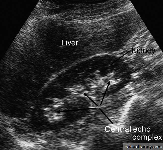
In most instances, the normal pelvicaliceal system is not visible within the renal sinus.
The normal adult renal length, measured by ultrasound,is 9–12 cm. Renal length varies with age, being maximal in the young adult. There may be a difference between the two kidneys, normally of less than 1.5 cm.Normal ureters are not usually visualized due to overlying bowel gas.
The urinary bladder should be examined in the distended state: the walls should be sharply defined and barely perceptible .The bladder may also be assessed following micturition, to measure the post micturition residual volume of urine.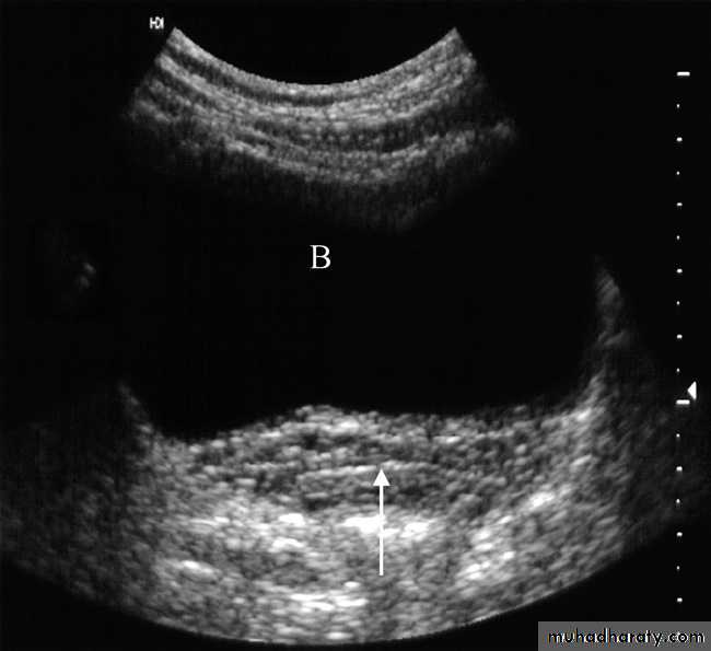
Urography
Urography is the term used to describe the imaging of the renal tract using intravenous iodinated contrast medium.There are 2 types :
1.The traditional intravenous urogram ( IVU )
2. CT urography .
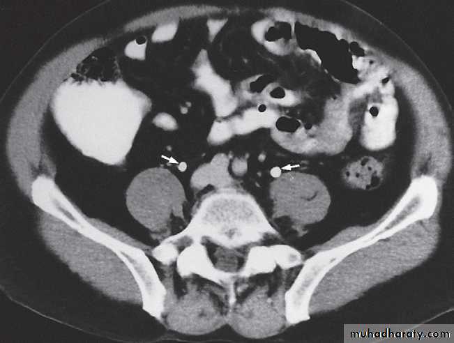
CT has the advantage of being highly sensitive for the detection of stones, including those that may be radiolucent on plain film, allows the characterization of renal lesions and the detection of ureteric lesions, and demonstrates the surrounding retroperitoneal and abdominal tissues. In addition , CT overcomes the overlap of superimposed tissues, which can cause difficulty when interpreting traditional IVU.
The principles of both techniques are similar. Firstly, ‘non-contrast’ imaging of the renal tract is required, in order to identify all renal tract calcifications. In some case, where the clinical question relates to renal calculi, the non contrast CT may be sufficient (known as the ‘CT KUB’).
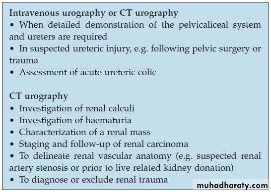
The main indications for urography are:
IVU study
-Plain film in intravenous urogram: it used to Identify all calcifications. Decide if they are in the kidneys by relating them to the renal outlines , calcification seen in the line of the ureters or bladder must be reviewed with post contrast scans, to determine whether the calcification lies in the renal tract. Note that calcification can be obscured by contrast medium and stones are missed if no plain film is taken.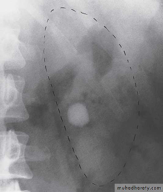
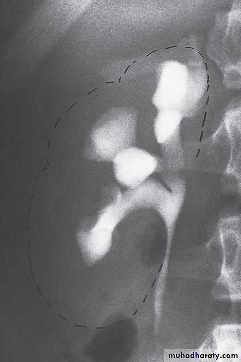
-Films taken after injection of contrast medium Kidneys
1 .Check that the kidneys are in their normal positions . The left kidney is usually higher than the right.
2 .Identify the whole of both renal outlines. If any indentations or bulges are present they must be explained.
• Local indentations .
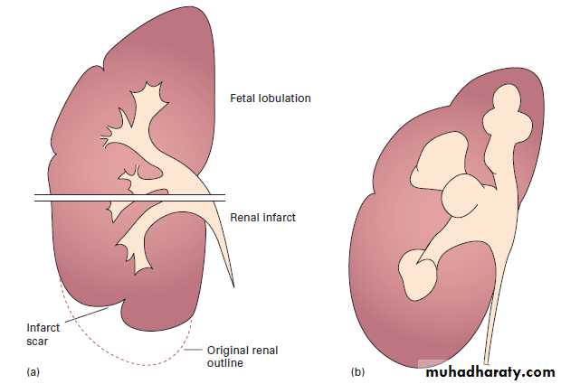
• Local bulges of the renal outline.
3 .Measure the renal lengths.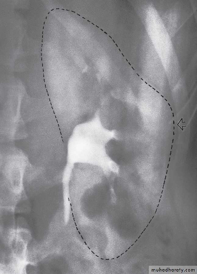
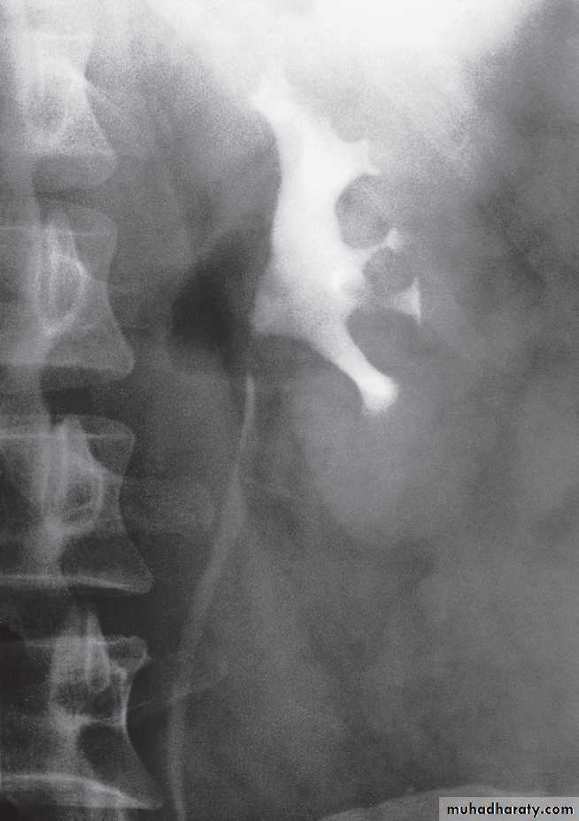
Calices
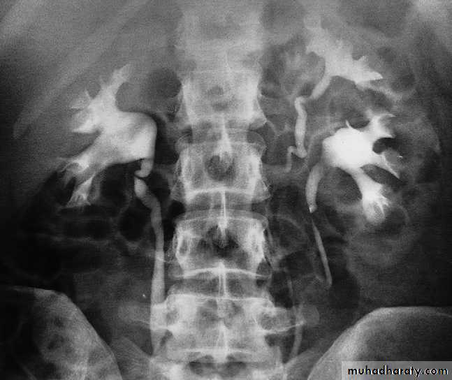
The calices should be evenly distributed and reasonably symmetrical. The shape of a normal calix is ‘cupped’ and when it is dilated it is described as ‘clubbed .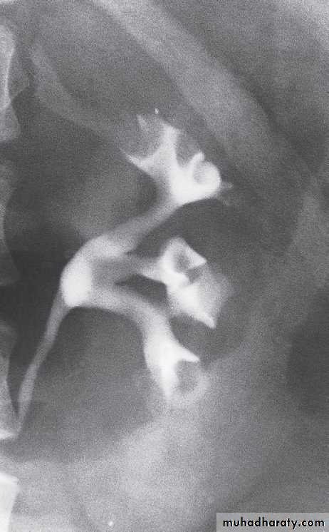
causes of dilated calcies
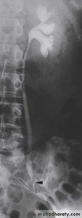
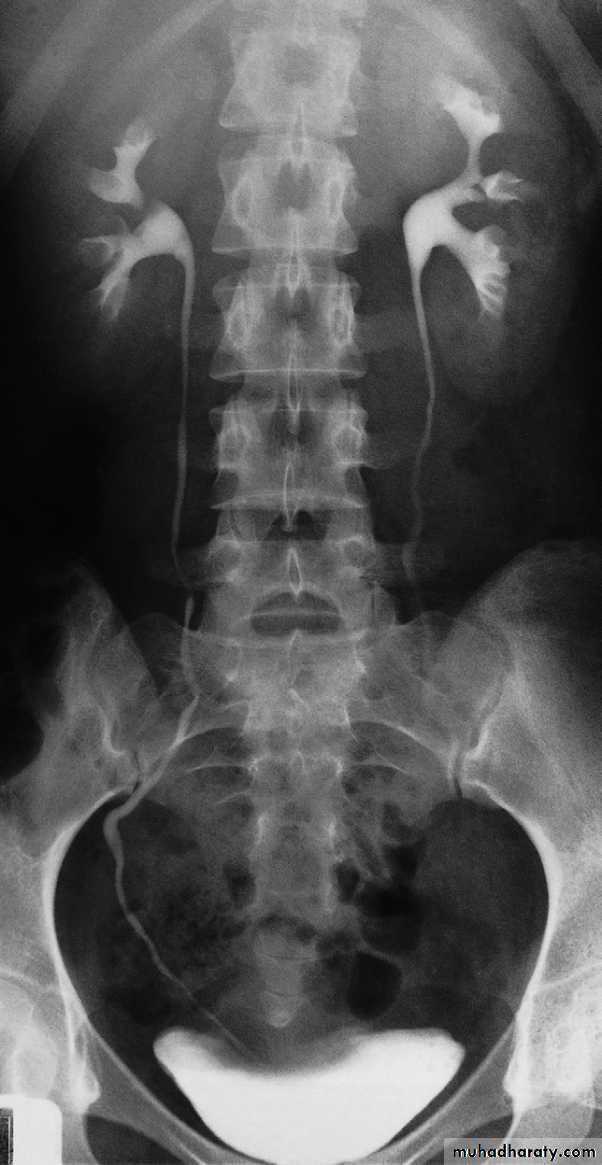
pelvis and ureters
The normal renal pelvis and pelvi-ureteric junction are funnel shaped. The ureters are usually seen in only part of their length on any one film of IVU because of obliteration of the lumen by peristalsis.Congenital variations of the renal collecting system are relatively common .
Bladder
The bladder is a centrally located structure that should have a smooth outline. It often shows normal smooth indentations from above owing to the uterus or the sigmoid colon, and from below by muscles of the pelvic floor .After micturition the bladder should
be empty, apart from a little contrast
trapped in the folded mucosa.
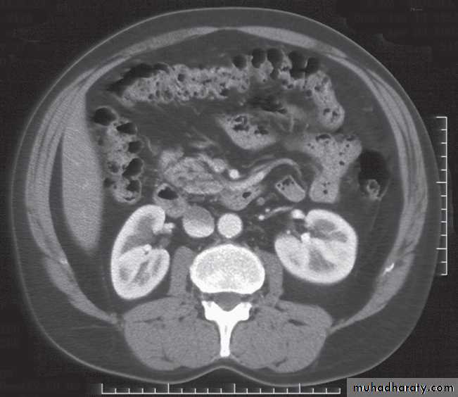
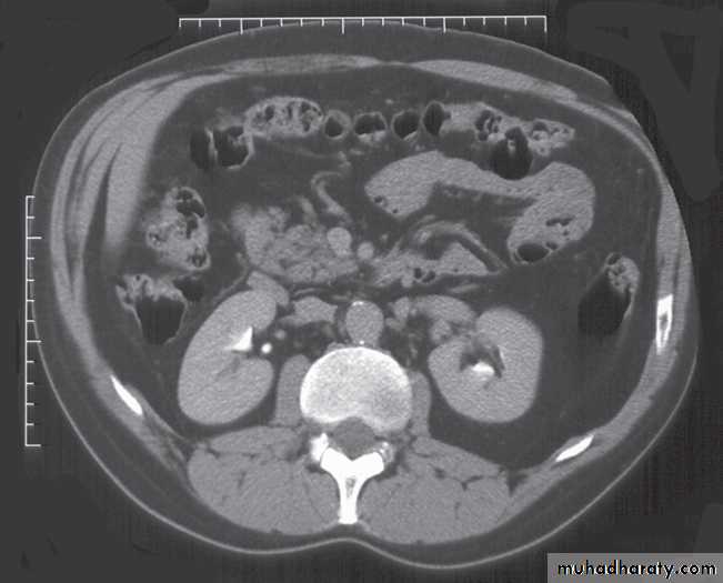
Computed tomography urography
CT is initially performed without intravnous contrast medium (non-contrast CT or ‘CT KUB’) to identify calcification .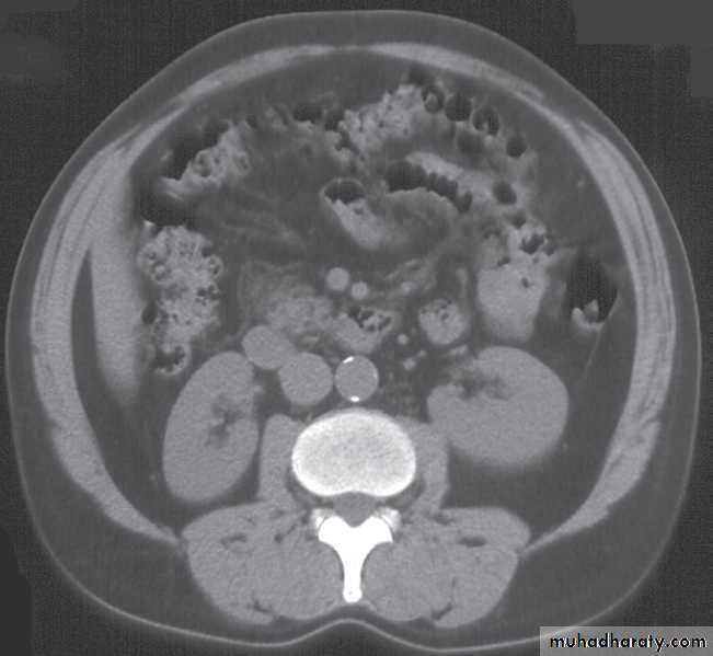
indication and include:
(i) The early renal cortical enhancement phase.(ii) The homogeneous nephrogramphase; and
(iii)The delayed urographic phase, obtained
several minutes later to demonstrate
contrast within the collecting systems.
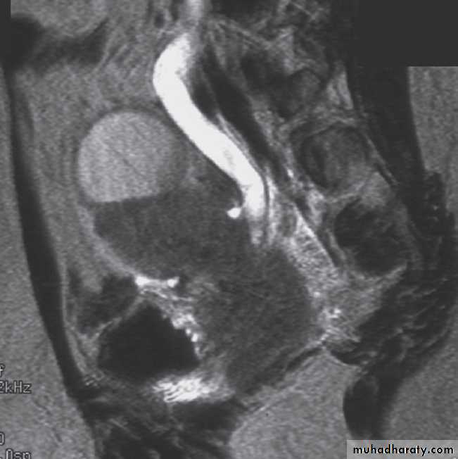
Magnetic resonance imaging
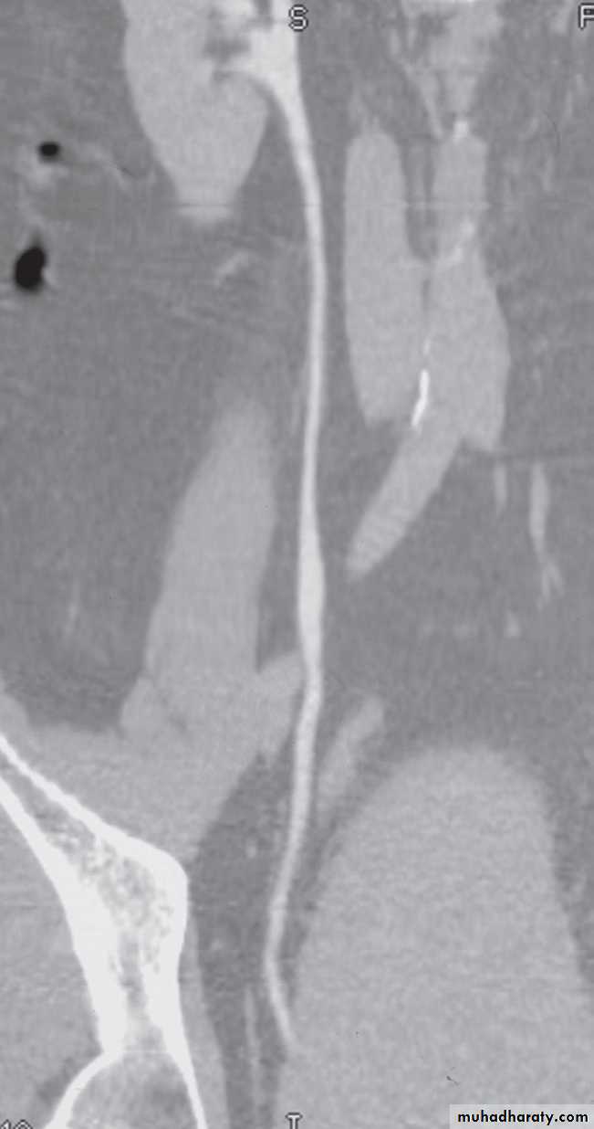
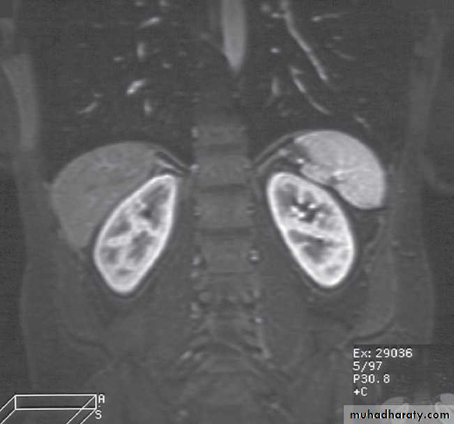
Magnetic resonance imaging gives similar anatomical information to CT, with the advantage of being able to obtain scans directly in multiple planes. It is generally used in selected circumstances , including :-To demonstrate renal artery stenosis .
- Inferior vena caval extension of renal tumours.
- To clarify problems not solved by ultrasound or CT.
-To assess the extent of bladder or prostate cancer prior to consideration for surgery.
Calcification is not visible on MRI, which is one of the main disadvantages of the technique for renal tract imaging.
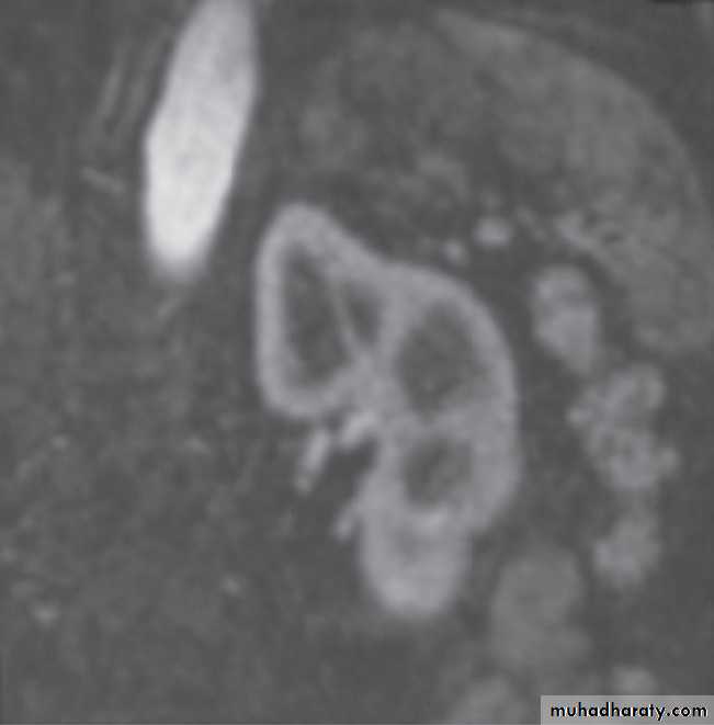
Normal magnetic resonance imaging
As with CT and ultrasound, the renal contours should be smooth. Corticomedullary differentiation is best seen on T1-weighted images and immediately following intravenous contrast enhancement with gadolinium .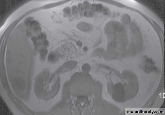
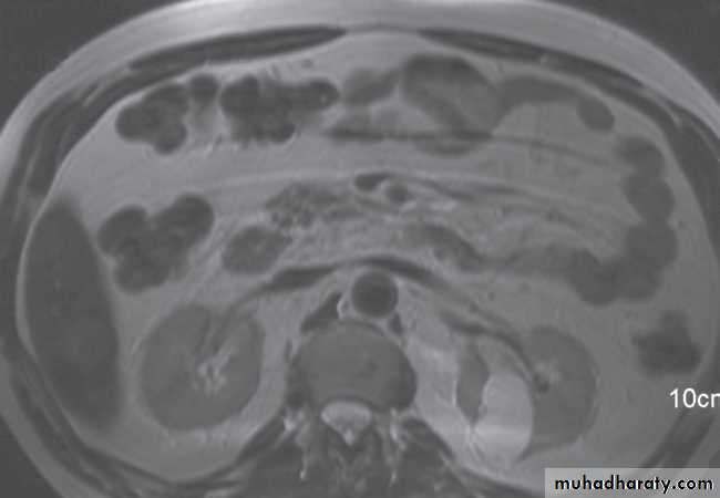
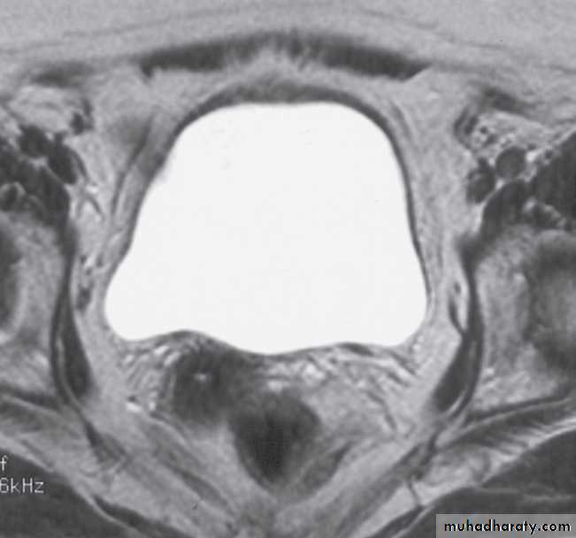
The renal collecting systems, ureters and bladder are best seen on T2-weighted images, as the fluid returns a high signal intensity .
Some normal variants are well demonstrated on MRI:
-Fetal lobulation & a column of Bertin (which is normal renal parenchyma that may look mass-like) .Special techniques
Retrograde and antegrade pyelographyThe techniques of retrograde and antegrade pyelography (the term pyelography means demonstrating the pelvicaliceal system and ureters) involve direct injection of contrast material into the pelvicaliceal system or ureters through catheters placed via cystoscopy (retrograde pyelography) or percutaneously into the kidney via the loin (antegrade pyelography).
The indications are limited to those situationswhere the information cannot be achieved by less invasive means, e.g. IVU, CT or MRI to confirm a possible transitional cell carcinoma in the renal pelvis or ureter.
Voiding cystourethrogram (micturating cystogram) and videourodynamics
In voiding cystourethrography, the bladder is filled with iodinated contrast medium through a catheter and films are taken during voiding. The entire process is observed fluoroscopically to identify vesicoureteric reflux.The bladder and urethra can
be assessed during voiding
to demonstrate strictures or
urethral valves .
Videourodynamic examination
Combines voiding cystourethrography with bladder pressur measurements, which necessitate bladder and rectal pressure lines.It is indicated in the investigation
- Patient with incontinence to distinguish detrusor instability from sphincter weakness (stress incontinence).
-In elderly men with obstructive symptoms, to differentiate true
obstruction from bladder instability
- In patients with a neurogenic bladder.
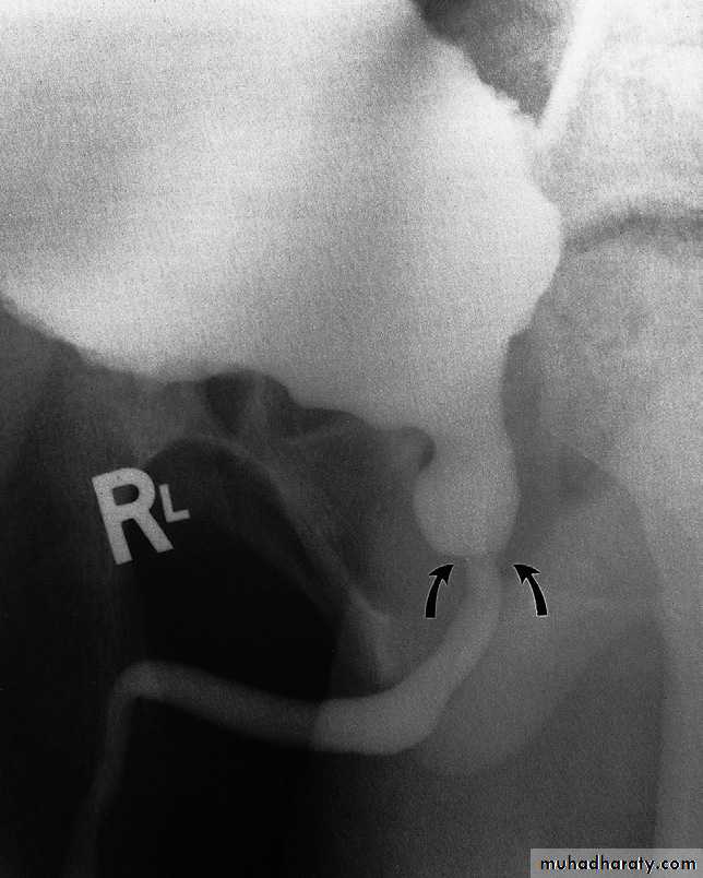
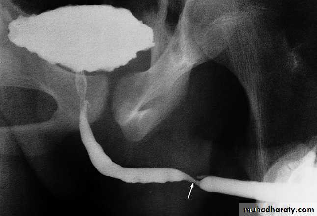
Urethrography
The urethra is visualized during voiding cystourethrography.For full visualization of the male urethra, however, an ascending urethrogram with contrast medium injection via the external urethral meatus is necessary .The usual indications for the examination are:
- The identificationof urethral strictures
- To demonstrate extravasation from
the urethra or bladder neck
following trauma.
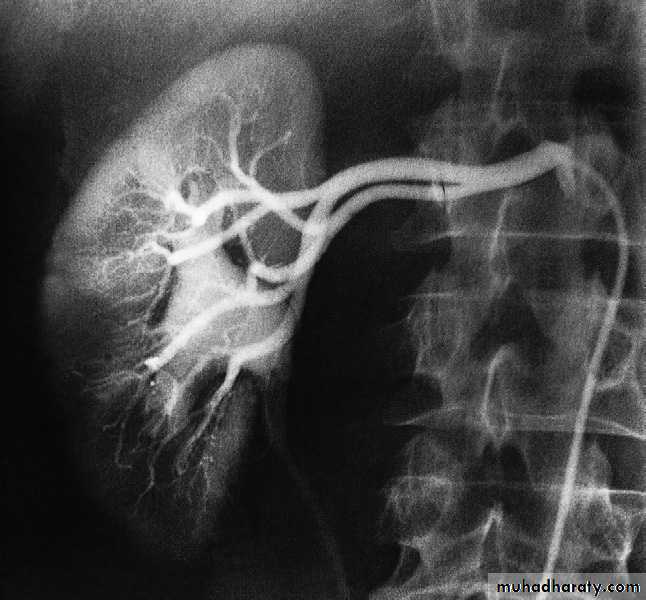
Renal arteriography
Renal arteriography is performed via a catheter introduced into the femoral artery by the Seldinger technique . Selective injection are made into one or both renal arteries . It is mainly used:- To confirm the CT or MRI findings of vascular anatomy prior to renal surgery.
-To confirm renal artery stenosis prior to percutaneous balloon angioplasty.