Blood Cells
Session 2 9 October 2017Prof. Dr.Baybeen Selevany
Objectives
Types of blood cells2. Hemopoiesis or Hematopoiesis
3. Red Blood Cells(RBC)
Blood cells are specialized cells that makes about 45 % of the total blood volume (5600ml) i.e. 2520 ml.. Blood cells are the solid phase of blood surround by liquid phase the plasma. Types:
a- red blood cells (RBC’s) or erythrocytes.
b- white blood cells (WBC’s) or leukocytes.
c.cell fragments called platelets or thrombocytes.
Hemopoiesis or Hematopoiesis:
Hemopoiesis is the process of blood cells production.Erythropoiesis: is the proliferation & differentiation of RBC’s or erythocytes.
Leukopoiesis: is the development of WBC’s or leukocytes.
Thrombopoiesis: is the development of platelets or thrombocytes.
All the blood cells are derived from a single population of stem cells located in red bone marrow. The bone marrow contains multipotent uncommitted stem cells (pluripotential stem cells ) that differentiate into one or other type of committed stem cells ( progenitor cells ) which is differentiated into various differentiated types of blood cells:
Proerythroblasts (pronormoblast ): from which erythrocytes develop.
Myeloblasts: from which granulocytes (neutrophil, eosinophils & basophiles ) develop.
Lymphoblasts: From which lymphocytes (T & B-lymphocytes) develop.
Monoblast: from which monocytes develop.
Megakaryoblasts: from which platelets or thombocytes develop.
Red Blood Cells (Erythrocytes)
About 95 % of the volume of the blood cells consists of erythrocytes. No nuclei in RBC.
Erythropoiesis: is the process of RBC’s production.Regulation of red blood cell production: Role of erythropoietin.
The rate of erythropoiesis is regulated by a humoral agent known as erythropoietin. This hormone was first discovered in the blood & urine, produced mainly in the kidneys. In the normal person 90% of the erythropoietin is formed in the kidneys & 10% in the liver. The function of this hormone is to stimulate committed stem cells to differentiate into proerythroblasts i.e stimulate the synthesis of messenger RNA. The main factor that cause the release of erythropoietin is hypoxia i.e. low blood o2 level. The causes of hypoxia are: 1- anemia. 2- decreased Hb. 3- lung diseases. 4- high altitude(normal). 5- low blood volume. 6- destruction of bone marrow by any means such as x-ray therapy. Hypoxia stimulates erythrocyte production by increasing the formation of circulating glycoprotein hormone erythropoietin by kidneys. Erythropoietin stimulates red bone marrow to produce more erythrocytes by increasing the number of proerythroblasts & by decreasing the time required for erythrocytes to mature. The increased number of erythrocytes increases the ability of the blood to transport o2.This mechanism return blood o2 levels to normal & maintain homeostasis by increasing the delivery of o2 to tissues. Conversely if blood o2 levels increase less erythropoietin is released & few RBC’s are formed by the bone marrow, when large quantities of erythropoietin are formed & if there is plenty of iron available the rate of RBC production can rise to ten or more times normal.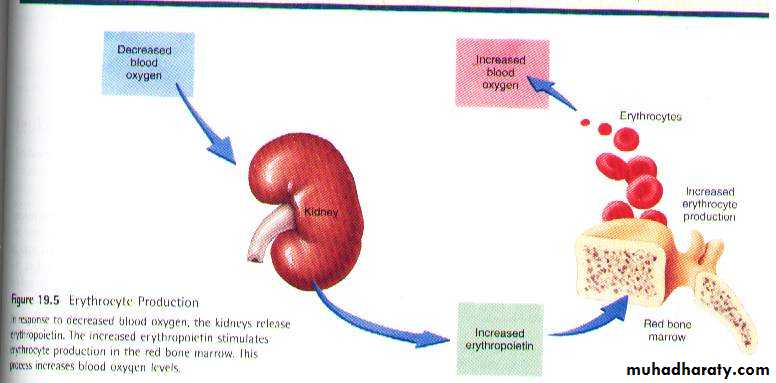
Maturation of Erythrocytes:
Cell division requires two vitamins:Vitamin B12 ( cyanocobalamine ).
Folic acid ( pteroylglutamic acid ).
Both of them are essential for the synthesis of DNA & are important for final maturation of the RBC. Therefore lack of either vit. B12 or folic acid causes:
diminished DNA & consequently failure of nuclear maturation & division.
The erythroblastic cells of the bone marrow failing to proliferate rapidly, produce mainly large than normal RBC’s called macrocytes.
The cells are irregular, large & oval instead of the usual biconcave disc & have a flimsy membrane. Cells after entering circulation are capable of carrying O2 normally but their fragility causes them to have a short life. Therefore it is said that vit. B12 or folic acid deficiency causes maturation failure in the process of erythropoiesis.
Deficiency of vit. B12: a common cause of maturation failure is failure to absorb vit.B12 from gastrointestinal tract (GIT). This often occurs in pernicious anemia in which the basic abnormality is an atrophic gastric mucosa that fails to secrete normal gastric secretion which contain intrinsic factor which is combining with vit.B12 of food & enhance the absorption of vit.B12 in ileum. Intrinsic factor is glycoprotein secreted by parietal cells of gastric glands. This factor combines with vit. B12 & combines to specific receptors sites in the ileum membrane then vit. B12 is transported into the blood. Once vit. B12 has been absorbed from GIT; it is stored in the liver & then released slowly as needed to the bone marrow & other tissues of the body. The daily requirement of vit. B12 is 1-3 micrograms.
Deficiency of folic acid: people with gastrointestinal absorption abnormalities such as sprue have serious difficulty in absorbing both folic acid & vit. B12. Folic acid is a normal constituent of green vegetables, some fruits, liver & meats, it is easily destroyed by cooking.
The type of anemia due to lack of folic acid & vit. B12 is called megaloblastic anemia (it is also called macrocytic normochromic anemia).
Morphology of mature Erythrocyte: The three basic features of RBC’s are: its size, its shape & its content.
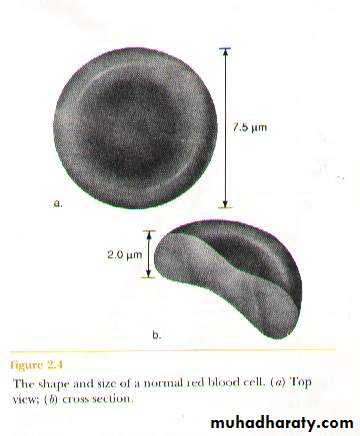
a. Size of erythrocytes: Normal mature erythrocyte is nearly uniform in size with diameter of 7.2 to 7.9 micrometers with edges which are about 2.5 micrometers thicker than the center which is about 1 micrometers.
.
b.Shape of erythrocytes: they are biconcave discs which are thick at their edges. This shape: 1- gives the cell great flexibility & elasticity. 2- Allow it to fold when it has to move through very narrow blood capillaries. 3- Increases the surface area of the RBC’s . So all mature RBC’s are perfectly round, flat & smooth.
c. Content of erythrocytes: The main component of erythrocyte is the pigmented protein hemoglobin which occupies about 1/3 of the total cell volume & account for its red color. The erythrocyte is thicker at the edges than in the middle, more hemoglobin is found on the outside of the cell & for that reason a normal erythrocyte stain pink to red on the outside with a pale area on the center. RBC also contain lipids , ATPase, & enzyme carbonic anhydrase. The erythrocyte consists of 65% water & 35% solids about 90 – 95 % of the solids are hemoglobin.
Red blood cell Count: average normal blood red blood cell count in adult male is 5400000 (5.4 x 10⁶) RBC per cubic millimeter (c.mm).
In normal adult female average normal RBC count is 4700000 ( 4.7 x 10⁶ ) RBC/c-mm. If the RBC’s counts are less than normal value the condition is called anemia & if the RBC’s count is more than normal value the condition is called Polycythemia.
Life span of the RBC: In human RBC survive in the circulation or an average of 120 days in males & 110 days in females.
Packed Cell Volume (PVC) or hematochrit (Hct) Is the volume or amount of RBC’s present in 100ml (dl) of blood. It's expressed as %. Average normal value in male is 47% & average normal value in adult female is 42%. In anemia PCV is less than normal value, while in polycythemia RCB is greater than normal value.
Hemolysis of RBC: process in which erythrocyte ruptures & hemoglobin is released.
Total blood Volume (TBV):The plasma and the cellular elements of the blood, principally red blood cells, fill the vascular system, and together they constitute the TBV i.e. total blood volume = plasma volume + red cell volume or packed red cell volume (volume occupied by all the circulating red cells in the body).The normal total circulating blood volume is about 8% of the body weight of a 70 Kg man (5600 ml in a 70 Kg man). About 55% of this volume is plasma (3080 ml), and about 45% of this volume is packed cell volume (2520 ml).The TBV in the average adult is 4-5 L in female & 5-6 L in adult males.