
Al-Mustansiriyah University
College of Medicine - Department of Medicine
Division of Radiology
IMAGING OF UPPER GIT BLEEDING
DR. ABDULLATEEF AL-BAYATI
Teaching Board Member
CABMS-RAD

Definition: Upper GIT bleeding is defined as bleeding from the esophagus to the ligament of
Treitz.
Clinical presentation:
hematemesis and/or melena.
Causes of acute upper GIT bleeding include:
1) Peptic ulcer disease.
2) Erosive gastritis.
3) Varices
4) Mallory–Weiss tear.
5) Malignancy of esophagus, stomach or duodenum.
An upper GIT source for GIT bleeding is diagnosed by emergency upper GIT endoscopy in the
majority of cases.
Endoscopic hemostatic therapies include:
Injection of sclerosants,
Injection of vasoconstrictors,
Thermal coagulation
Mechanical methods.
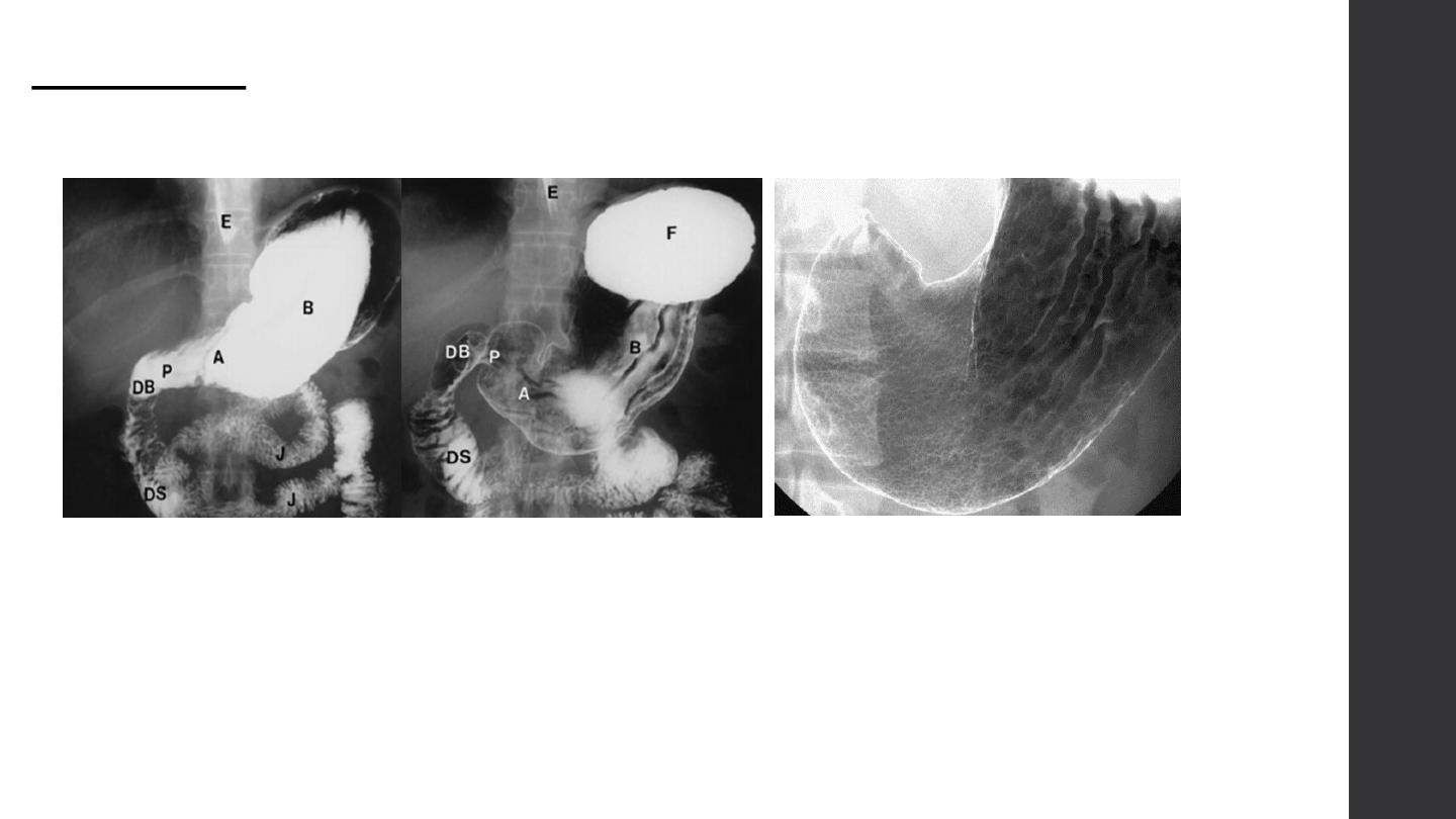
Barium meal
• Technique of examination.
• Normal anatomy

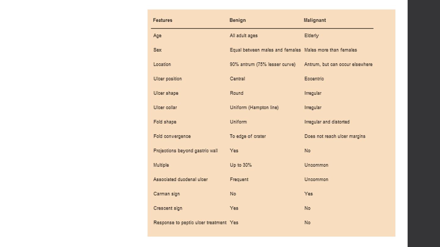
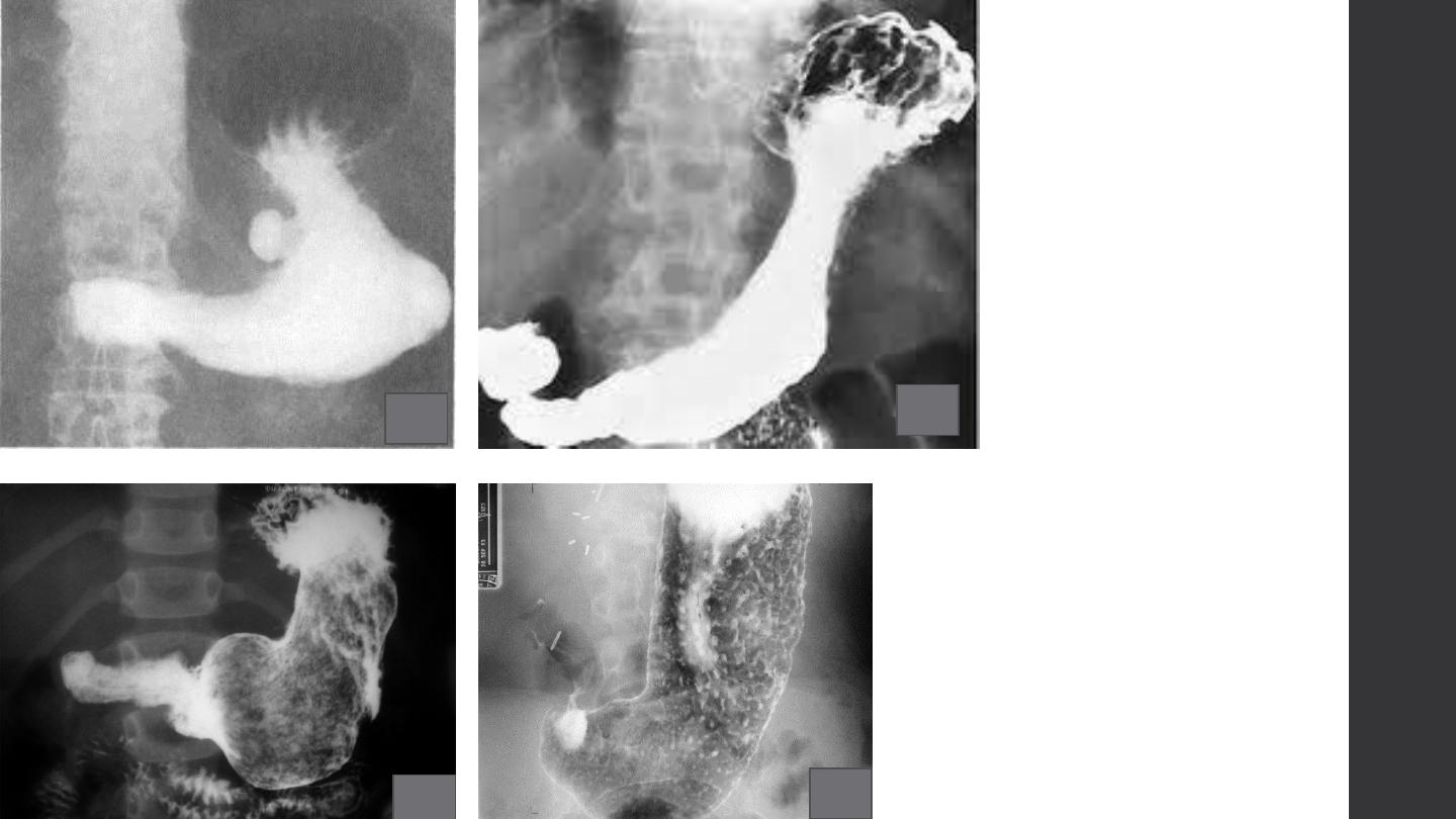
1. Ulcer:
benign vs. malignant
2. Filling defect:
types and causes
3. Stricture:
benign and malignant / causes
4. Contour changes:
5. Mucosal changes:
types and causes
6. Diverticulum:
types and causes
Ulcer
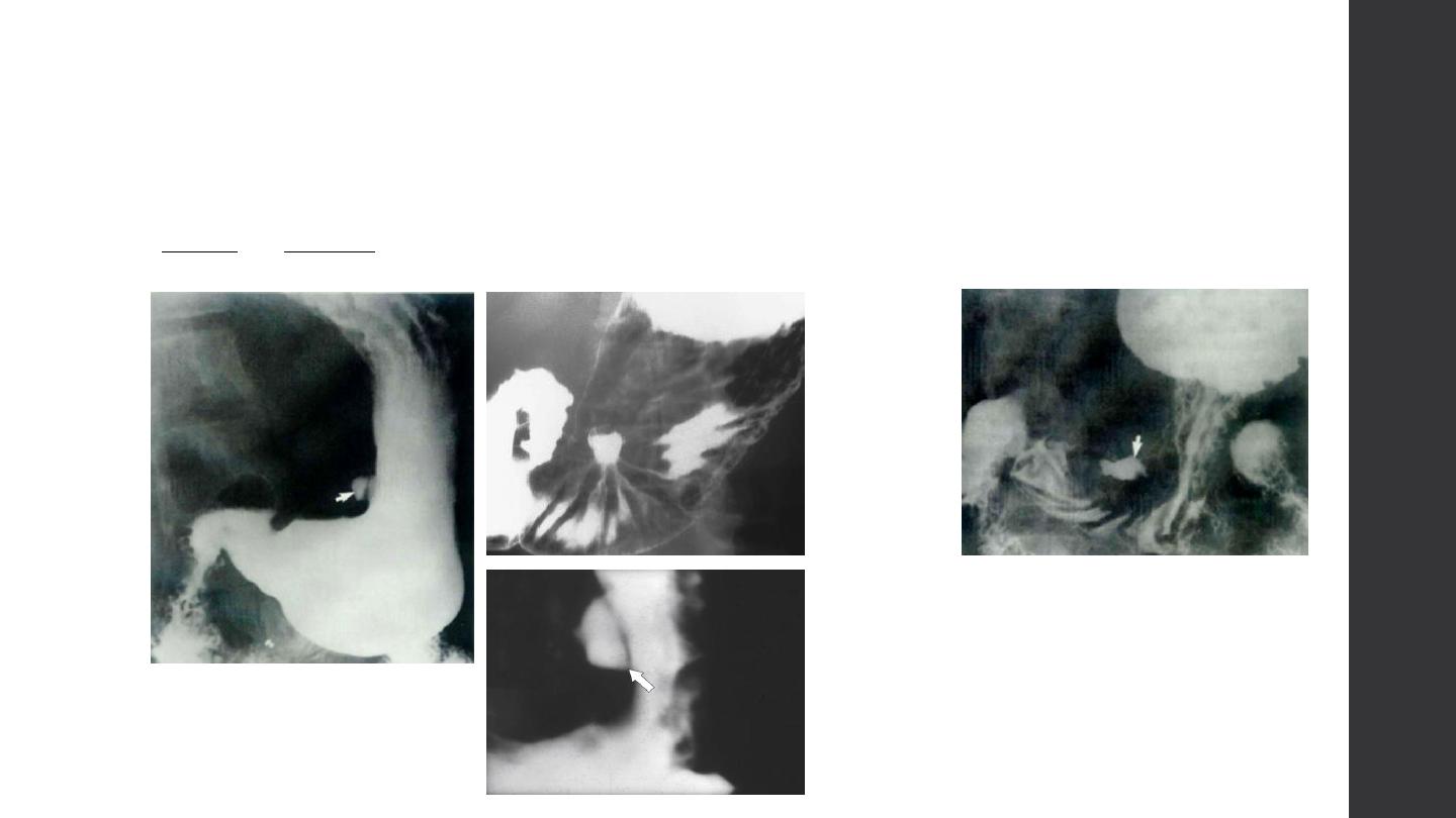
Ulcer
Types
Causes
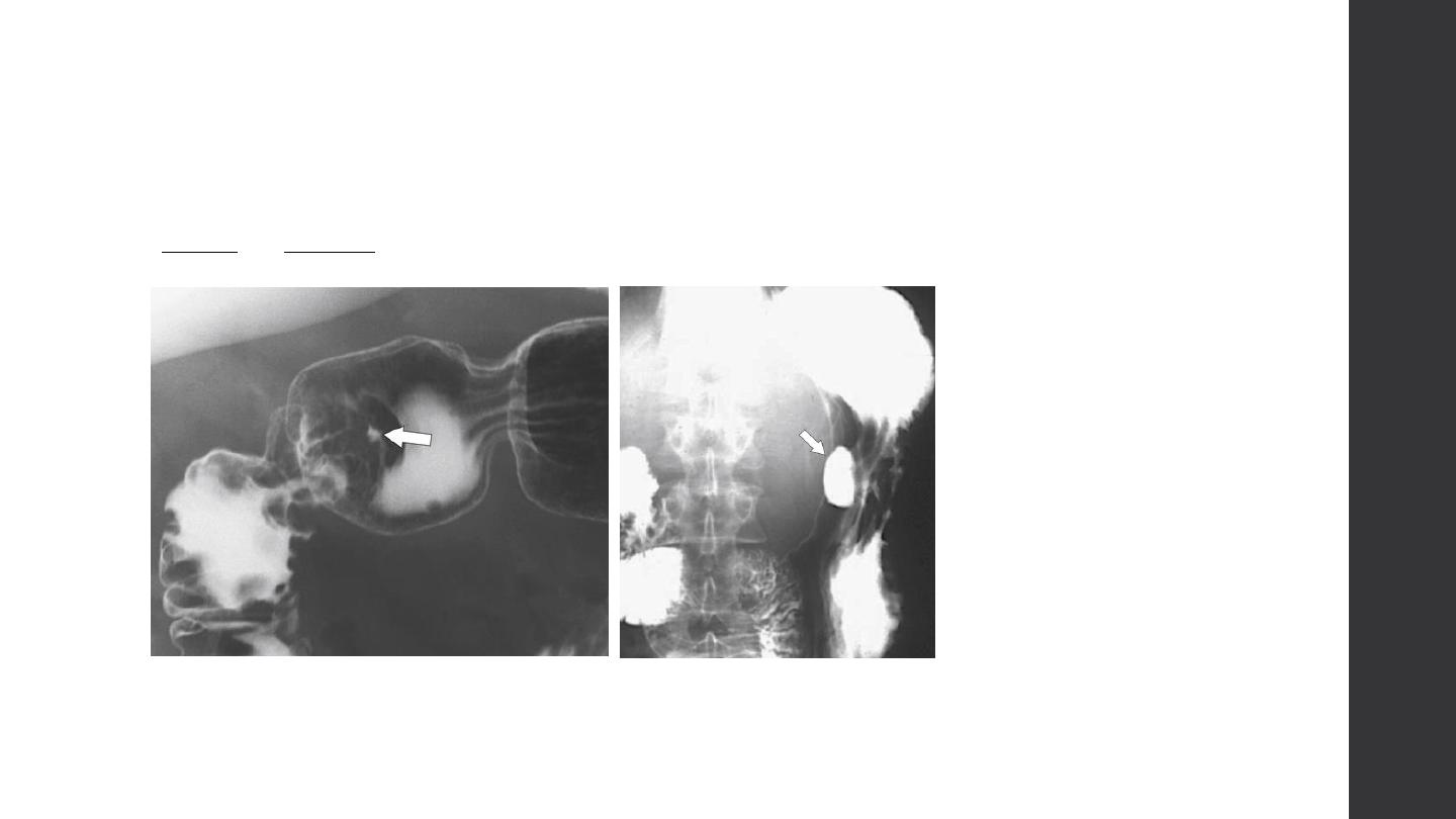
Ulcer
Types
Causes
Ulcer
Types
Causes
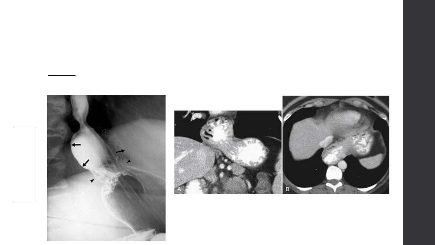
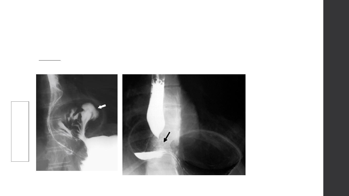
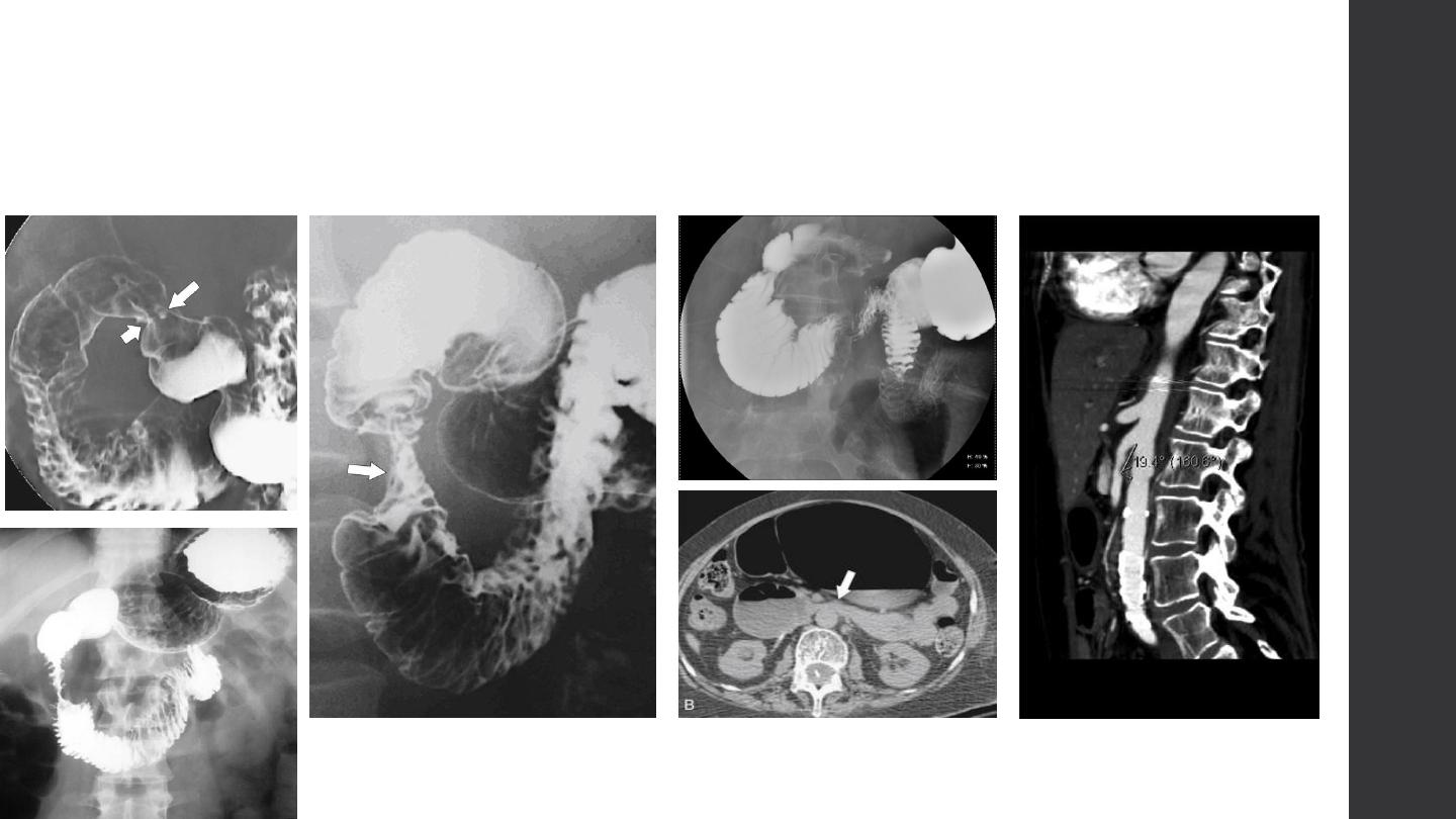
Stricture
Benign vs. malignant
Causes
Stricture
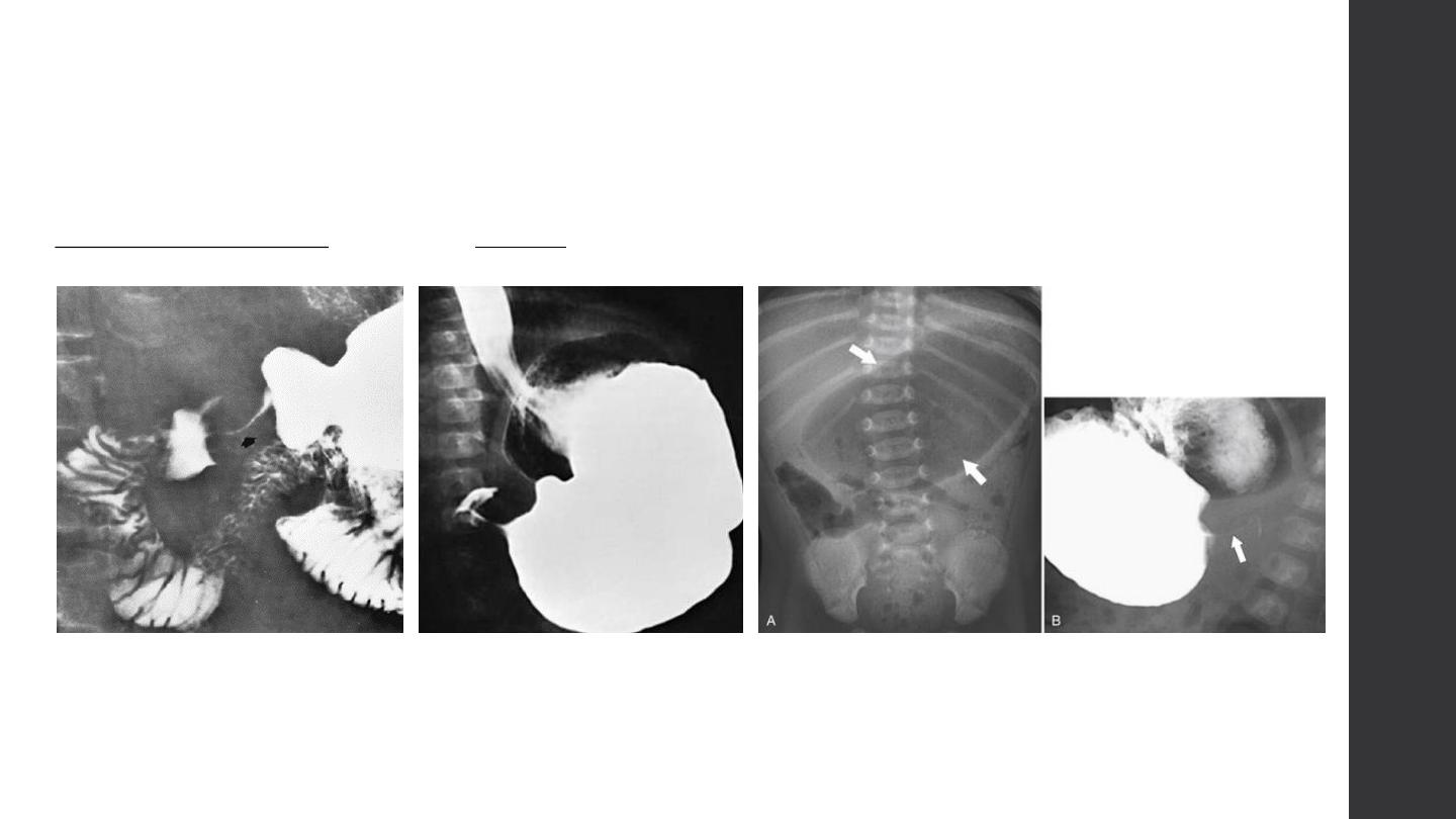
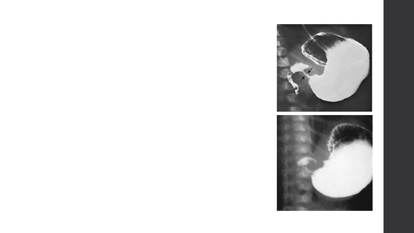
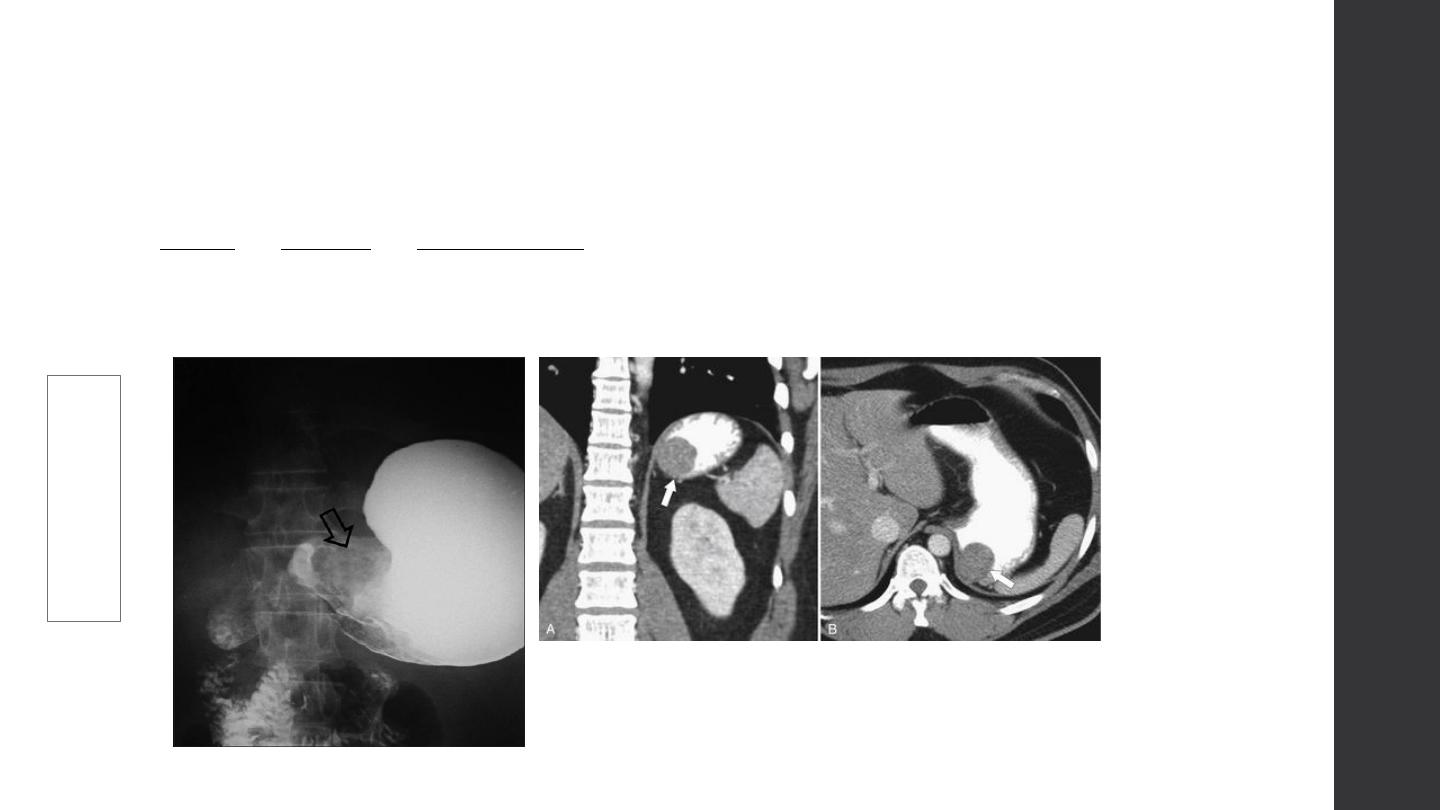
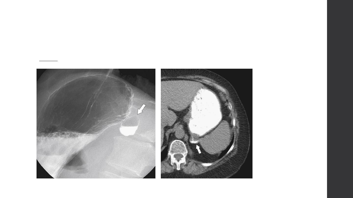
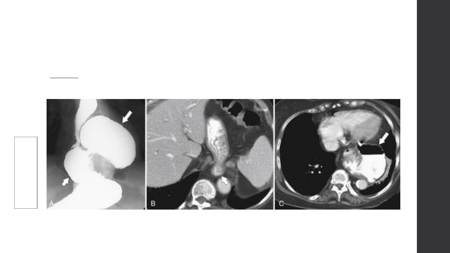
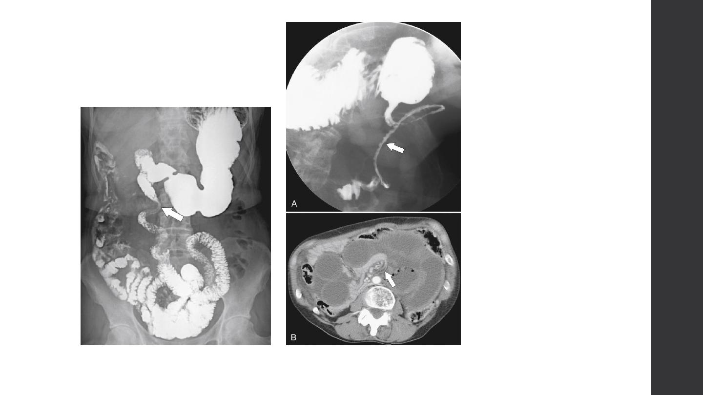
Gastric outlet obstruction
Gastric outlet obstruction result in gastric dilatation .
The causes are:
1. chronic duodenal ulceration
2. carcinoma of antrum
3. duodenal, ampullary and pancreatic carcinoma
4. acute or chronic pancreatitis including pseudocyst formation
5. poor functional patency of a gastroeneterostomy
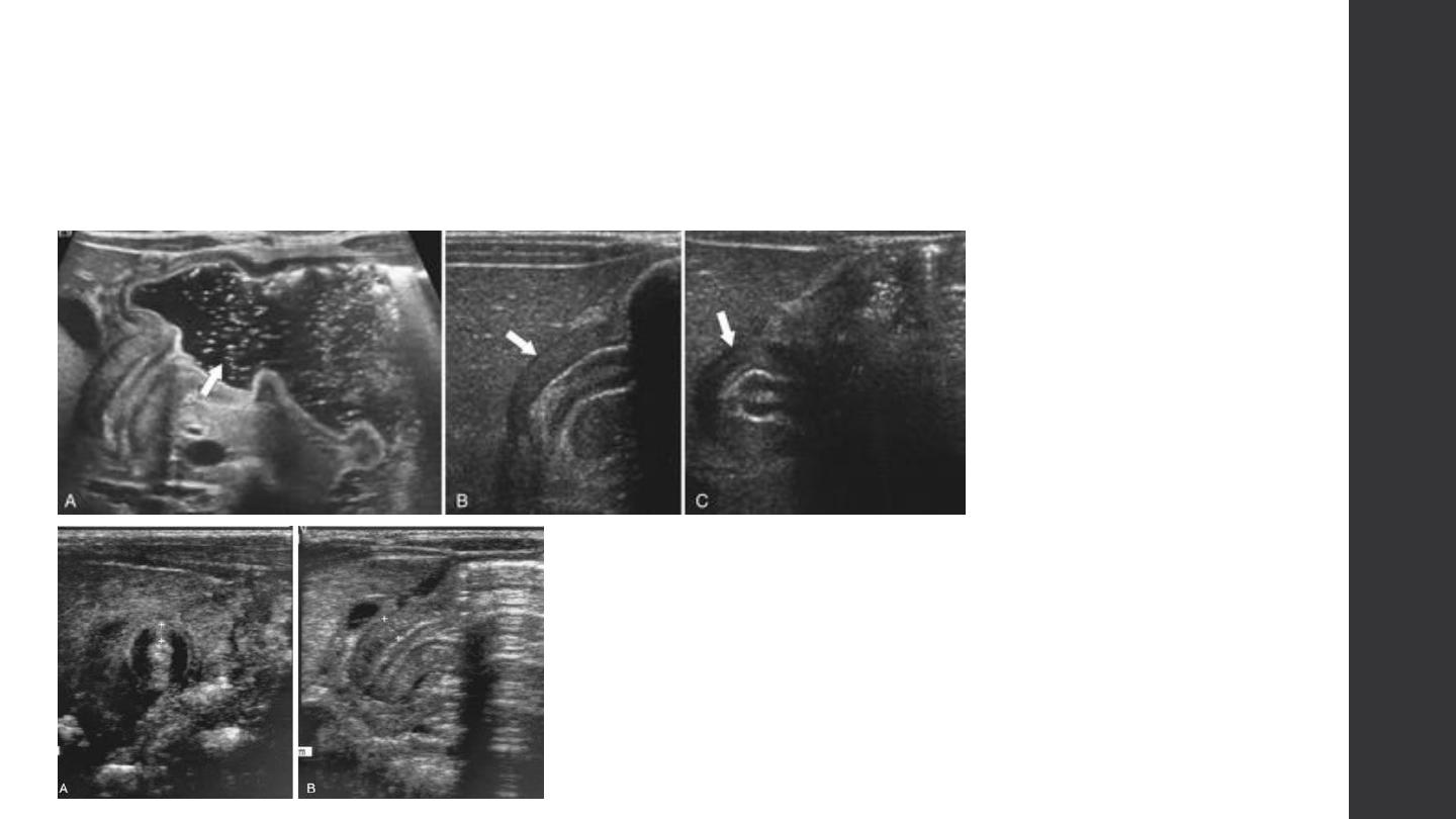
6. pyloric stenosis in infants (US shows a thickened, elongated
pyloric canal)
Stricture
Stricture
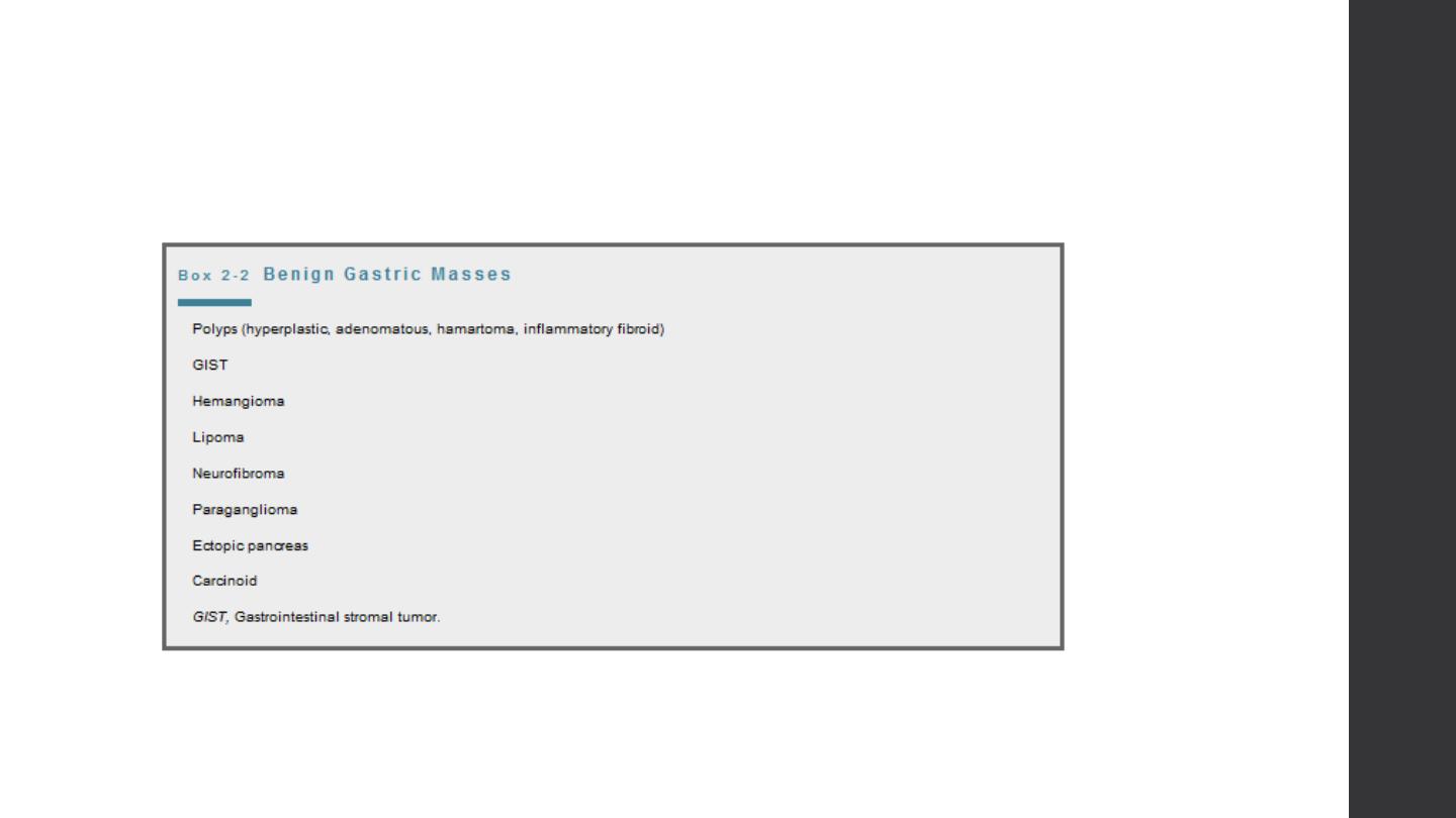
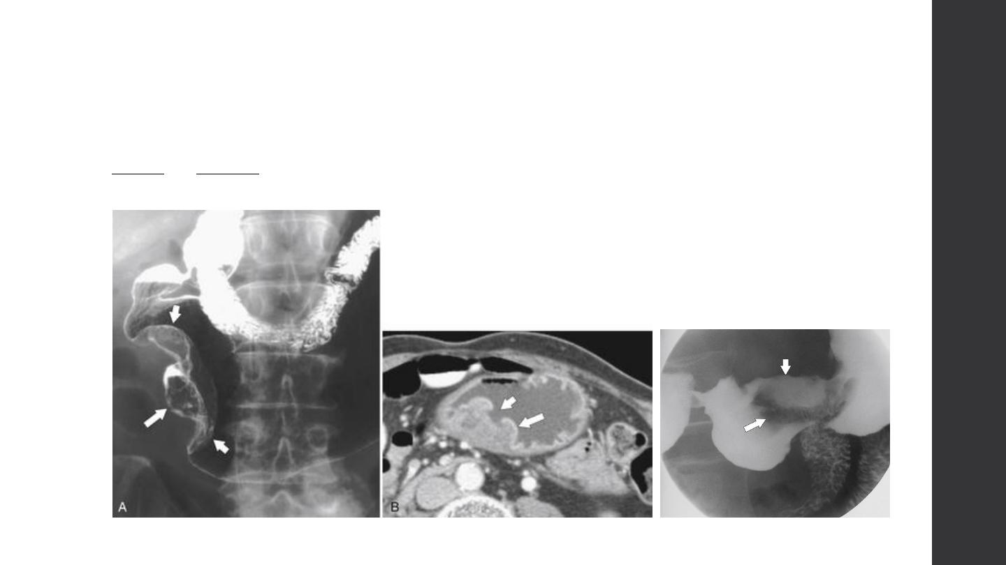
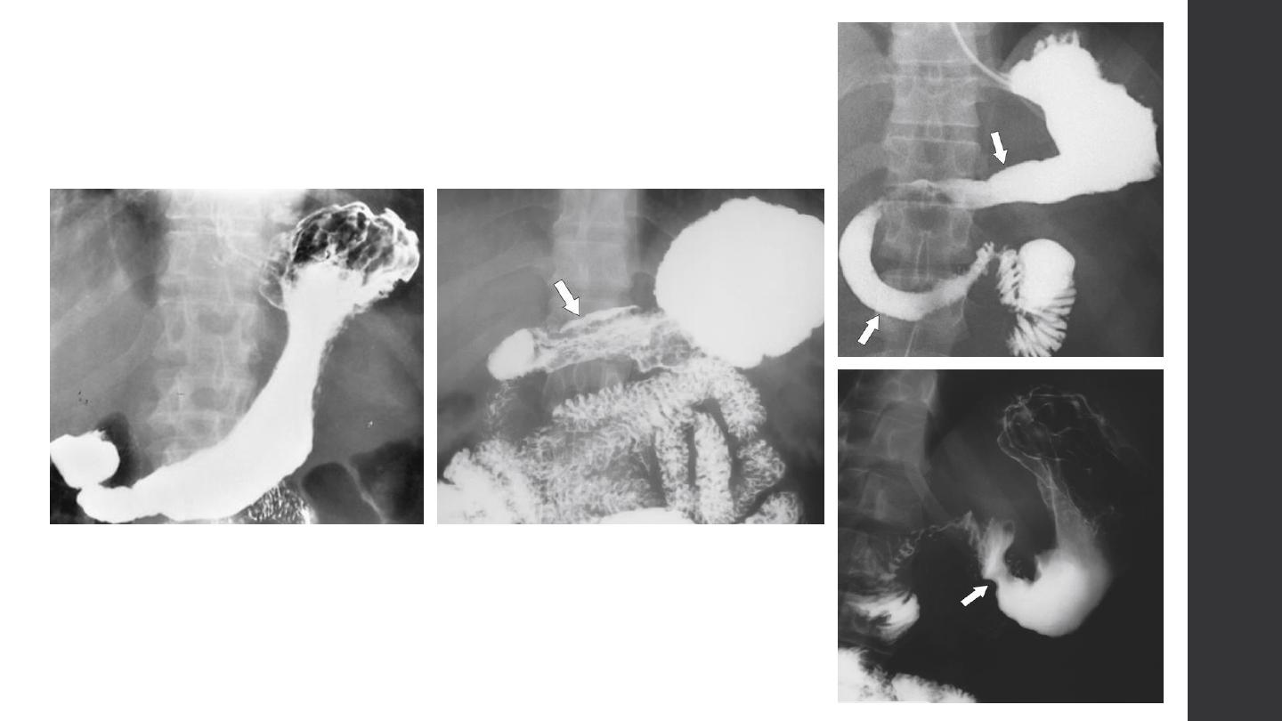
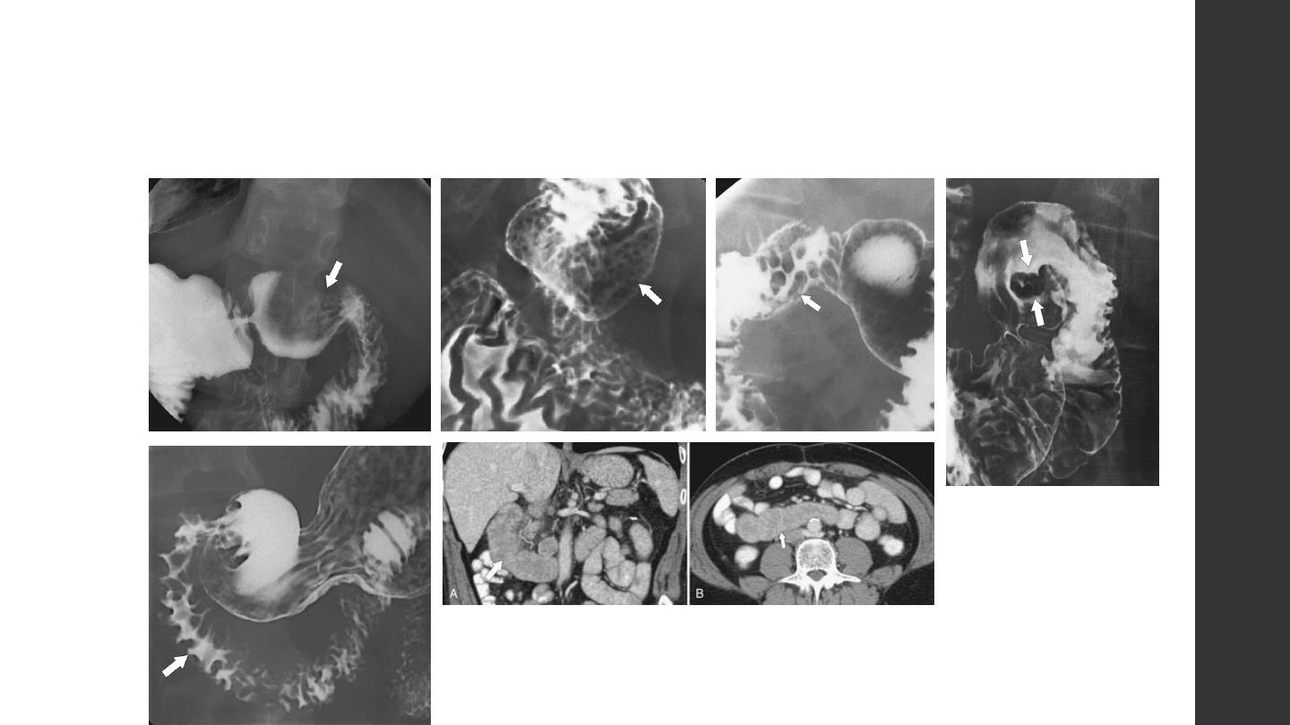
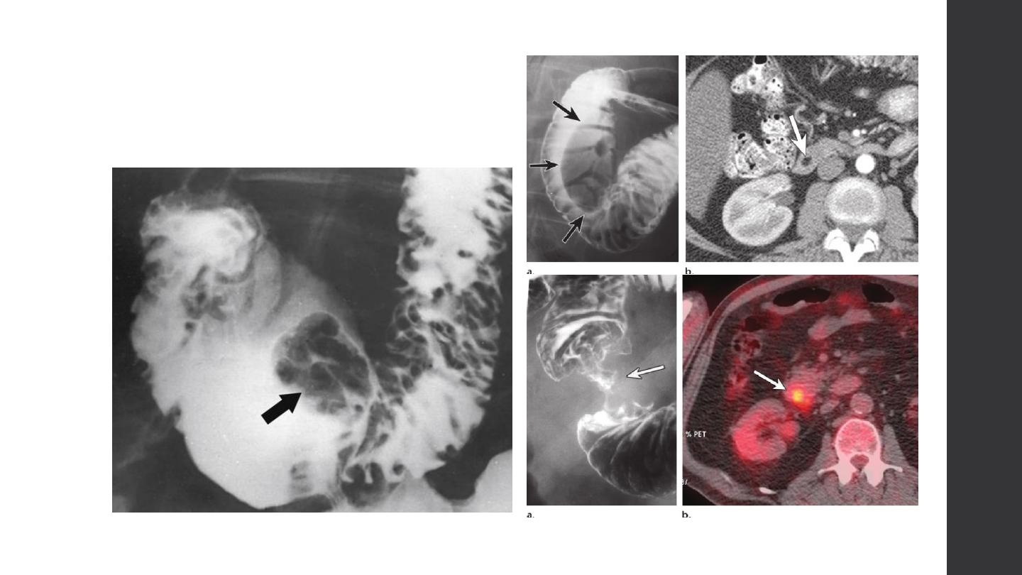
Filling defect

Filling defect
Filling defect
Types
Causes
Intra
-lu
min
al
Filling defect
Types
Causes
Intra
-mur
al
Filling defect
Types
Causes
Specific Type
Int
ra
-mur
al
Diverticulum
Types
Contour changes
Causes
Slidi
ng
type
Contour changes
Causes
Rolli
ng
type
Contour changes
Causes
Mixed t
ype
Contour changes
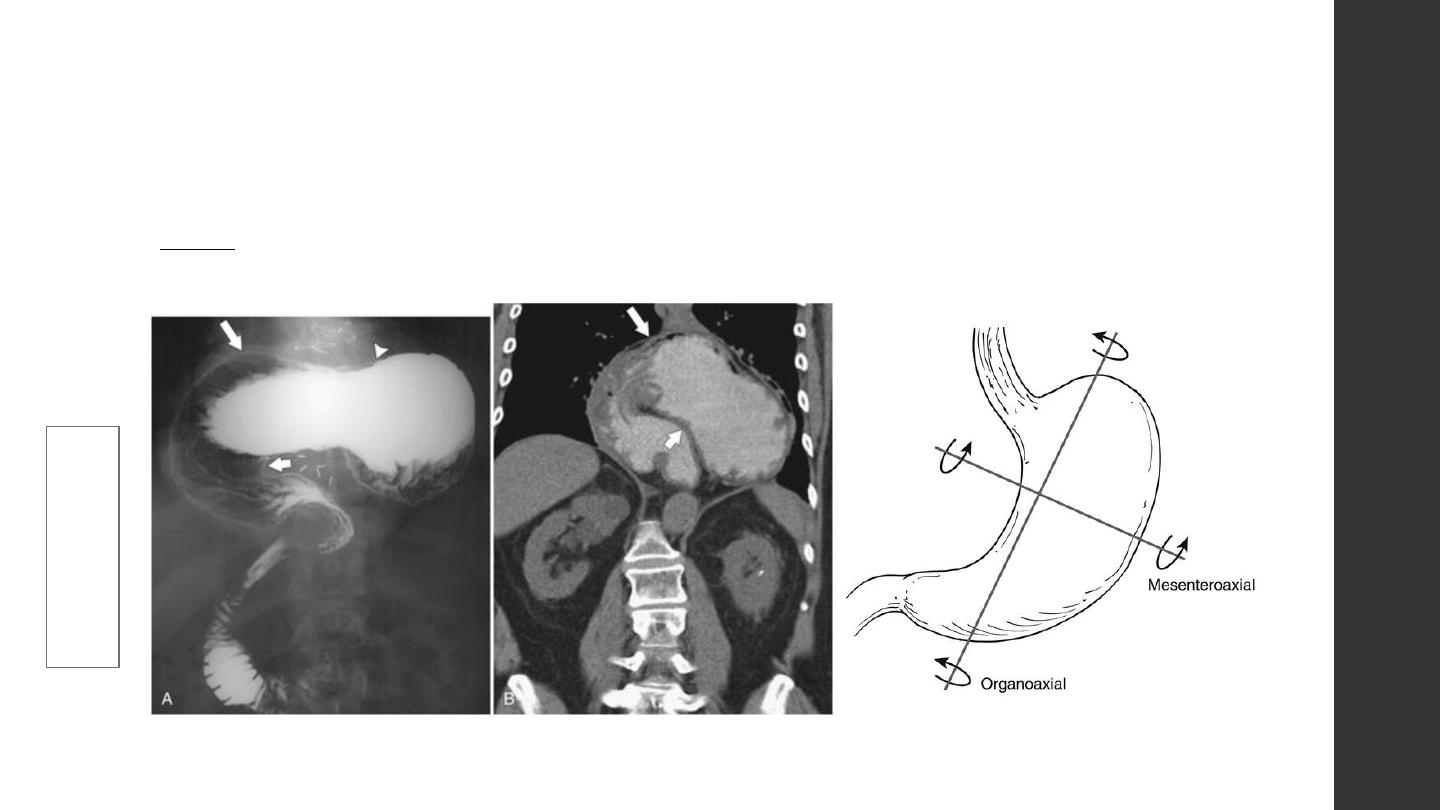
Types
organoa
xial
Contour changes
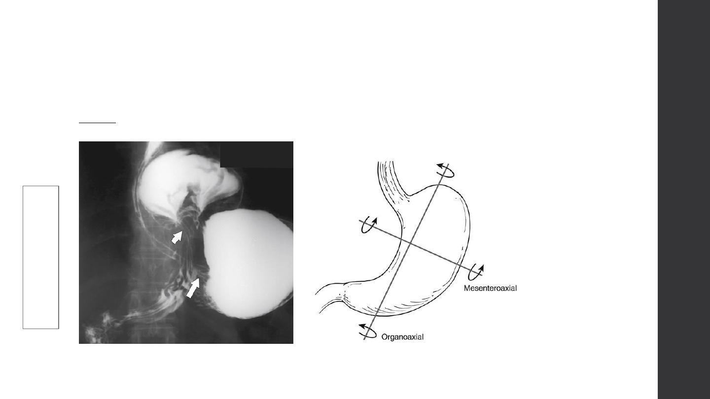
Types
mese
ntroaxial
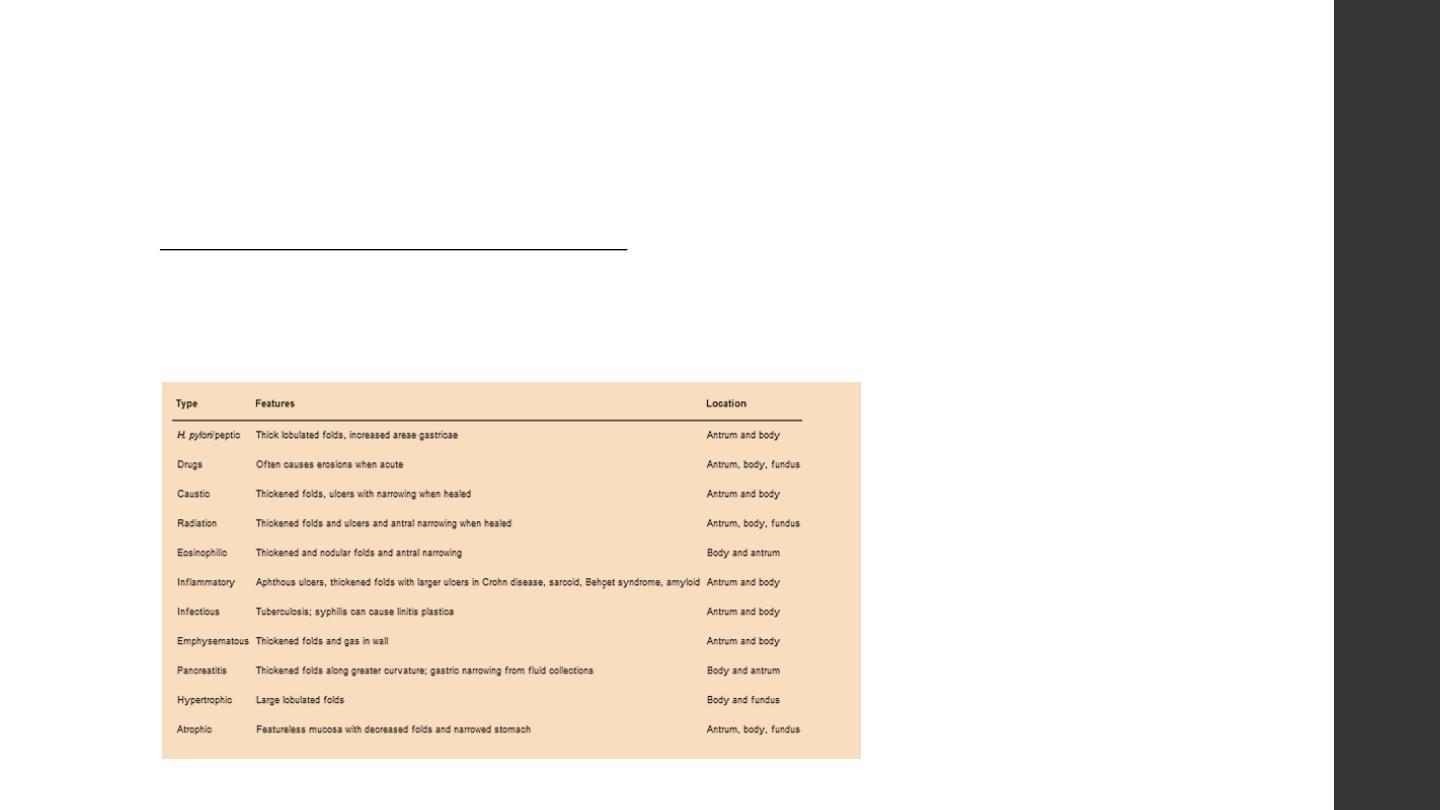
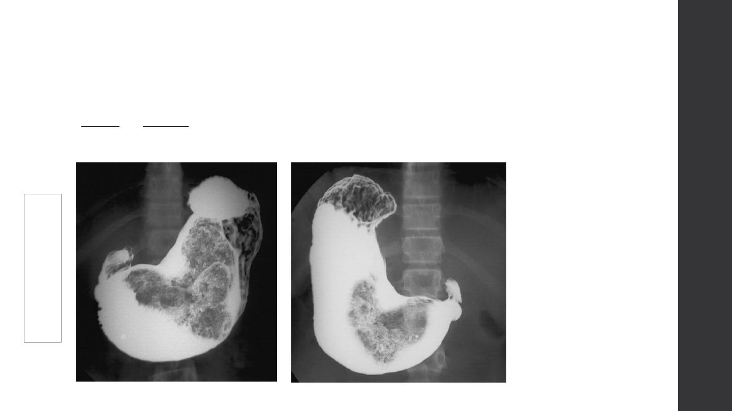
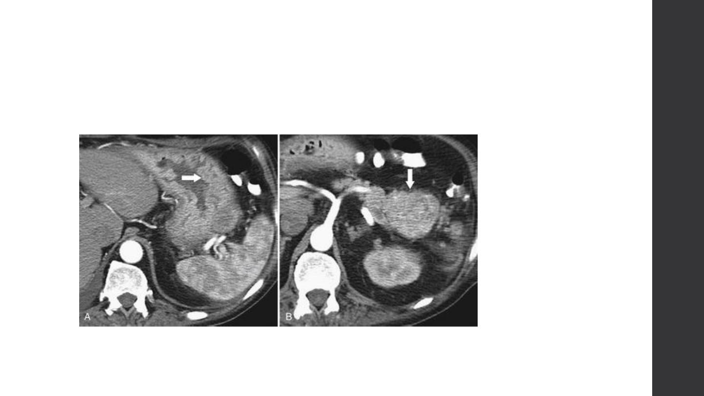
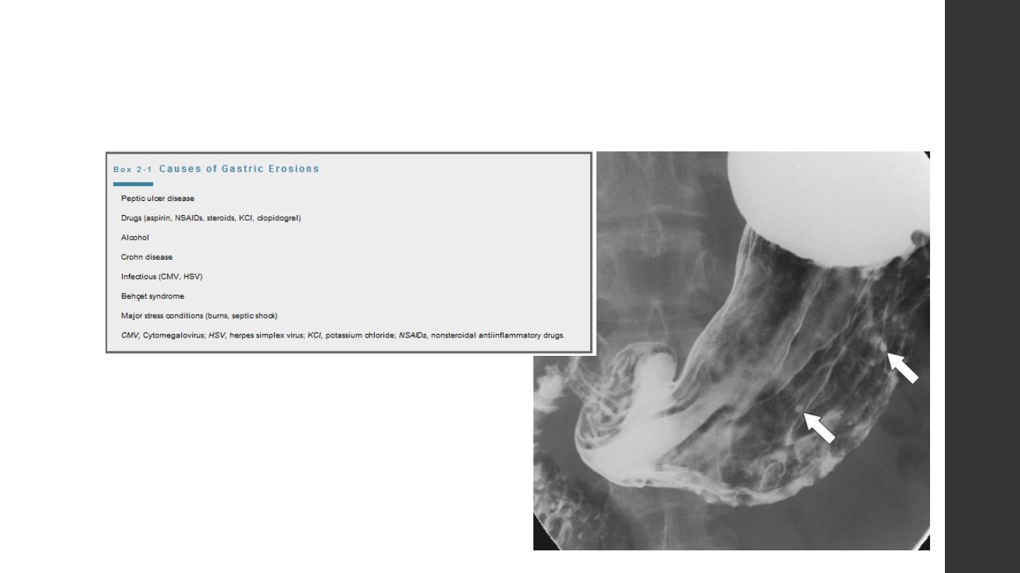
Mucosal alteration
Causes of diffuse mucosal thickening
Benign gastritis [erosive, granulomatous, caustic, radiation], varices
Malignant ca. lymphoma metastasis
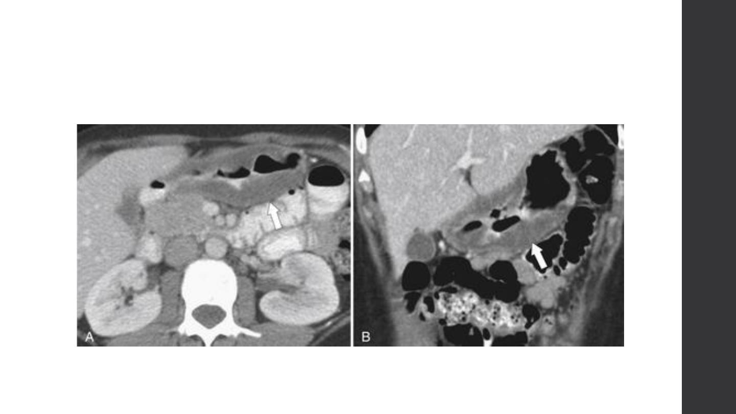
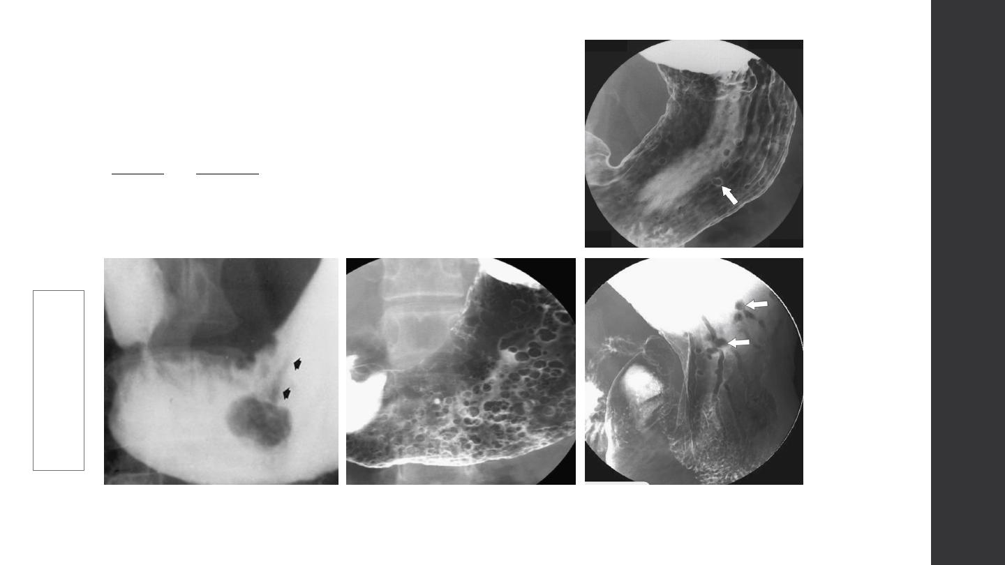
Mucosal alteration
Mucosal alteration
hypertrophic gastritis
Ménétrier Disease
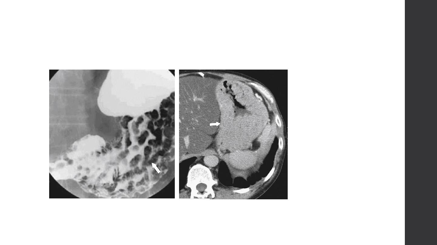
Mucosal alteration
Zollinger-Ellison Syndrome
Mucosal alteration
Mucosal alteration
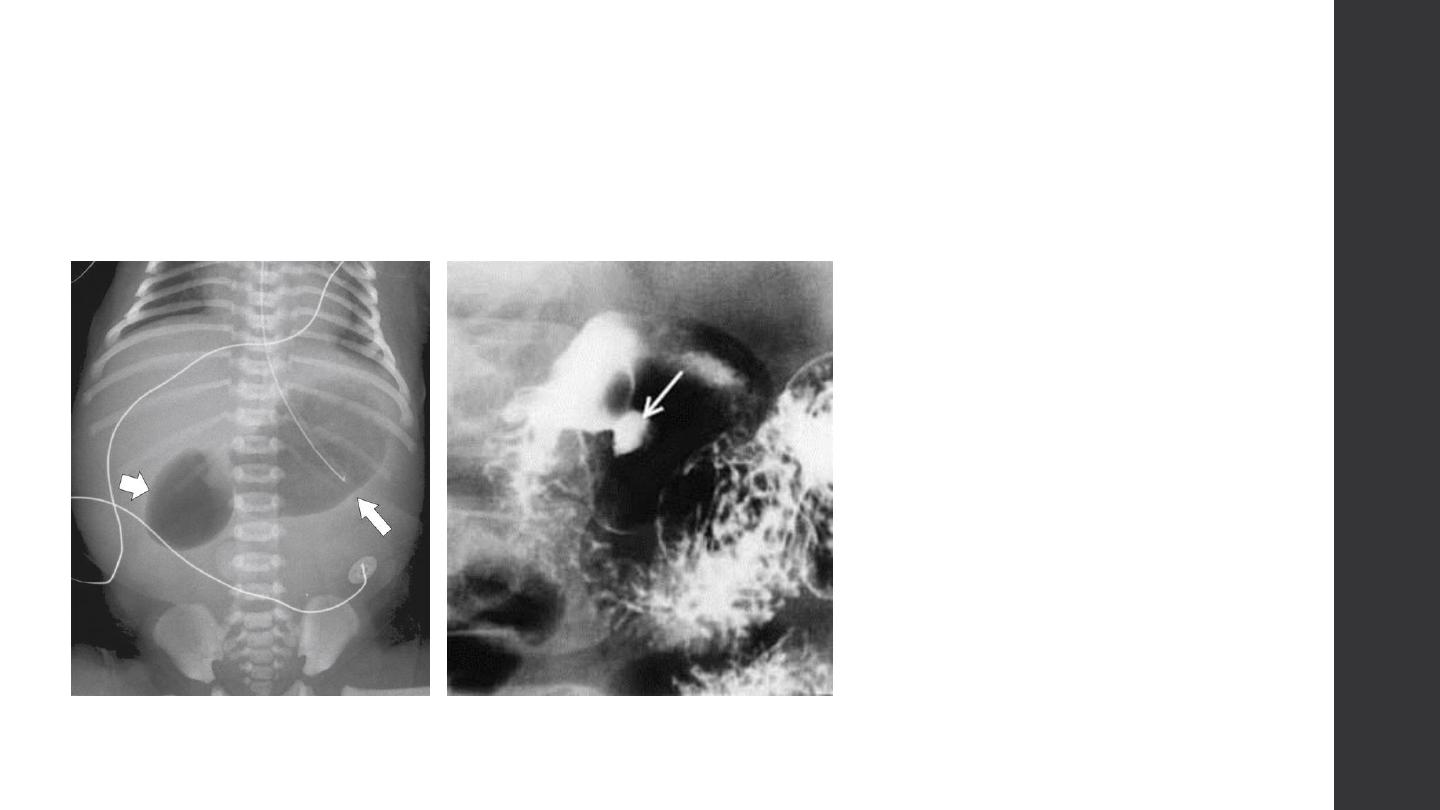
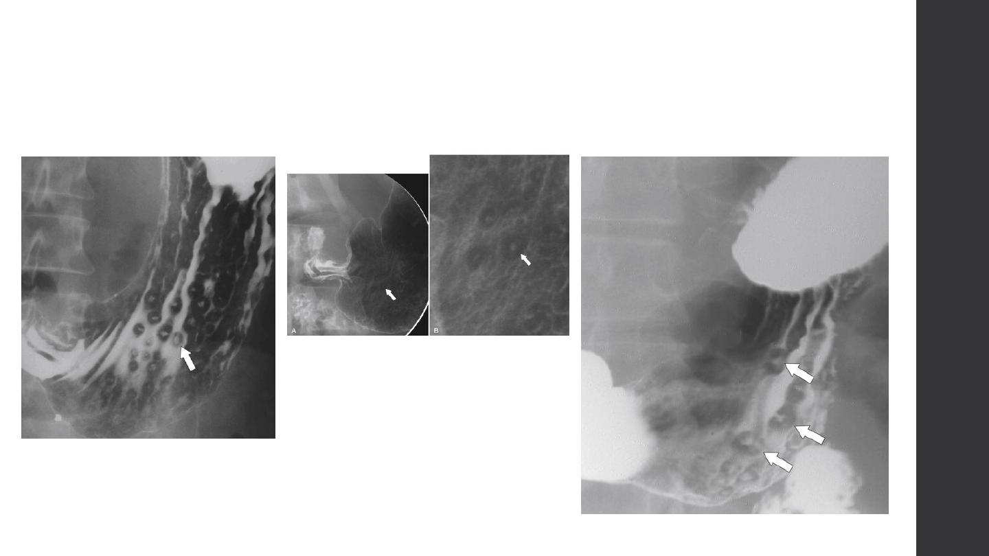
Duodenum
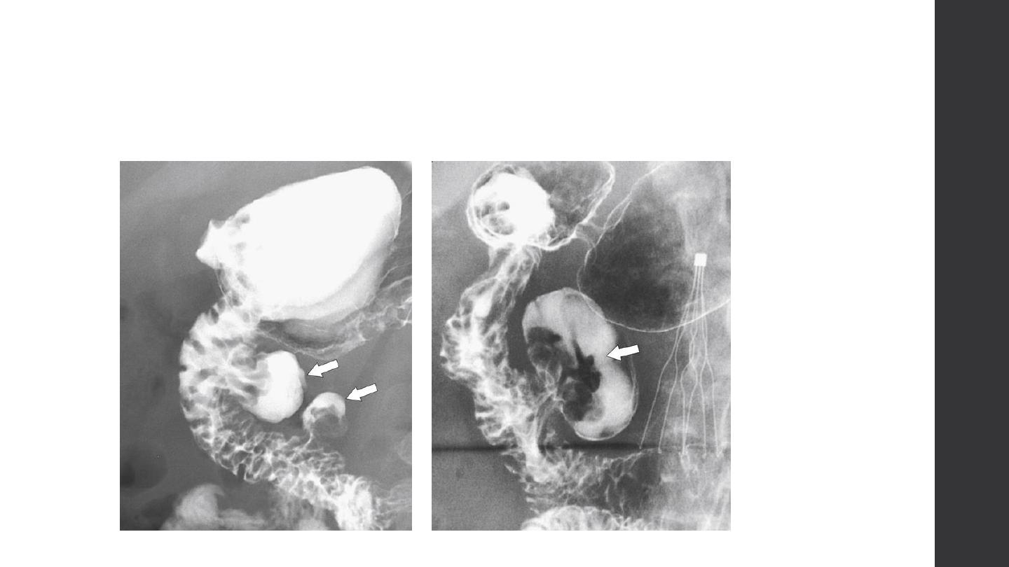
Duodenum
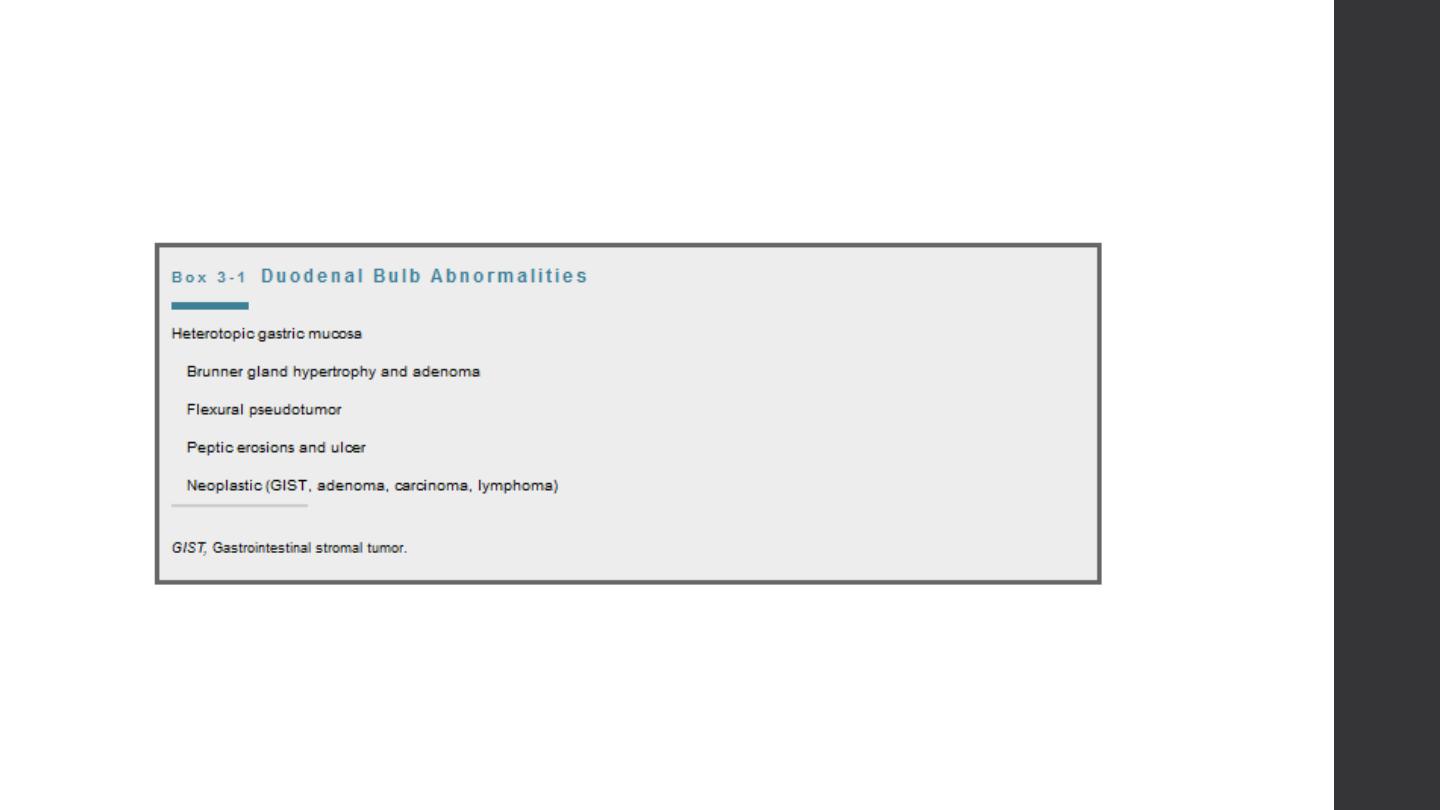
Duodenum
Duodenum
Duodenum
Duodenum
Duodenum
1
2
3
4