Contraction of Skeletal Muscle
About 40 per cent of the body is skeletal muscle, and perhaps another 10 per cent is smooth and cardiac muscle.Physiologic anatomy of skeletal muscle fiber
All skeletal muscles are composed of numerous fibers ranging from 10 to 80 micrometers in diameter, each fiber is usually innervated by only one nerve ending, located near the middle of the fiber.
Sarcolemma.
The sarcolemma is the cell membrane of the muscle fiber.The sarcolemma consists of a true cell membrane, called the plasma membrane, the sarcolemma fuses with a tendon fiber, and the tendon fibers in turn collect into bundles to form the muscle tendons that then insert into the bones.
Myofibrils
Actin and myosin filaments.
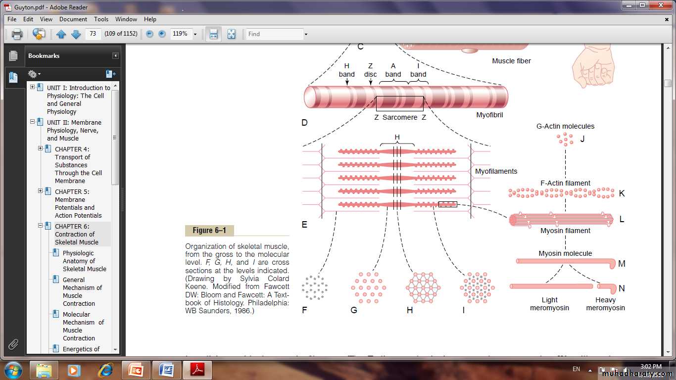
Each muscle fiber contains several hundred to several thousand myofibrils, it composed of about 1500 adjacent myosin filaments and 3000 actin filaments, which are large polymerized protein molecules that are responsible for the actual muscle contraction.
The ends of the actin filaments are attached to a so-called Z disc. From this disc, these filaments extend in both directions to inter digitate with the myosin filaments. it attaching the myofibrils to one another all the way across the muscle fiber. The entire muscle fiber has light and dark bands.
These bands give skeletal and cardiac muscle their striated appearance. The portion of the myofibril that lies between two successive Z discs is called a sarcomere.
Titin filamentous molecules.
It Keeps the Myosin and Actin Filaments in Place . it is very springy. It act as a frame work that holds the myosin and actin filaments in place.Sarcoplasm. The spaces between the myofibrils are filled with intracellular fluid called sarcoplasm, containing large quantities of potassium, magnesium, and phosphate, plus multiple protein enzymes. Also present are tremendous numbers of mitochondria that lie parallel to the myofibrils. These supply the contracting myofibrils with large amounts of energy.
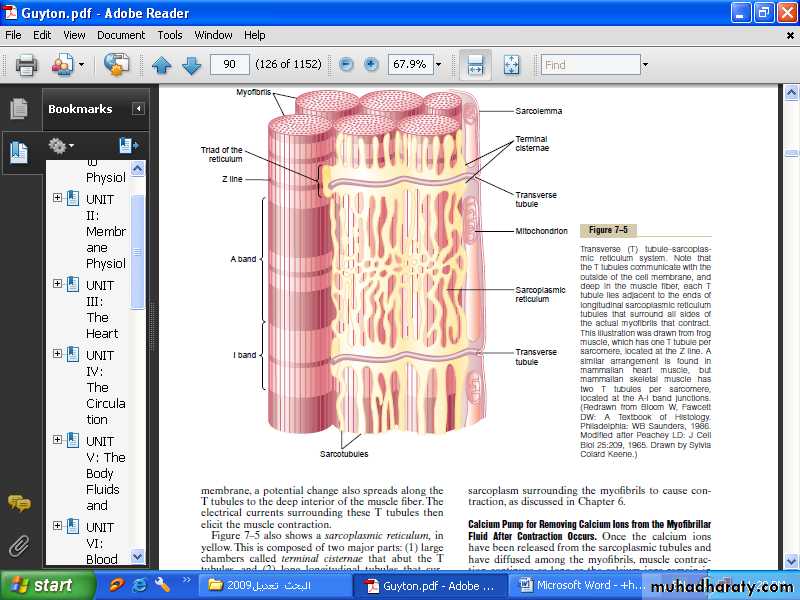
Sarcoplasmic Reticulum.
Also in the sarcoplasm surrounding the myofibrils of each muscle fiber is an extensive reticulum called the sarcoplasmic reticulum. It is extremely important in controlling muscle contraction. The very rapidly contracting types of muscle fibers have especially extensive sarcoplasmic reticula.
Sarcoplasmic Reticulum
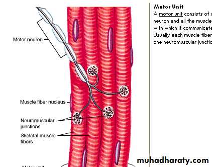
Motor unit:Each motor neuron innervates multiple muscle fiber. All muscle fiber innervated by a single nerve fiber are called motor unit. Small muscle that reacts rapidly and whose control must be exact have more nerve fiber for fewer muscle fiber as few as 2-3 muscle fiber per motor unit in some of laryngeal muscle. Conversely large muscle that does not require fine contraction such as soleus muscle have several hundred muscle fiber in a motor unit. In general 80-100 muscle fiber is provided by to a motor unit.
Motor unit
General mechanism of muscle contractionThe initiation and execution of muscle contraction occur in the following sequential steps.
1. An action potential travels along a motor nerve to its endings on muscle fibers.
2. At each ending, the nerve secretes a small amount of the neurotransmitter substance acetylcholine.
3. The acetylcholine acts on a local area of the muscle fiber membrane to open multiple “acetylcholinegated” channels through protein molecules floating in the membrane. 4. Opening of the acetylcholine-gated channels allows large quantities of sodium ions to diffuse to the interior of the muscle fiber membrane. This initiates an action potential at the membrane.
5. The action potential travels along the muscle fiber membrane in the same way that action potentials travel along nerve fiber membranes.
6. The action potential depolarizes the muscle membrane, and much of the action potential electricity flows through the center of the muscle fiber. Here it causes the sarcoplasmic reticulum to release large quantities of calcium ions that have been stored within this reticulum.
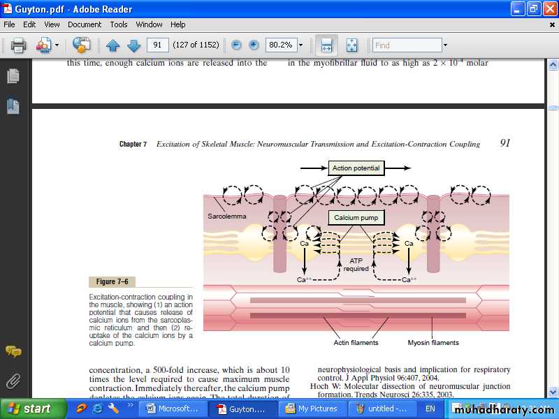
Excitation of skeletal muscle: neuromuscular transmission and excitation-contraction coupling.
7. The calcium ions initiate attractive forces between the actin and myosin filaments, causing them to slide alongside each other, which is the contractile process.
8. After a fraction of a second, the calcium ions are pumped back into the sarcoplasmic reticulum by a Ca++ membrane pump, and they remain stored in the reticulum until a new muscle action potential comes along; this removal of calcium ions from the myofibrils causes the muscle contraction to cease.
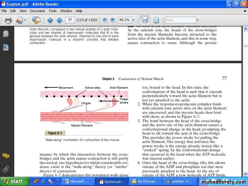
“Walk-along”mechanism for contraction of the muscle.
Effect of amount of actin and myosin filament overlap on tensiondeveloped by the contracting muscle
At point D on the diagram, the actin filament has pulled all the way out to the end of the myosin filament, with no actin-myosin overlap. At this
point, the tension developed by the activated muscle is zero. Then, as the sarcomere shortens and the actin filament begins to overlap the myosin filament, the tension increases progressively until the sarcomere length decreases to about 2.2 micrometers. At this point, the actin filament has already overlapped all the cross-bridges of the myosin filament but has not yet reached the center of the myosin filament. With further shortening, the sarcomere maintains full tension until point B is reached, at a sarcomere length of about 2 micrometers. At this point, the ends of the two actin filaments begin to overlap each other in addition to overlapping the myosin filaments. As the sarcomere length falls from 2 micrometers down to about 1.65 micrometers, at point A, the strength of contraction decreases rapidly. At this point, the two Z discs of the sarcomere abut the ends of the myosin filaments. Then, as contraction proceeds to still shorter sarcomere lengths, the ends of the myosin filaments are crumpled and, as shown in the figure, the strength of contraction approaches zero, but the entire muscle has now contracted to its shortest length.
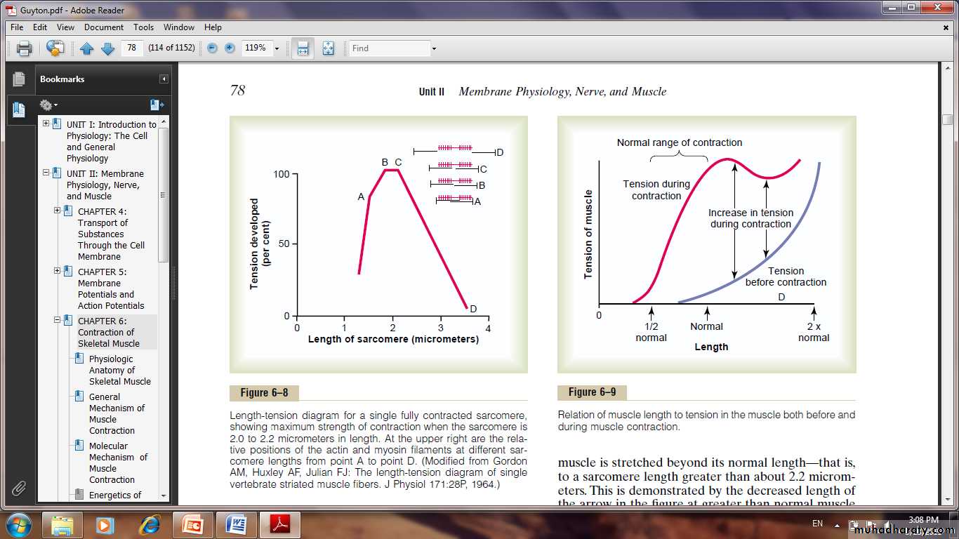
Length-tension diagram for afully contracted sarcomer
Fast Versus Slow Muscle Fibers.Every muscle of the body is composed of a mixture of so-called fast and slow muscle fibers. The muscles that react rapidly are composed mainly of “fast” fibers with only small numbers of the slow variety. Conversely, the muscles that respond slowly but with prolonged contraction are composed mainly of “slow” fibers.
Fast Fibers.
(1) Large fibers for great strength of contraction.
(2) Extensive sarcoplasmic reticulum for rapid release of calcium ions to initiate contraction.
(3) Large amounts of glycolytic enzymes for rapid release of energy by the glycolytic process.
(4) Less extensive blood supply because oxidative metabolism is of secondary importance.
(5) Fewer mitochondria, also because oxidative metabolism is secondary.
(6) It called white muscle due to the deficit of red myoglobin.
Slow Fibers.
(1) Smaller fibers.
(2) Also innervated by smaller nerve fibers.
(3) More extensive blood vessel system and capillaries to supply extra amounts of oxygen.
(4) Greatly increased numbers of mitochondria, also to support high levels of oxidative metabolism.
(5) Fibers contain large amounts of myoglobin, an iron containing protein similar to hemoglobin in red blood cells. Myoglobin combines with oxygen and stores it until needed; this also greatly speeds oxygen transport to the mitochondria. The myoglobin gives the slow muscle a reddish appearance and the name red muscle,
Multiple Fiber Summation.
When the central nervous system sends a weak signal to contract a muscle, the smaller motor units of the muscle may be stimulated in preference to the larger motor units. Then, as the strength of the signal increases, larger and larger motor units begin to be excited as well, with the largest motor units often having as much as 50 times the contractile force of the smallest units. This is called the size principle.
Frequency Summation and Tetanization.
The twitch contractions occurring one after another at low frequency of stimulation.Then, as the frequency increases,there comes a point where each new contraction occurs before the preceding one is over. As a result, the second contraction is added partially to the first, so that the total strength of contraction rises progressively with increasing frequency. When the frequency reaches a critical level, the successive contractions eventually become so rapid that they fuse together, and the whole muscle contraction appears to be completely smooth and continuous. This is called tetanization. At a slightly higher frequency, the strength of contraction reaches its maximum, so that any additional increase in frequency beyond that point has no further effect in increasing contractile force.This occurs because enough calcium ions are maintained in the muscle sarcoplasm, even between action potentials, so that full contractile state is sustained without allowing any relaxation between the action potentials.
Skeletal Muscle Tone. Even when muscles are at rest, a certain amount of tautness usually remains, called muscle tone. This results from :
a low rate of nerve impulses coming from the spinal cord. signals transmitted from the brain to the appropriate spinal cord anterior motor neurons. partly by signals that originate in muscle spindles located in the muscle itself.