Contraction and excitation of
smooth muscleSmooth muscle composed of far smaller fibers— usually 1 to 5 micrometers in diameter and only 20 to 500 micrometers in length.
Types of Smooth Muscle
smooth muscle can generally be divided into two major types :
multi-unit smooth muscle .
unitary (or single-unit) smooth muscle.
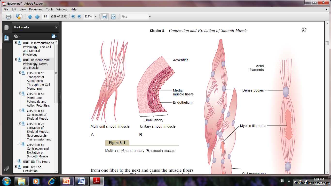
Multi-Unit Smooth Muscle.
This type of smooth muscle is composed of discrete, separate smooth muscle fibers. Each fiber operates independently of the others and often is innervated by a single nerve ending. Further, the outer surfaces of these fibers are covered by a thin layer of basement membrane–like substance, ( insulate the separate fibers from one another).
In contrast, a major share of control of unitary smooth muscle is exerted by non-nervous stimuli.
Some examples of multi-unit smooth muscle are the ciliary muscle of the eye, the iris muscle of the eye, and the piloerector muscles that cause erection of the hairs when stimulated by the sympathetic nervous system.
Unitary Smooth Muscle.
It means a mass of hundreds to thousands of smooth muscle fibers that contract together as a single unit. The fibers usually are arranged in sheets or bundles, and their cell membranes are adherent to one another at multiple points so that force generated in one muscle fiber can be transmitted to the next. The cell membranes are joined by many gap junctions through which ions can flow freely from one muscle cell to the next so that action potentials or simple ion flow without action potentials can travel from one fiber to the next and cause the muscle fibers to contract together.This type of smooth muscle is also known as syncytial smooth muscle because of its syncytial interconnections among fibers. It is also called visceral smooth muscle because it is found in the walls of most viscera of the body, including the gut, bile ducts, ureters, uterus, and many blood vessels.
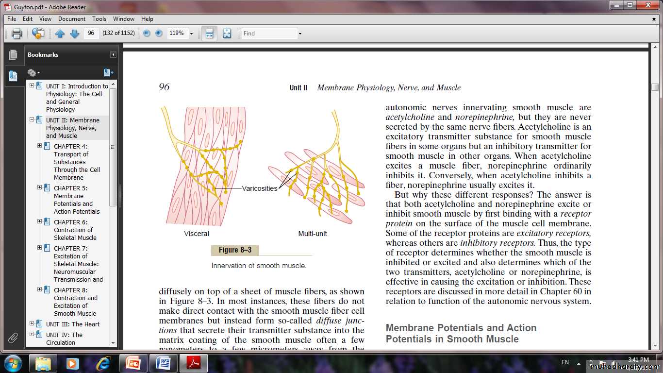
Innrevation of smooth muscle
Contractile Mechanism in Smooth Muscle
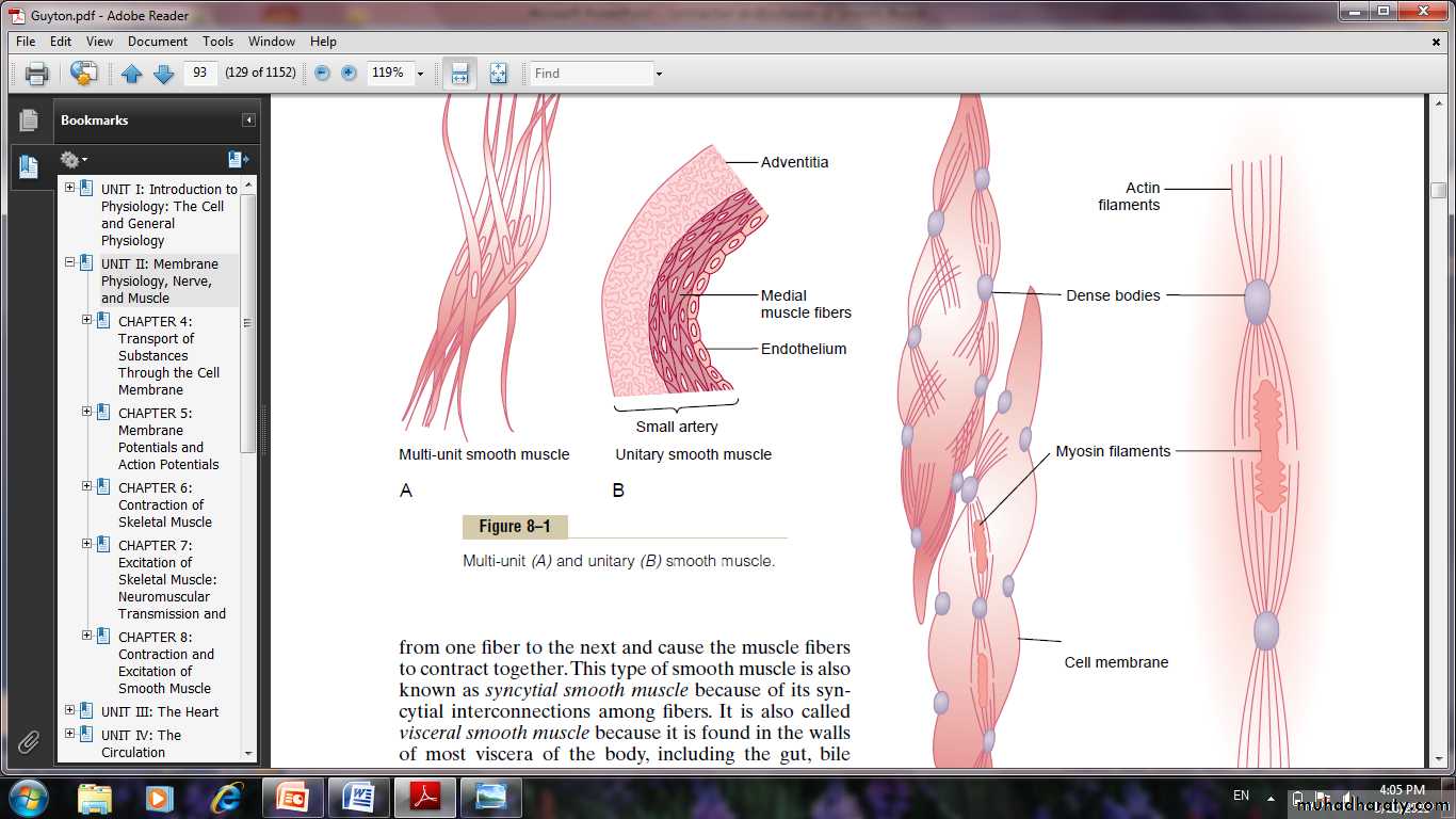
Smooth muscle contains both actin and myosin filaments, having chemical characteristics. They interact with each. The contractile process is activated by calcium ions.actin filaments attached to so-called dense bodies. Some of these bodies are attached to the cell membrane.Others are dispersed inside the cell. Some of the membrane dense bodies of adjacent cells are bonded together by intercellular protein bridges. It is mainly through these bonds that the force of contraction is transmitted from one cell to the next.
Interspersed among the actin filaments in the muscle fiber are myosin filaments. These have a diameter more than twice that of the actin filaments. In the smooth muscle there are large numbers of actin filaments radiating from two dense bodies; the ends of these filaments overlap a myosin filament located midway between the dense bodies. Most of the myosin filaments have what are called “side polar” cross-bridges.
Comparison of Smooth Muscle Contraction and Skeletal Muscle Contraction Slow Cycling of the Myosin Cross-Bridges.
The rapidity of cycling of the myosin cross-bridges in smooth muscle is much, much slower in smooth muscle than in skeletal muscle.
Energy Required to Sustain Smooth Muscle Contraction.
Only 1/10 to 1/300 as much energy is required to sustain the same tension of contraction in smooth muscle as in skeletal muscle.
Slowness of Onset of Contraction and Relaxation of the Total Smooth Muscle Tissue.
A typical smooth muscle tissue begins to contract 50 to 100 milliseconds after it is excited, reaches full contraction about 0.5 second later, and then declines in contractile force in another 1 to 2 seconds, giving a total contraction time of 1 to 3 seconds. This is about 30 times as long as a single contraction of an average skeletal muscle fiber.
Force of Muscle Contraction. The maximum force of contraction of smooth muscle is often greater than that of skeletal muscle. This great force of smooth muscle contraction results from the prolonged period of attachment of the myosin cross bridges to the actin filaments.
Nervous and Hormonal Control of Smooth Muscle Contraction:
smooth muscle can be stimulated to contract by multiple types of signals: Nervous signals, Hormonal stimulation. Stretch of the muscle and Several other ways. The principal reason for the difference is that the smooth muscle membrane contains many types of receptor proteins that can initiate the contractile process. Still other receptor proteins inhibit smooth muscle contraction, which is another difference from skeletal muscle.
Neuromuscular Junctions of Smooth Muscle
The autonomic nerve fibers that innervate smooth muscle generally branch diffusely on top of a sheet of muscle fibers. In most instances, these fibers do not make direct contact with the smooth muscle fiber cell membranes but instead form so-called diffuse junctions that secrete their transmitter substance into the matrix coating of the smooth muscle often a few nanometers to a few micrometers away from the muscle cells; the transmitter substance then diffuses to the cells.
Excitatory and Inhibitory Transmitter Substances Secreted at the Smooth Muscle Neuromuscular Junction. The most important transmitter substances secreted by the autonomic nerves innervating smooth muscle are acetylcholine and norepinephrine. Acetylcholine is an excitatory transmitter substance for smooth muscle fibers in some organs but an inhibitory transmitter for smooth muscle in other organs. When acetylcholine excites a muscle fiber, norepinephrine ordinarily inhibits it. Conversely, when acetylcholine inhibits a fiber, norepinephrine usually excites it. Both acetylcholine and norepinephrine excite or inhibit smooth muscle by first binding with a receptor protein on the surface of the muscle cell membrane. Some of the receptor proteins are excitatory receptors, whereas others are inhibitory receptors.
Membrane Potentials in Smooth Muscle.
In the normal resting state, the intracellular potential is usually about -50 to -60 millivolts.
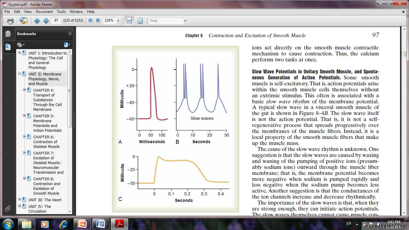
Action Potentials in Unitary Smooth Muscle.
Action potentials occur in unitary smooth muscle (such as visceral muscle) in the same way that they occur in skeletal muscle. They do not normally occur in many, if not most, multi-unit types of smooth muscle. The action potentials of visceral smooth muscle occur in one of two forms: (1) spike potentials. (2) action potentials with plateaus.
Spike Potentials. Typical spike action potentials, occur in most types of unitary smooth muscle. The duration of this type of action potential is 10 to 50 milliseconds.
Action Potentials with Plateaus.
The onset of this action potential is similar to that of the typical spike potential,but the repolarization is delayed for several hundred to as much
as 1000 milliseconds (1 second). The importance of the plateau is that it can account for the prolonged contraction that occurs in some types of smooth muscle, such as the ureter, the uterus,certain types of vascular smooth muscle and in cardiac muscle fibers .
Importance of Calcium Channels in Generating the Action Potential.
The smooth muscle cell membrane has far more voltage-gated calciumchannels than does skeletal . Flow of calcium ions to the interior of the fiber is mainly responsible for the action potential. The calcium channels open many times more slowly than do sodium channels, and they also remain open much longer. This accounts in large measure for the prolonged plateau action potentials of some smooth muscle fibers.
The calcium ions act directly on the smooth muscle contractile mechanism to cause contraction. Thus, the calcium performs two tasks.
Source of Calcium Ions That Cause Contraction:
(1) Through the Cell Membrane
(2) From the Sarcoplasmic Reticulum
Effect Of Extracellular Calcium Ion Concentration changing.
Although changing the extracellular fluid calcium ion concentration from normal has little effect on the force of contraction of skeletal muscle, this is not true for most smooth muscle. When the extracellular fluid calcium ion concentration falls to about 1/3 to 1/10 normal, smooth muscle contraction usually ceases. Therefore, the force of contraction of smooth muscle usually is highly dependent on extracellular fluid calcium ion concentration.