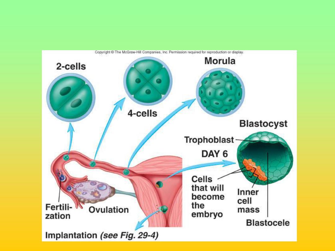
Blastocyst
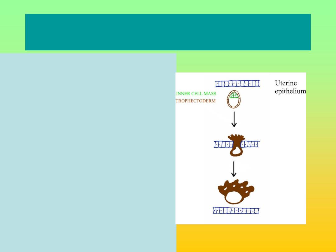
Attachment and implantation into uterine
wall
• Blastocyst passes between
uterine epithelial cells
• Inner cell mass give rise to
embryo
• Trophoblast cells (from
trophectoderm around outside
of blastocysts) proliferate
• Migrate into uterine wall to
establish placenta
• Uterine epithelium closes
behind
• Early placental (trophoblast)
cells secrete HCG
– to
maintain corpus luteum

How does placenta form?
• Proliferated trophoblast on embryonic
pole differentiates into outer layer of
syncytio-trophoblast and inner cyto-
trophoblast
• Lacunae develop on syncytiotrophoblast
layer into which maternal blood passes
• Villi of syncytiotrophoblast with inner core
of cytotrophoblast develop floating in
sinuses(lacunae)
Placenta
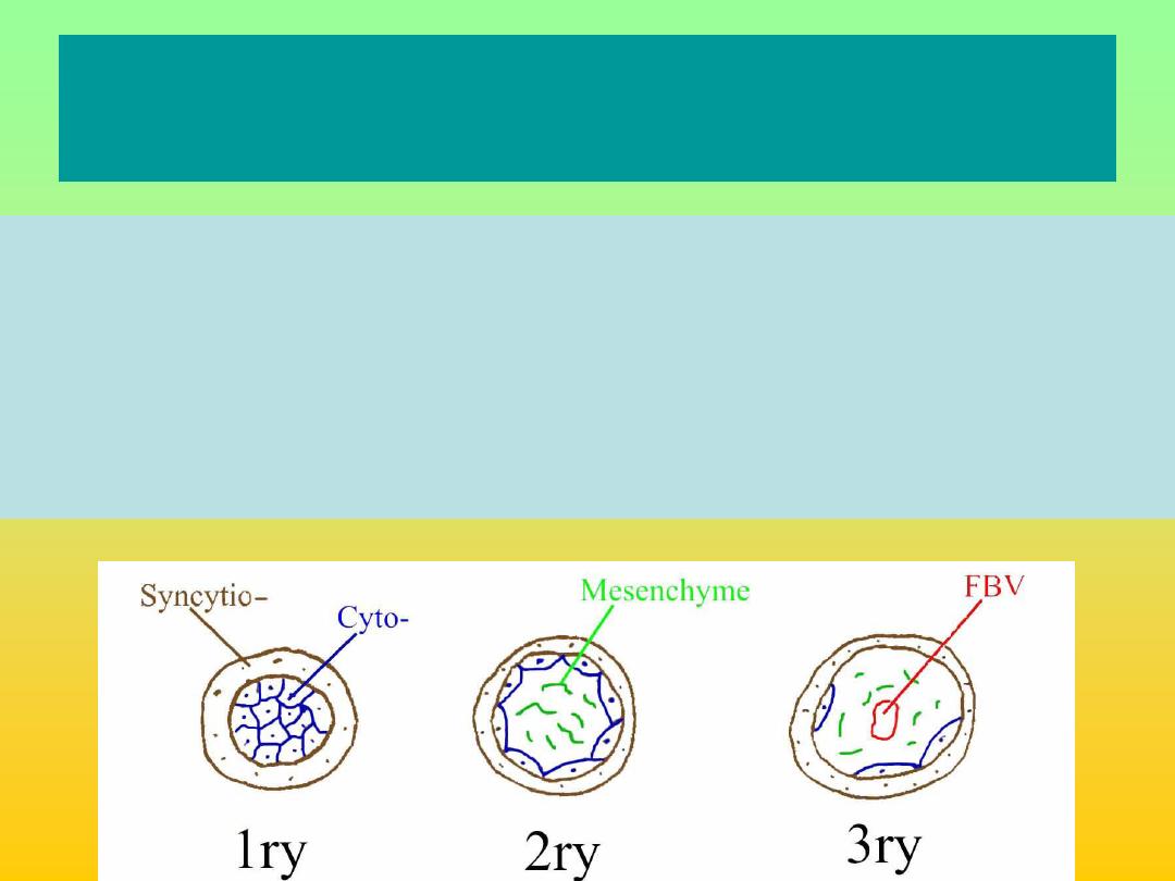
Floating villi
• Primary to secondary to tertiary villi by end of 3
rd
week
of gestation at the beginning consist of 2 layers of
trophoblast; cytotrophoblast and syncytiotrophoblast,
then an inner core of mesoderm inside which fetal
blood starts running. The thickness will decrease after
20 th weeks because of disappearance of mesoderm
and cytotrophoblast.
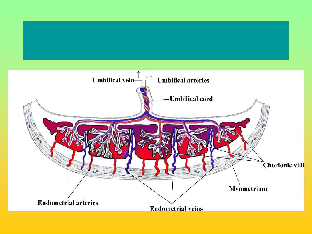
Mature Placenta and Fetus

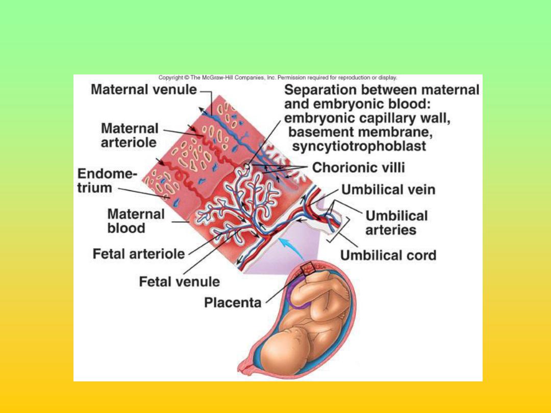
What is a placenta?
• Generally regarded as the site at which
there is exchange of gasses and nutrients
between maternal and foetal blood
• Site has separate maternal and foetal
blood circulations which do not mix :
• It is a discoid shaped organ of 15 -20
lobules. It‘s fetal surface covered with
amnion while maternal surface consist of
lobules of compressed villi separated from
each
other by sulci.

Placental function
• 1-Placenta provides anchorage, establishes fetal
vascular network in association with maternal
blood supply but without mixing.
• 2-Gaseous exchange(Respiratory function) umb
.vein carry O2 from placenta to the fetus while
umb. Artery carry deoxygenated blood from the
fetus to the mother :
• 3-Nutritional function: transferring substance
important for fetal growth e.g glucose ,a.a , lipids,
Ab also synthesize and store some substances
like glycogen.

Placental function
• 4-Placenta act as endocrine gland :secraeting
hormones & enzymes
• 5-Barrier function: the placental filters out some
substances which could harm the foetus e.g blood
borne pathogens, Drugs but not all (alcohol,
soscial drugs &some viruses e.g CMV)
• 6-Immunological protection?:it protects fetus from
rejection .

Placenta as a filter/transfer
organ
Receives nutrients, oxygen, antibodies and
hormones from the mother and passes out
waste .There are many factors influencing
the placental transfer as:
1- Nature of the substance whether fat- solube
or not &their molecular size.
2- Thickness of the barrier which depend on
gestational age.
3- Surface area available for transfer.
4- Rate of circulation across the barrier.

The Placenta
• Permeability of the placenta increases
constantly
– reaches a peak in the last
month
– sharp decrease in permeability.
Pe
rmea
bi
lity
0
8
20
40

Mechanism of transfer
The transfer of substance across the
placenta occurs by several mechanisms
1- Simple diffusion : transport from high
to lower pressure e.g CO2, O2 H2O and
electrolyte.
2- Facilitated diffusion : by carrier system
as glucose, vitamins & minerals .
3- Active transport : in which high energy
is required to carry substance against
pressure gradient e.g. Heavy materials ,
Iron& amino acids..

Placenta as an endocrine gland
• HCG (Human chorionic gonadotropin) - maintains
ovariean corpus luteum
• Progesterone – maintains pregnancy (especially after
1
st
trimester)
• Sommatomammotropin (Placental lactogen –
increases maternal blood glucose and lipids
• Oestrogen
• Relaxin
• Prostaglandins
• Enzymes & proteins e.g alkaline phosphates,
oxytokinase, lactate dehydrogenase and Insulinase.

HCG
• It is produced by cytotrophoblast detected in
maternal blood from 10 th day of fertilization
&peaks at 60-90ds then decline to amoderate
constant level
• 1-For the 1
st
6-8w of pregnancy it maintains the
corpus luteum to ensure continued
progesterone out put until production shifts to
the placenta
• 2-regulate steroid biosynthesis in the placenta
& fetal adrenal gland &stimulates the fetal
testicle to secrete testosterone .

• Human placental lactogen (HPL)
It is protein produced by syncytotrophoblas.
It’s level rise slowly up to 40 weeks of
pregnancy.
It decreases maternal insulin sensitivity &
promotes release of FFA from maternal stores
–
alternative source of energy for her metabolism
.
HPL level is low in case of; threatened
abortion & IUGR

16
It is steroid produced by feto-placental units from
fetal liver ,
adrenals and placental
cytotrophoblast.
The level increase in early pregnancy up to 38
weeks then after it decrease. It originates from
corpus luteum in early pregnancy then from the
placenta. Estriol is the most abundant form in
pregnancy &can be used as indicator for
placental function sudden decline of esteriol in
maternal circulation indicate fetal compromise.
. Oestrogen

17
•
Progesterone:
It is steroid produced by CL in 1
st
6-7w of
pregnancy thereafter by placental
syncytiotrophoblast ( no fetal role ).
The level of progesterone increase in the
pregnancy from early stage until the onset of
labour.
It is important for support of pregnancy and
increased vascularity of the placental bed.It
prevents uterine contraction &play acentral
role in maintaining uterine quiescence
throught pregnancy.

Amniotic fluid
It surrounds the fetus, produced by:
1- In early pregnancy secreated by amnion,but
by 10
th
w it is mainly atransudate of fetal
serum via the skin & umbilical cord .
2- From 16w the net increase in AF is through
asmall imbalance between urine & lung fluids
secreation& removal by fetal swallowing.
It’s volume increases progressively(10w:30ml,
20w:300ml,38w:1000ml) but from term there
is rapid fall in volume(40w:800ml,42w:350ml)
It
t

Function of amniotic fluid
.
1-It protect fetus mechanical injury
2-Allows room for fetal growth, movement
&development while preventing limb contracture
3-Is of value for assessing fetal well being e.g in
renal agenesis, cystic kidneys or fetal growth
restriction oligohydramnios results but when
there is reduced removal of fluid in conditions like
anencephaly & esophageal/ duodenal artesia
polyhydramnios results.
4-Permits fetal lung development as there is two-
way movement of fluide into the fetal bronchioles
& its absence in 2
nd
trimester is associated with
pulmonary hypoplasia.
It
t