هستو نظري / ثاني اسنان كركوك
د. ايوب23\ 10\ 2017
19
3rd Lecture (22nd -26th October 2017)
Connective tissue (Co.T):
The connective tissue (Co.T) is one of the four types of biological tissue that support, connect, or separate different types of tissues and organs in the body. In contrary to epithelial cells the cells of Co.T are separated by non-living materials to provide cohesion and internal support. The Co.T is found in between other tissues everywhere in the body, including the nervous system (NS). In the central nervous system (CNS),
the outer membranes, the HYPERLINK "https://en.wikipedia.org/wiki/Meninges" \o "Meninges"meninges which cover the brain and spinal cord are composed of connective tissue. All Co.T, apart from blood and lymph, consists of three main components: fibers (elastic, reticular and HYPERLINK "https://en.wikipedia.org/wiki/Collagen" \o "Collagen"collagenous fibers), ground substance (matrix) and cells while blood and lymph lack the fiber component:
Fibroblasts: Secrete both fibers and ground substance of the matrix (wandering).
Macrophages: Phagocytes that develop from Monocytes (wandering or fixed).
Plasma Cells: Antibody secreting cells that develop from B-Lymphocytes (wandering)
Mast Cells: Produce histamine that help dilate small blood vessels in reaction to injury (wandering)
Adipocytes: Fat cells that store triglycerides, support, protect and insulate (fixed).
All above components are immersed in the body water. The Co.T include several types of fibrous tissue that vary only in their density and cellularity, as well as the more specialized and recognizable variants-bone, ligaments, tendons, cartilage, and adipose (fat) tissue.
In general, all types of the Co.T originate from the mesenchyme cells which are loosely associated cells that (1). lack polarity and (2). are surrounded by a large extracellular matrix (ECM). Mesenchymal cells are able to develop into the tissues of the lymphatic and circulatory systems, and within the body i.e. bone and cartilage. Mesenchymal cells can migrate easily. Mesenchyme stem cells, or MSCs, are multipotent HYPERLINK "https://en.wikipedia.org/wiki/Stromal_cell" \o "Stromal cell"stromal cells that can differentiate into a variety of cell types, including: osteoblasts (bone cells), HYPERLINK "https://en.wikipedia.org/wiki/Chondrocyte" \o "Chondrocyte"chondrocytes (cartilage cells), HYPERLINK "https://en.wikipedia.org/wiki/Myocyte" \o "Myocyte"myocytes (muscle cells) and HYPERLINK "https://en.wikipedia.org/wiki/Adipocyte" \o "Adipocyte"adipocytes (fat cells). This phenomenon has been documented in specific cells and tissues in living animals and their counterparts growing in tissue culture.
General Functions of the Co.T:
It performs several functions including: (1). Binds and supports body parts; (2). Protects; (3). Fills spaces; (4). Stores fat (for energy); and (5). Transports materials.
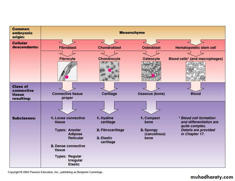
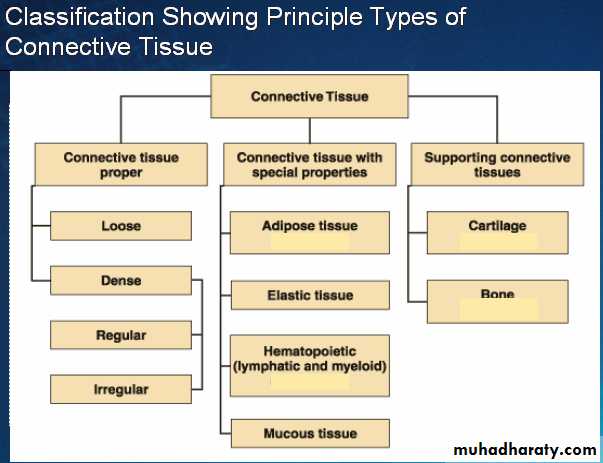
(Fig. 1 & 2): Demonstration of origin and classification of Connective tissues in human and animal embryos.
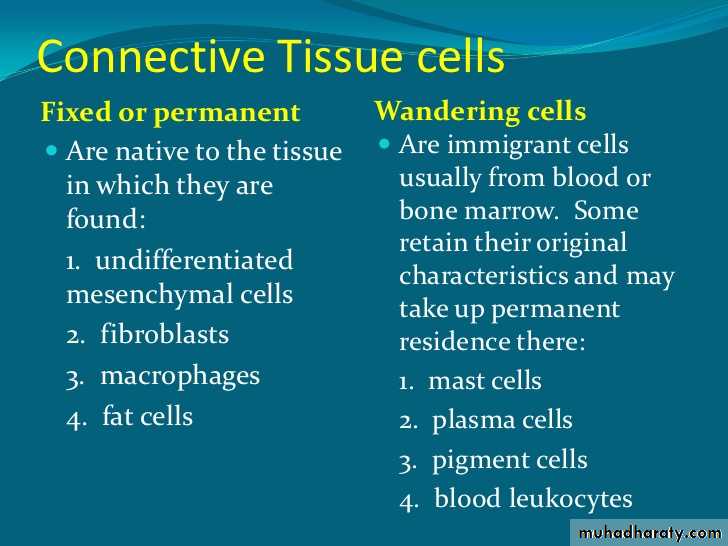
(Fig. 3): Nature and characters of CT cells e.g. fixed and wandering cells.
(I): Connective Tissue Proper:
Structure of Loose and Dense CT:
Both loose Co.T and dense Co.T contain three kinds of fibers. (1). Collagen fibers provide strength and flexibility. Collagen is the most abundant protein in animal and human bodies; (2). Elastic fibers provide elasticity i.e. when stretched, they return to their original shape and (3). Reticular fibers are small and branched. They provide a support framework for organs such as the liver and lymph nodes. The cells of loose and dense connective tissue are called fibroblasts. Fibroblasts produce both the fibers and nonliving matrix material. Macrophages are cells specialized for phagocytizing foreign materials, bacteria, and cleaning up debris. Macrophages will be discussed in the chapter on the immune system.
(a): Loose Connective Tissue: Loose connective tissue includes areolar, adipose, and reticular connective tissue
1). Areolar Connective Tissue:
The fibroblasts (cells) of areolar connective tissue are separated by a nonliving, jellylike matrix. The tissue contains collagen fibers for flexibility and strength, and numerous elastic fibers that enable it to be stretched. Areolar connective tissue is found in the skin and in most internal organs of vertebrates, where it allows the organs to expand; it also forms a protective covering for muscles, blood vessels, and nerves.
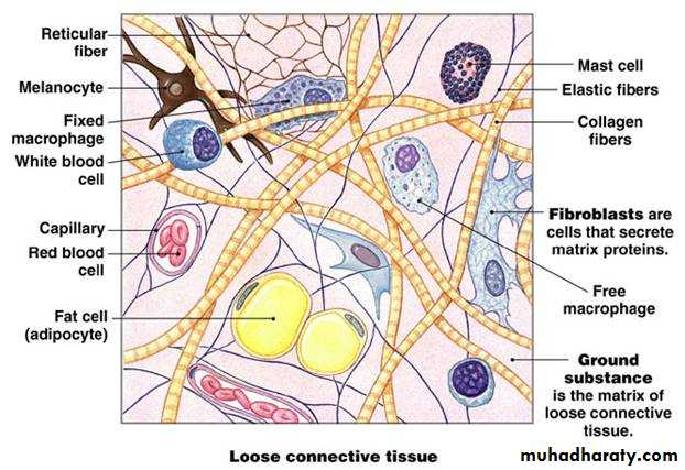
(Fig. 4): Schematic diagram of Areolar CT where main components are shown.
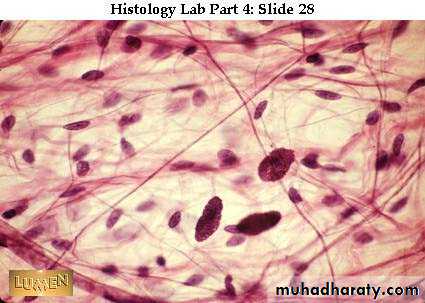
(Fig. 5): A typical Areolar CT taken from human reveals the above components in figure 4.
2). Adipose tissue: Is a type of loose connective tissue which has reduced matrix material and contains enlarged fibroblasts (cells) that store fat. Adipose tissue functions to store energy, insulate, and provide padding, especially in the skin and around the kidneys and heart. In routine histological slides the nucleus is shifted at periphery due to a large amount of fatty vacuoles. Between adipose cells, intracellular matrix is found which may contain blood capillaries. Adipose tissue could easily be identified in histological slides which are characterized by their large vacuoles and the peripheral nuclei.
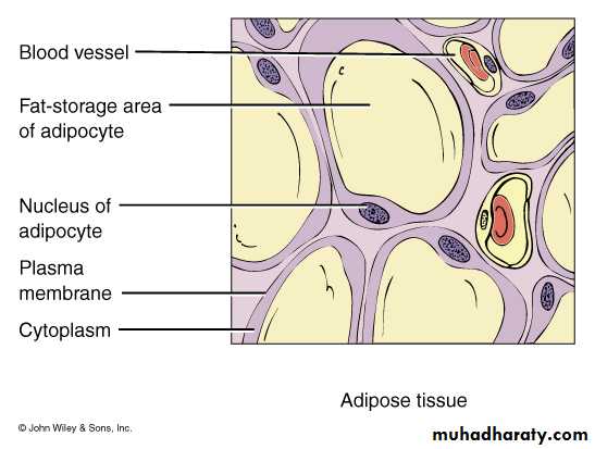
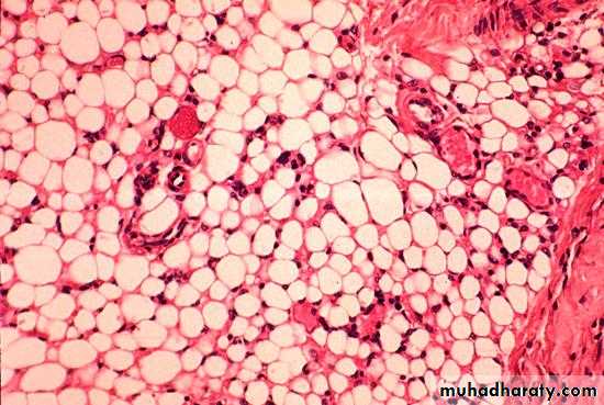
(Fig. 6&7): Schematic diagram (a) and normal adipose tissue (b)where nuclei are shifted to the periphery due to large sized central vacuoles.
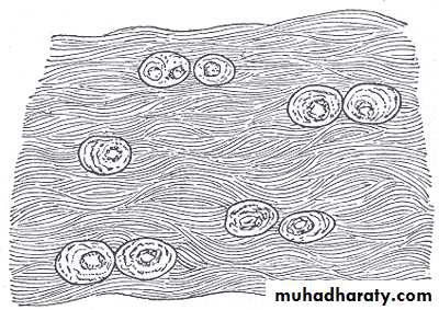
3). Reticular CT: Reticular connective tissue contains an abundance of reticular fibers. It provides a supporting framework for organs such as the lymph nodes, spleen, and liver. Found in liver, spleen and lymph nodes. Its function is to forms the framework (stroma) of organs and binds together smooth muscle tissue cells.
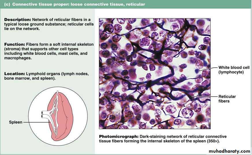
(Figs. 9&10): Reticular Co.T reveals the mesh of reticular fibers between the cells to provide a network and skeletal to the organs i.e. spleen, liver and lymphatic nodules.
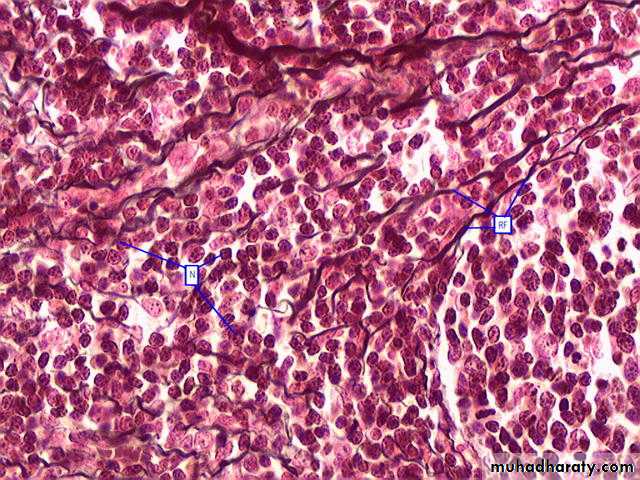
(b): Dense (Fibrous) Co.T.:
The collagen fibers of dense Co.T. are more closely packed than those of loose connective tissue.
(1): Regular dense Co.T contains collagen fibers oriented in one direction to provide strength in that direction. It is found in tendons and ligaments. Tendons connect muscle to bone; ligaments connect bone to bone.
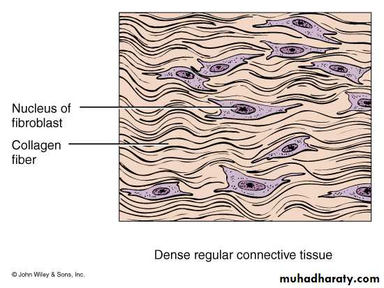
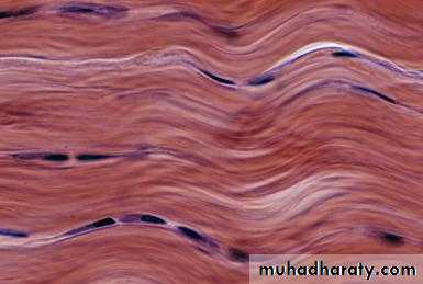
(Fig. 11&12): Dense CT show the packed collagen fibers where fibroblast nuclei could easily be seen in between. Very few ECM is visible.
(2): Irregular dense Co.T: Contains collagen fibers oriented in many different directions (randomly). It is found in the deep layers of the skin (dermis) in fasciae, joint capsules, and heart valves and the tough capsules that surround many of the organs such as the kidneys, adrenal glands, nerves, bones, and the covering of muscles. It provides support and strength.
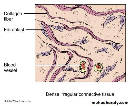
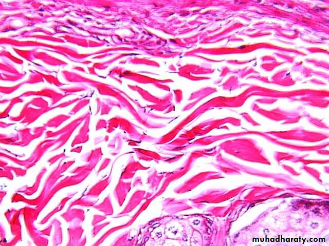
(Fig. 13&14): Irregular Dense CT shows the randomly run collagen fibers. Nuclei of fibroblast could still be easily seen in between them.
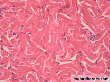
(Fig. 15): Another Irregular Dense Connective tissue. Blood capillaries are easily seen in between them as well as the nuclei of fibroblasts.
(II): Connective tissue Special:
A). Cartilage:
Cartilage is a non-vascular tissue. As such, the chondrocytes rely on blood vessels in the tissue surrounding the cartilage for nutrient supply and waste removal.
1). Hyaline Cartilage:
Cartilage is a somewhat elastic, compact type of Co.T characterized by three components: lacunae, chondrocytes, and a rigid matrix. The matrix is a firm gel material that contains fibers and other substances. There are three basic types of cartilage in the human body: (1): Hyaline cartilage; (2): Elastic cartilage and (3): Fibrocartilage. As the fetus ages, the cartilage is gradually replaced by more supportive bone. In the mammalian adult, hyaline cartilage is mainly restricted to the nose, trachea, bronchi, ends of the ribs, and the articulating surfaces of most joints. The function of the hyaline cartilage is to provide slightly flexible support and reduce friction within joints. It also provides structural reinforcement. The cells of cartilage are embedded in a protein-containing matrix that is strong but flexible.
(Fig. 16): Schematic diagram of hyaline cartilage shows the main components
It does not stretch and can resist compression. It is also flexible but maintains its shape. It is found in the ends of bones where it prevents friction within the joints. In the nose, external ear, and the walls of the trachea it functions to support the softer tissues.
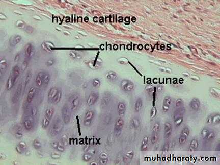
(Fig. 17): A typical hyaline cartilage covered with upper perichondrium Co.T
It is found in inter-vertebral disks which function as shock pads. The fetal skeleton of vertebrate animals is composed of cartilage before bone forms. The skeleton of cartilaginous fish is composed of cartilage. Cartilage is a non-vascular tissue (avascular). As such, the chondrocytes rely on blood vessels in the tissue surrounding the cartilage for nutrient supply and waste removal.2). Fibro-cartilage:
White fibro-cartilage consists of a mixture of white fibrous tissue and cartilaginous tissue in various proportions. It owes its flexibility and toughness to the former of these constituents, and its elasticity to the latter. It is the only type of cartilage that contains type-I collagen in addition to the normal type-II. Fibro-cartilage is found in the pubic symphysis and in the HYPERLINK "https://en.wikipedia.org/wiki/Anulus_fibrosus_disci_intervertebralis" \o "Anulus fibrosus disci intervertebralis"anulus fibrosus of inter-vertebral discs. It is also present at the tendon bone interface, where there is a transition from soft tendon to uncalcified then calcified fibrocartilage before becoming bone. During labor, relaxin loosens the pubic symphysis to aid in delivery, but this can lead to later joint problems.
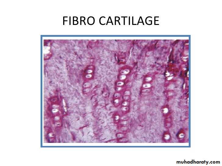
(Fig. 18&19): Fibro-cartilage, schematic (above) and normal. Note the parallel packed chondrocytes in between bundles of parallel collagen fibers.
3). Elastic cartilage:
It contains threadlike network of elastic fibers within the matrix and found in external ear, auditory tubes, epiglottis. Its (1): provides support, (2): maintains shape, and (3): allows flexibility.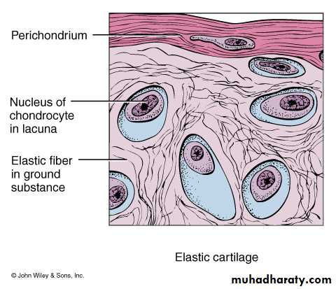
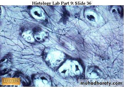
(Fig. 20&21): Schematic diagram and normal elastic cartilage where elastic fibers are found within the matrix and between the lacunae.
B). Bone
Bone forms when calcium salts are deposited around protein fibers. The calcium salts provide rigidity while the fibers provide elasticity and strength. Bone, or osseous tissue, is a connective tissue that has a large amount of two different types of matrix material. The organic matrix is materially similar to other connective tissues, including some amount of collagen and elastic fibers. This gives strength and flexibility to the tissue. The inorganic matrix consists of mineral salts, mostly calcium that gives the tissue hardness. Without adequate organic material in the matrix, the tissue breaks; without adequate inorganic material in the matrix, the tissue bends.
There are three types of cells in bone: osteoblasts, osteocytes, and osteoclasts. Osteoblasts are active in making bone for growth and remodeling. They deposit bone material into the matrix and, after the matrix surrounds them, they continue to live, but in a reduced metabolic state as osteocytes. Osteocytes are found in lacunae of the bone and assist in maintenance of the bone. Osteoclasts are active in breaking down bone for bone remodeling, providing access to calcium stored in tissues in order to release it into the blood. Osteoclasts are usually found on the surface of the tissue.
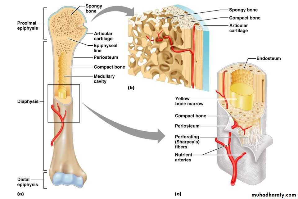
Bone can be divided into two types: compact and spongy. Compact bone is found in the shaft (or diaphysis) of a long bone and the surface of the flat bones, while spongy bone is found in the end (or epiphysis) of a long bone. Compact bone is organized into subunits called osteons. A blood vessel and a nerve are found in the center of the osteon within a long opening called the Haversian canal, with radiating circles of compact bone around it known as lamellae. Small spaces between these circles are called lacunae. Between the lacunae are microchannels called canaliculi; they connect the lacunae to aid diffusion between the cells. Spongy bone is made of tiny plates called trabeculae, which serve as struts, giving the spongy bone strength. HYPERLINK "https://www.boundless.com/biology/textbooks/boundless-biology-textbook/the-animal-body-basic-form-and-function-33/animal-primary-tissues-193/connective-tissues-bone-adipose-and-blood-739-11969/images/bone-structure/"
Bone structure:
(a). Compact bone is a dense matrix on the outer surface of bone. Spongy bone, inside the compact bone, is porous with web-like trabeculae. (b) Compact bone is organized into rings called osteons. Blood vessels, nerves, and lymphatic vessels are found in the central Haversian canal. Rings of lamellae surround the Haversian canal. Between the lamellae are cavities called lacunae. Canaliculi are microchannels connecting the lacunae together. (c) Osteoblasts surround the exterior of the bone. Osteoclasts bore tunnels into the bone and osteocytes are found in the lacunae.
The osteon or Haversian system is the fundamental functional unit of much compact bone. Osteons are roughly cylindrical structures that are typically several millimeters long and around 0.2mm in diameter. They are present in many bones of most mammals and some bird, reptile, and amphibian species.
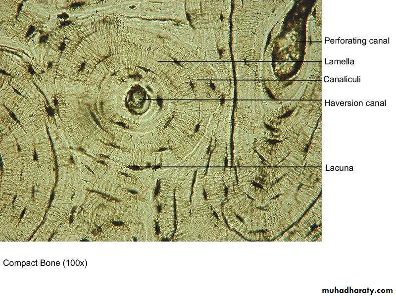
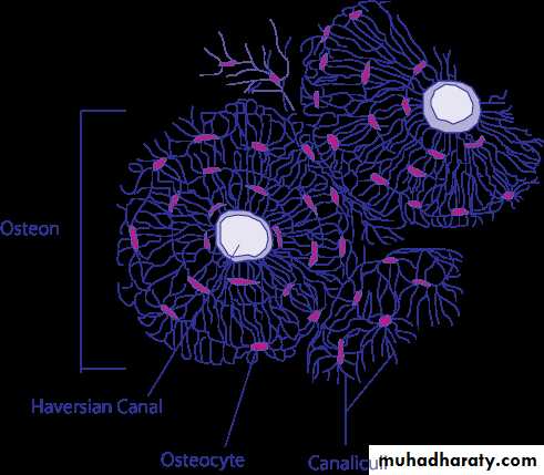
(Fig. 22,23&24): Anatomy of a compact bone, normal bonny tissue and Schematic diagram of compact bone from a transverse section of a long bone's cortex. Note the structure of the bonny tissue and the location of each part of it.
Function: (1): They Support and protect organs, (2):Provide levers and attachment site for muscles, (3): Stores calcium and other minerals, (4): Stores fat and (5): the bone Marrow is site for blood cell formation.