بسم الله الرحمن الرحيم
Anatomy of female reproductive system
Dr. Shaima Hazim Al- Tayar
The internal genital organs
Begins with :- vagina , cervix , the uterus , ovaries & ligamentsINTERNAL GENITALIA
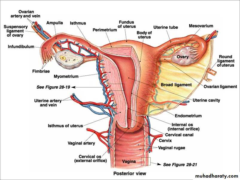
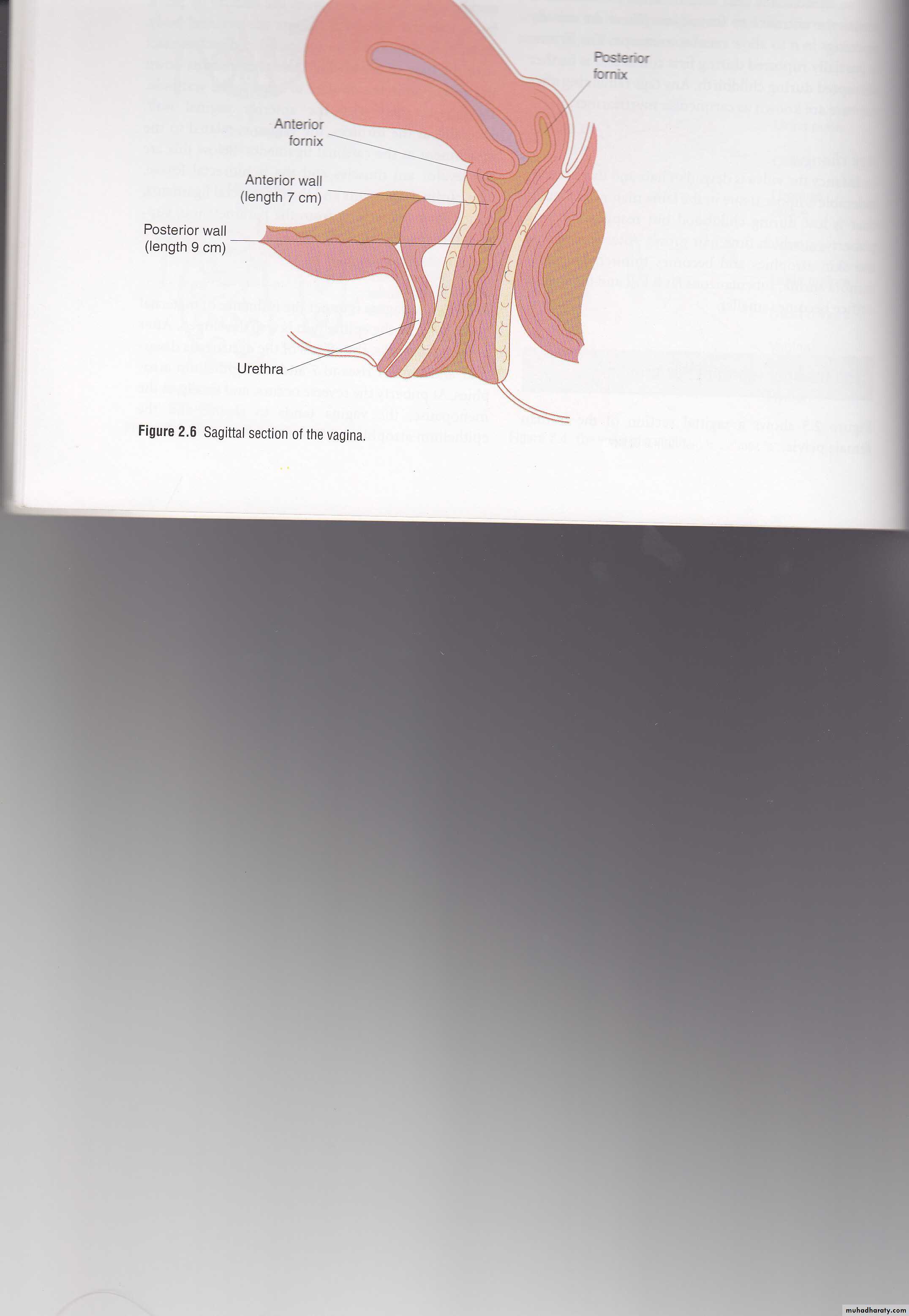
VaginaThe vagina is a musculomembranous tube which links the uterus to the vestibule. It is 8-12 cm in length Anteriorly, the vagina is closely related to the base of the bladder and the urethra, and posteriorly, to the pouch of Douglas, rectum and anal canal .
Vagina
The lining of the vagina is thrown into folds known as rugae .The uterine cervix projects into the upper 1-2 cm vagina, outlining the 4 fornices-anterior, posterior and lateral (left and right).
Blood supply of the vagina
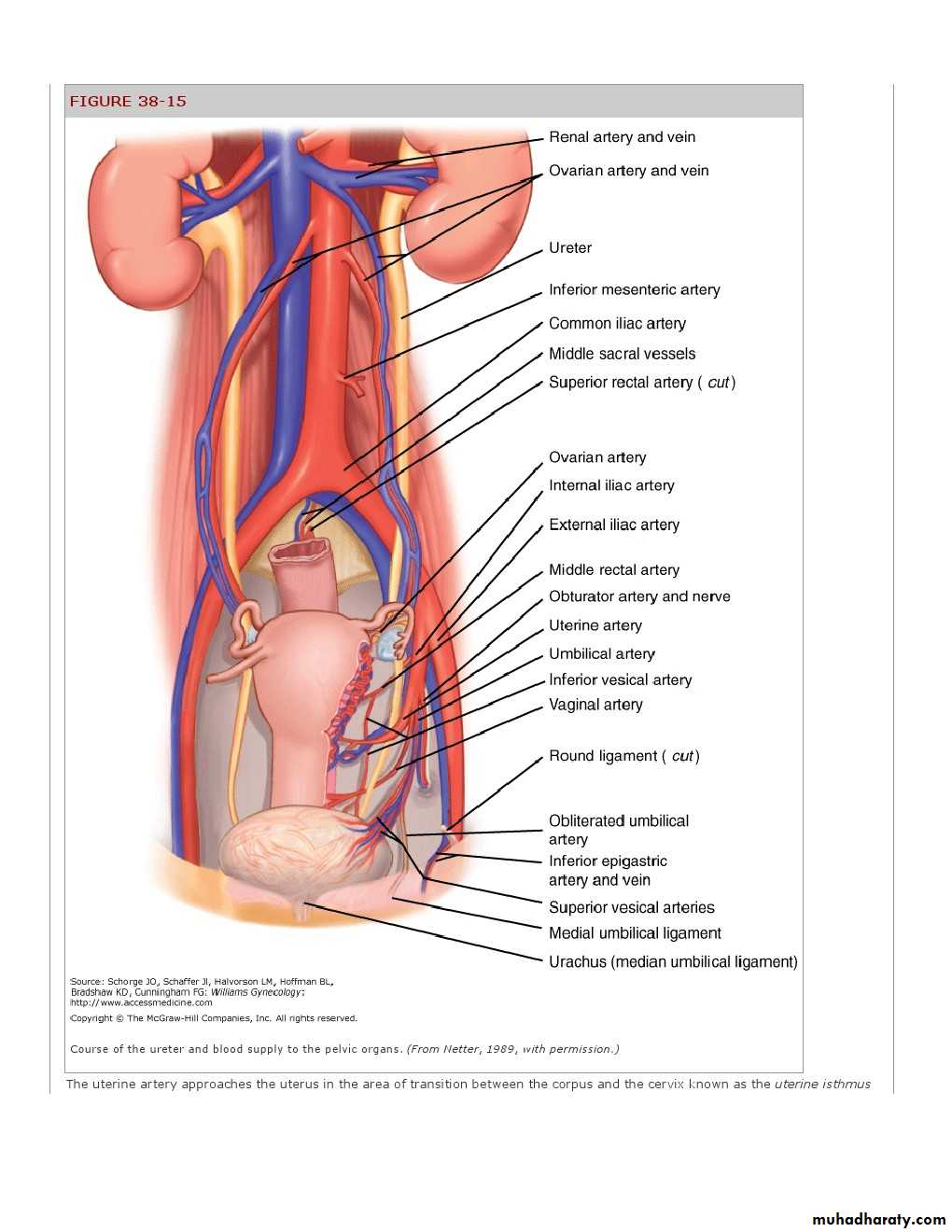
Is from the vaginal arteries which arise from the internal iliac arteries and the descending branches of the uterine arteriers aided by the arteries to the bladder , middle rectal &internal pudendal arteries..
Cervix

The cervix is 2-4cm length and delinated by the external os inferiorly and by internal os superiorly attached to pelvic walls by ligaments
-pubocervical anteriorly ,
uterosacral posteriorly,and
tansverse cervical(Mackenrodt) laterally

2-
Uterine bodyThe uterus is a hollow muscular organ,the interior being roughly tringular in shape.The upper angles of the tringle are formed by the interstitial portions of the Fallopian tubes.The uterus is covered externally by peritoneum except the lower part anteriorly,where the peritoneum is reflected onto the bladder.
The lower segment lies at the junction of uterus &cervix . during pregnancy and labour, expand to10 cm in length. The uterus is globular in shape ; both anteverted (rotated forward) & anteflexed(bent forward on itself). In 20%of women, the uterus is rotated backwards.
.
The structure of the uterus
1-The endometrium or lining is composed of columnar epithelium which dip into submucosa to form branched tubular glands .
The thickness of the lining depends on the stage of menstrual cycle
2-The myometrium- It is the middie muscular layer composed of several interlacing layers of smooth muscle.In the cervix muscles form 10% only .
3-The serosa - is formed by the peritoneal covering and its associated blood vessels, lymphatics and nerves.

Blood supply of the uterus
Is from the uterine arteries on each side which arise form the internal iliac, pass down to the junction of the cervix &uterus where cervical &vaginal branches given off then the vessels continue up ward at the sides of uterus in a tortuous manner ; linking up with ovarian arteries at upper part of broad ligament.Many branches arise from uterine artery pass deep in anterior &posterior surfaces of the uterus
Fallopian tubes
are delicate tubular structures which carry the ovum or sperm between the ovary and uterine cavity.The tubes are divided into named regions,most medially the interstial portion within the uterine wall, then the isthmus followed by the infundibulum ,ampulla and finally fimbrial ends which wrap themselves around the ovary at the time of ovulation. .
They are lined by columnar epithelium and cilia which together with the peristaltic action of the surrounding smooth muscle propel the fertilized ovum towards the uterine cavity.
The bloodsupply of the fallopian tubes arises from both the uterine and ovarian arteries through the mesosalpinx which is covered by peritoneum.
Ovaries-Gonads
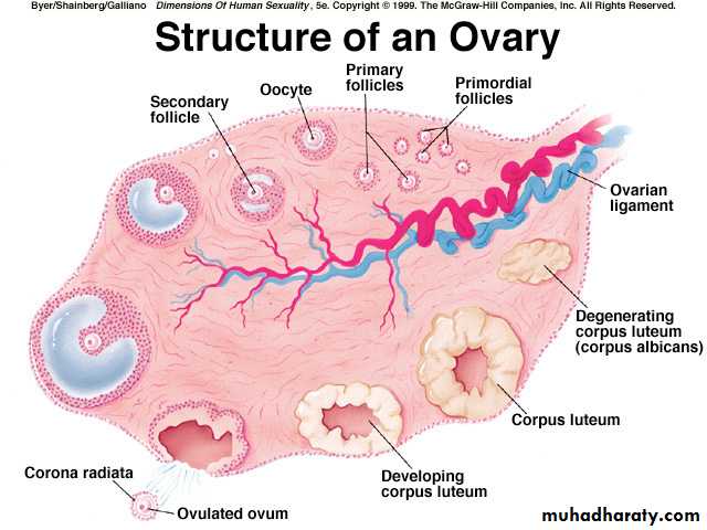
Are paired structures on the back of broad ligament attached by mesentry(mesovarium).each ovary 2-4 cm length;have yellowish-white irregular surface ,often charcterized by developing graffian follicle or active or regressing corpora lutea.It possess an outer cortex in which are follicle &specialized connective tissue (theca)&the inner medulla composed of loose connective tissue &blood vessels there are 400,000 primary oocyte.Its blood supply from ovarian arterie and branches of uterine arteries.
Round and ovarian ligaments
These are 2 continuous ligaments.The round ligaments provide some anterior support for uterus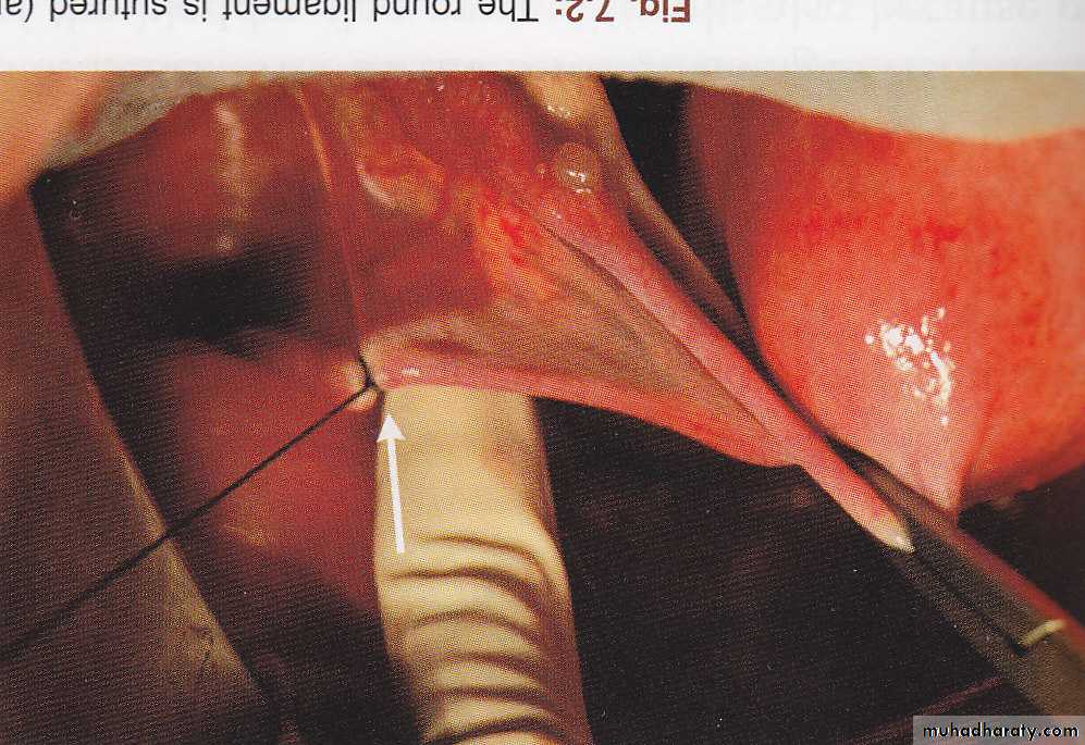
The lymphatic drainage
-uterus , cervix, upper 2/3 vagina : internal iliac, obturator & external iliac L.N.,commen iliac ¶ –aortic L.N.-few vessels from the fundus follow the ovarian drainage, & some through the round ligament to the inguinal L.N.
-ovaries, fallopian tubes : para -aortic L.N.
Nerve supply of the pelvic viscera
Nerve fibres of the pre aortic plexus of the sympathetic nervous system are continuous with those of the superior hypogastric plexus which divides, on each side its fibres continuous with the uterovaginal plexusParasympathetic fibres from S2,S3,S4 join
the uterovaginal plexus. Fibres from (or to)
the bladder, uterus, vagina & rectum join the
plexus.
The Genital organs
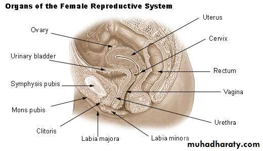
External GenitaliaThese are visible on inspection&include :
Vulva
The vulva is the area of the perineum including the Mons pubis, labia majora and minora and the opening into both the vagina and urethra
Vestibule
This is the area enclosed by the labia minora.The urethra , vagina ,Bartholin and Skene ducts open in the vestibule. The bartholin duct open at the posterolateral aspect, just outside the hymen . The ducts are 1.5-2.0 cm long and run up to the paired Bartholin glands
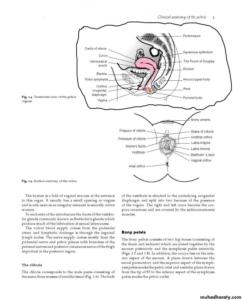
Mons veneris or mons pubis
This is a fibrofatty cushion lying anterior and superior to the junction of the 2 pubic bones (symphysis pubis) . It is covered by hair .The hair-covered labia majora are areas of skin with underlying fat pads which bound the vagina. Medial to these, are the labia minora which devoid of hair
. Anteriorly the labia minora come together to form the prepuce of the clitoris and posteriorly they form the forchette.
The glitoris is arranged in a central corpus with 2 crura
The vulval blood supply comes from the pudendal
arteryThe nerve supply mostly from the:-
1- pudendal nerve (S2,S3,S4) which divide into
- perineal n. supply vulva
-dorsal nerve of the clitoris
2-pelvic plexus
3-and posterior cutaneous nerve of the thigh important in the posterior region.
The lymphaic drianage : vulva, lower 1/3 vagina :superficial inguinal & femoral L.N.
The nerve supply
Nerve supply of the vulva& perineum- pudendal nerve(S2,S3,S4) Divide into
perineal n. supply vulva
the dorsal nerve of the clitoris
- sensory fibers from the mons & labia also
pass in the ilioinguinal & genitofemoral n.
to the 1st Lumbar n.
- posterior femoral cutaneous n. carries
sensation from perineum to the small sciatic n.
(S1,S2,S3).
Bony pelvis
The bony pelvis consists of two hip bones (consisting of the ileum and ischium) which are joined together by the sacrum posteriorly and the symphysis pubis anteriorly
In addition, the coccyx lies on the inferior
aspect of the sacrum.
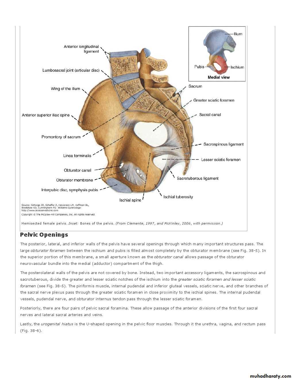
Sacrum and Coccyx
The sacrum and coccyx are an extension of the vertebral column resulting from the five fused sacral vertebrae and the four fused coccygeal vertebrae..Sacral promontory— the most prominent and anterior projection of the sacrum.
The bony pelvis
The dimensions of the true pelvis appreciated by studying three planes
1-The brim(inlet)
2-The mid pelvis
• 3-The outlet
A plane drawn between the sacral promontory and the superior aspect of the symphysis pubis marks the pelvic inlet and a similar plane drawnfrom the tip of S5 to the inferior aspect of the symphysis pubis marks the pelvic outlet
The pelvic brim-inlet
Is bounded infront by the symphysis pubis on each side by the superior aspects of the pubic bones,the iliopectineal lines,the ala and promontory of acrum.There are 4 pelvic types.
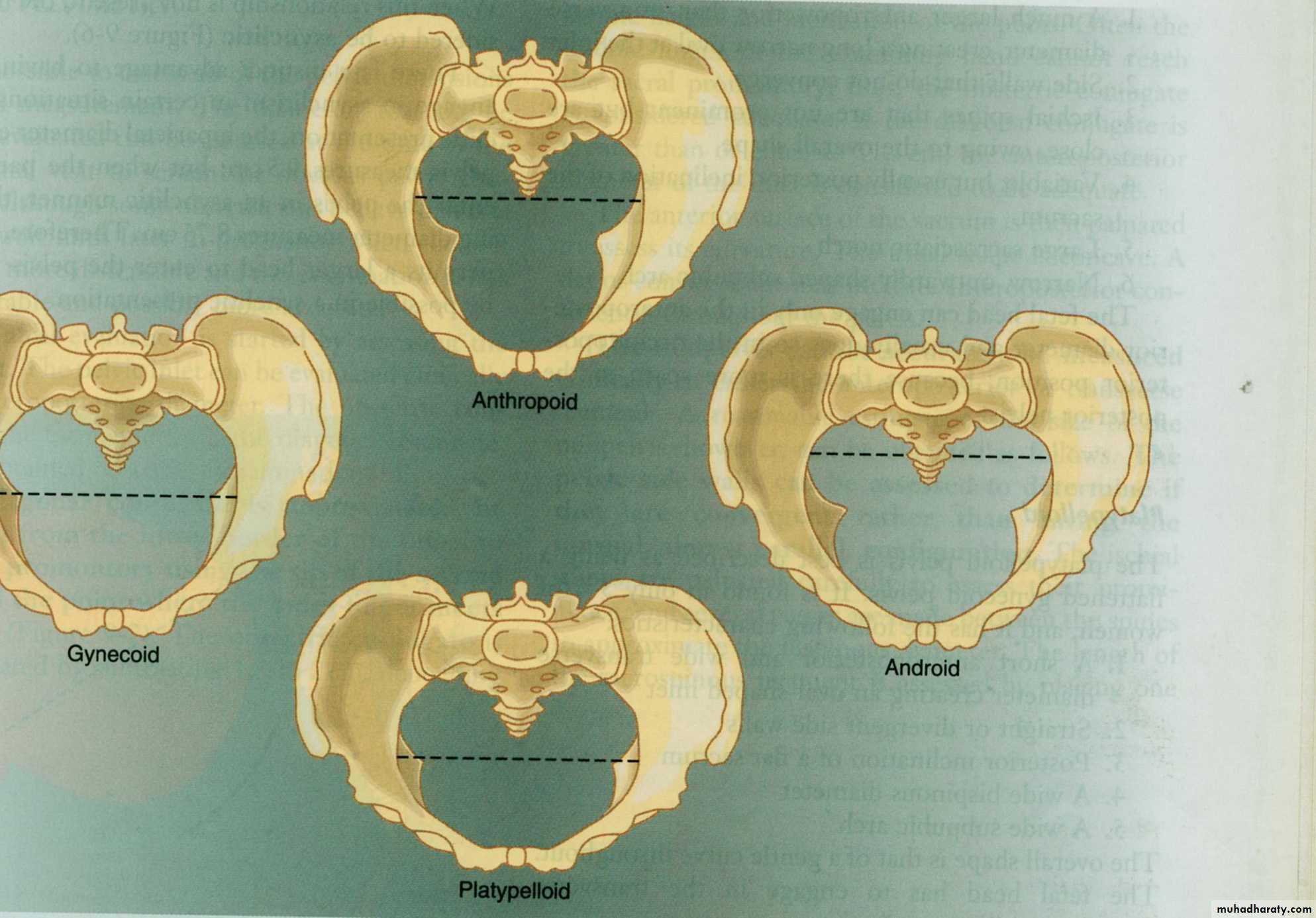
Pelvic brim
The nteroposterior diameter (obstetrical conjugate) 11cmThe trasverse diameter13.5 is the largest one.
The outlet
This outlined by the lower margin of the symphysis pubis , on each side by the descending ramus of the pubic bone ,ischial tuberosities,the sacrotuberous ligaments and the coccyx.The subpubic arch is 90 .
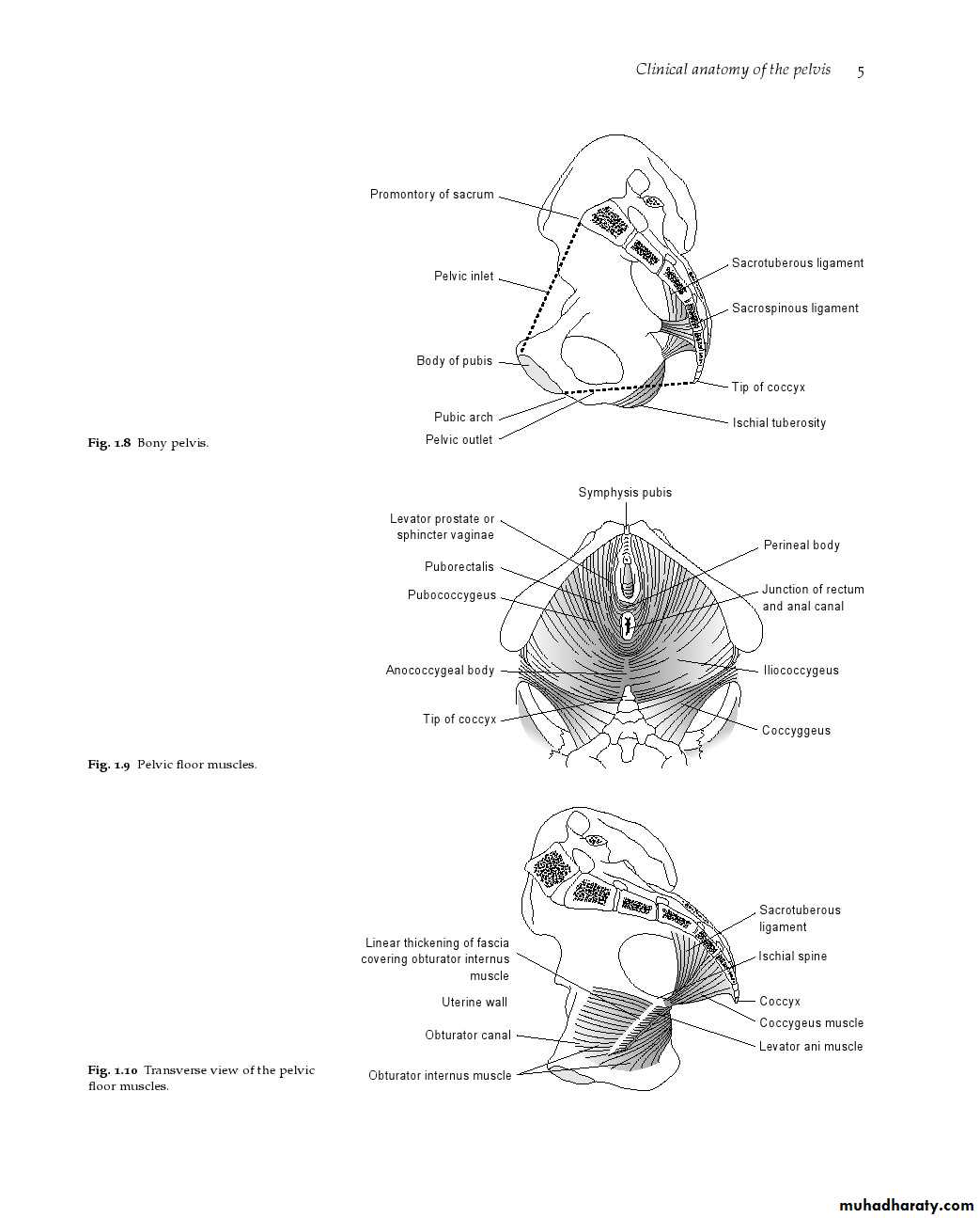
Pelvic floor
The bony pelvis is clothed by a number of muscles, the chief of which from the floor of the pelvis and perineum.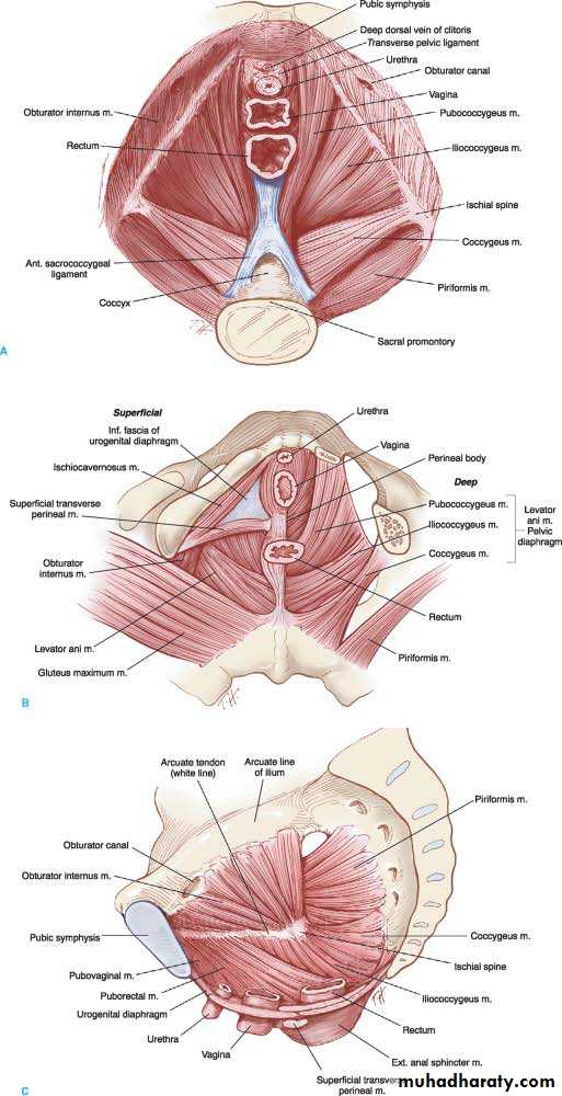
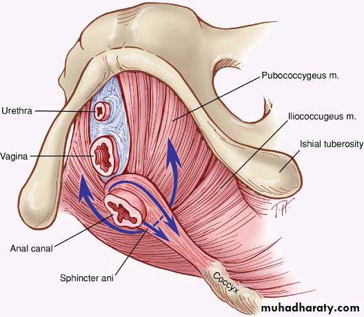
Pelvic floor / Levator Ani
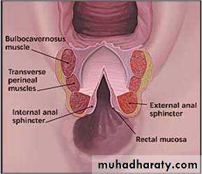
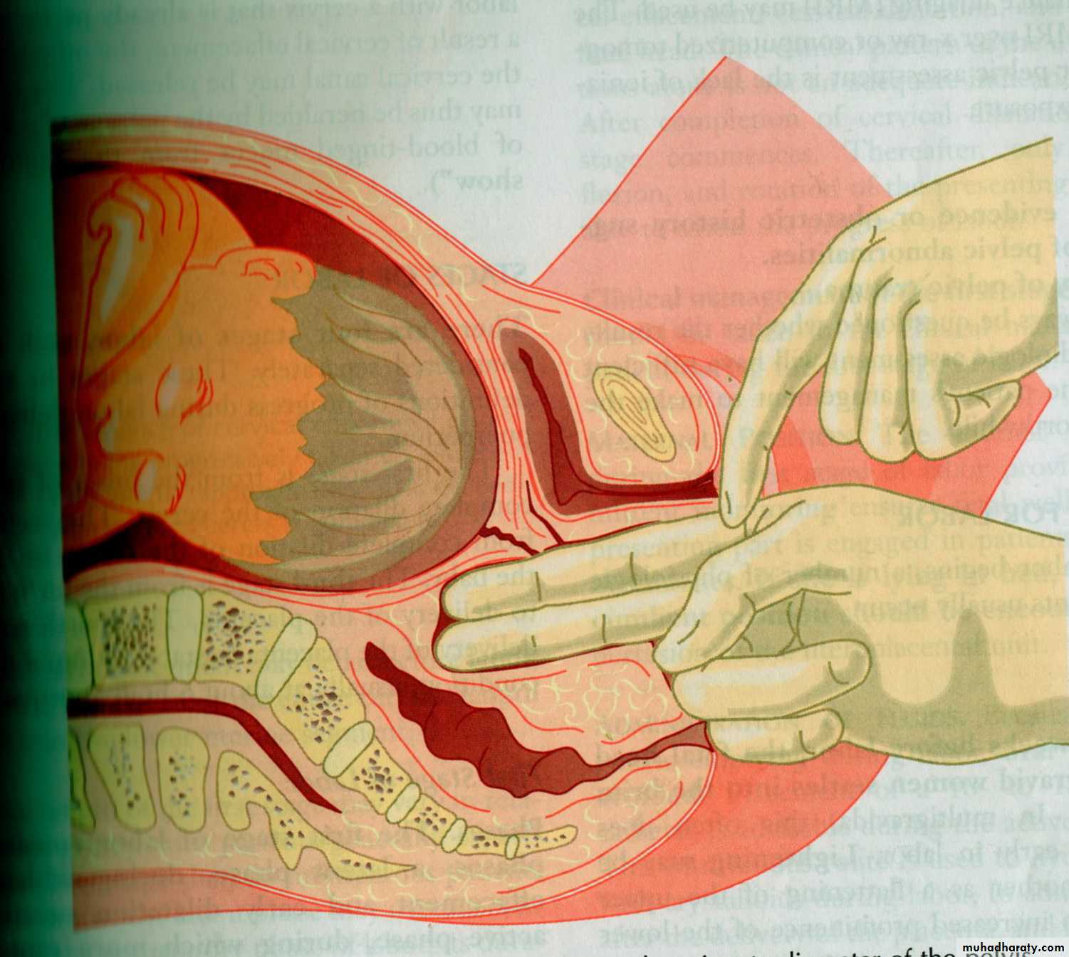
sling of various muscles which are pierced by the urethra, the vagina and the anal canal .Posterior to the vagina these muscles form the perineal body. The levator ani muscles are composed of the pubococcygeus (including the pubovaginalis, pubourethralis, puborectalis ) and the iliococcygeus.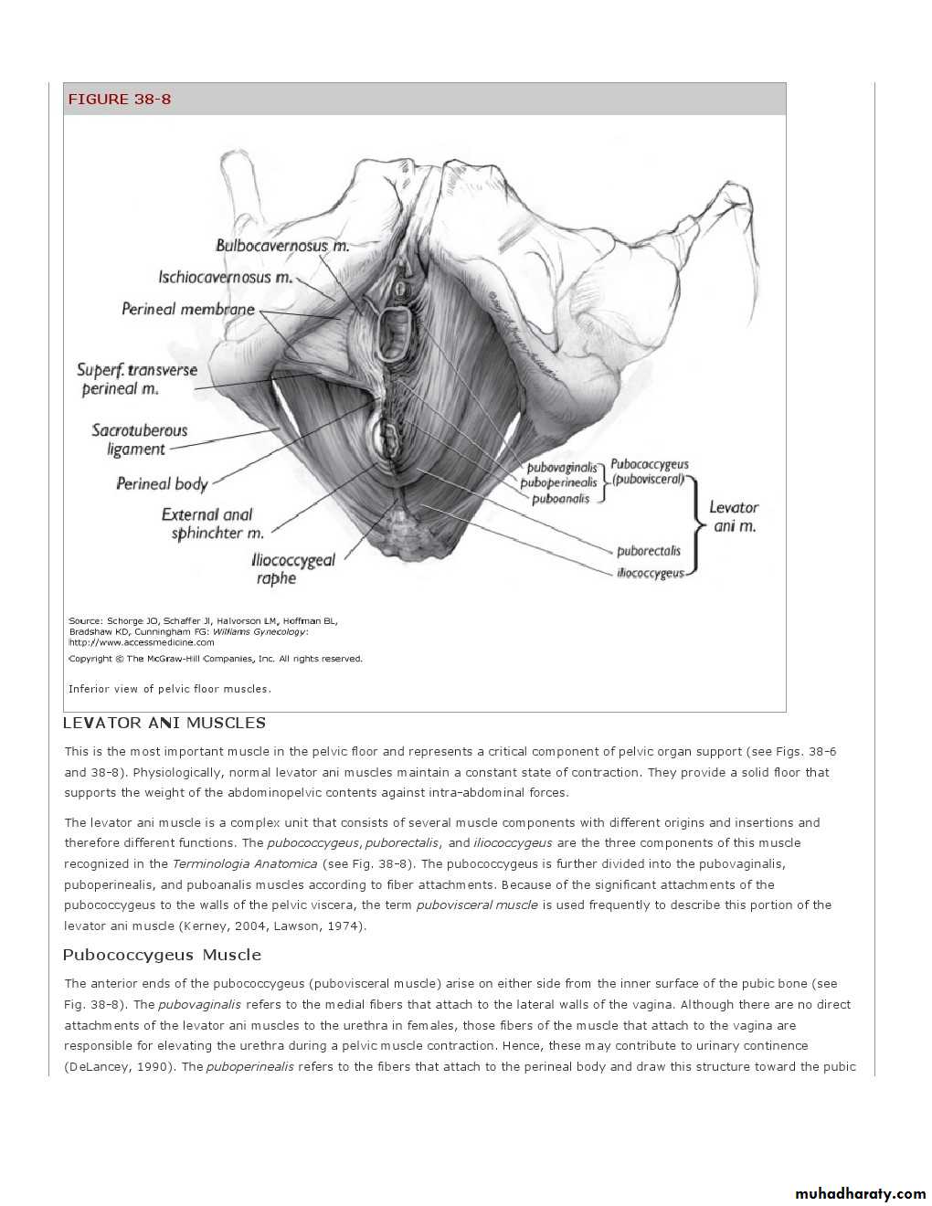
Action : assists the anterior abdominal wall muscles in containing the abdominal and pelvic contents. During parturition, the levator ani supports the fetal head while the cervix dilates.
1
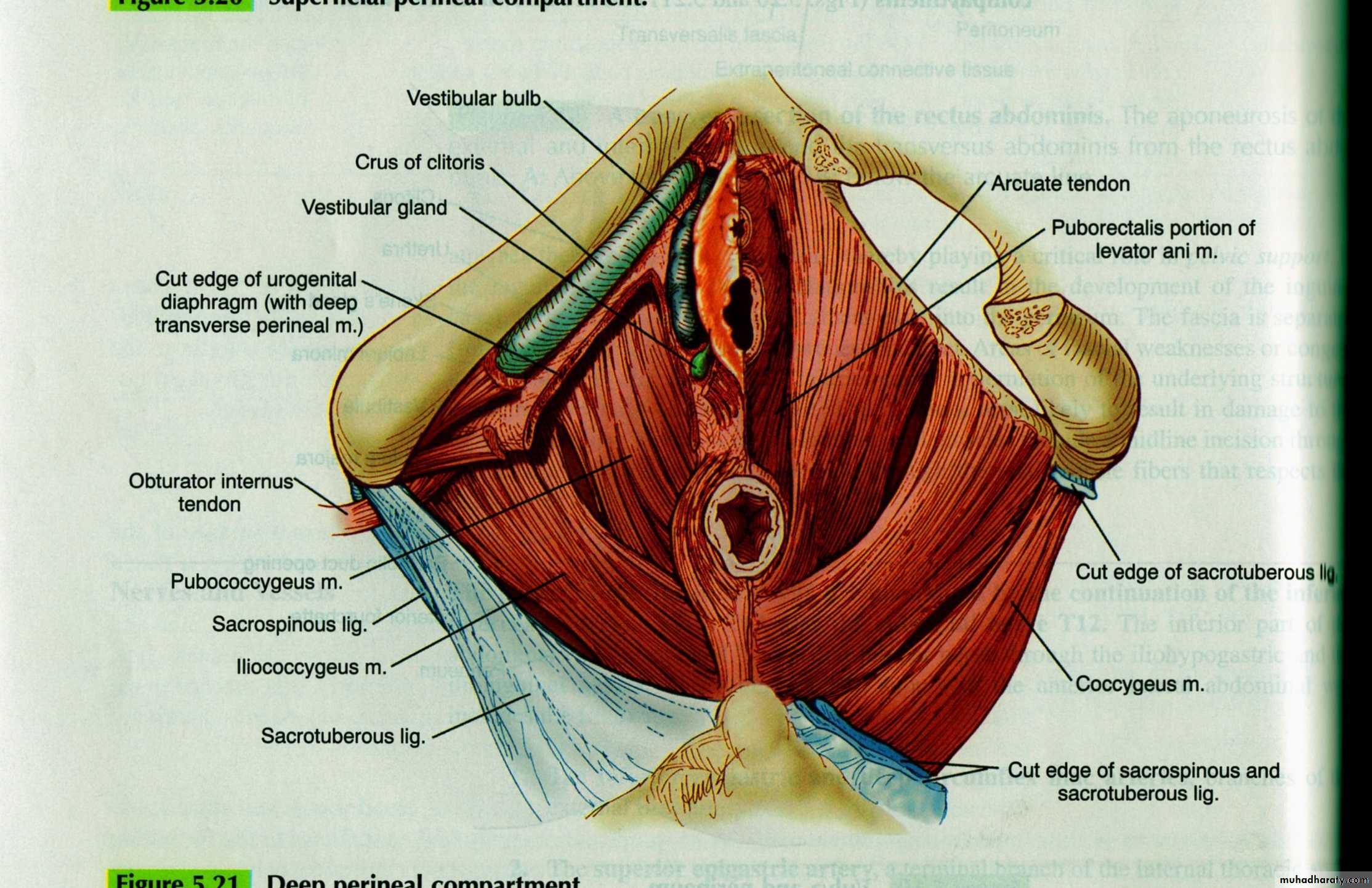
The urogenital diaphragm
This is a triangular-shaped diaphragm through it pass the urethra and vagina. The muscles of the urogenital diaphragm reinforce the pelvic diaphragm anteriorly.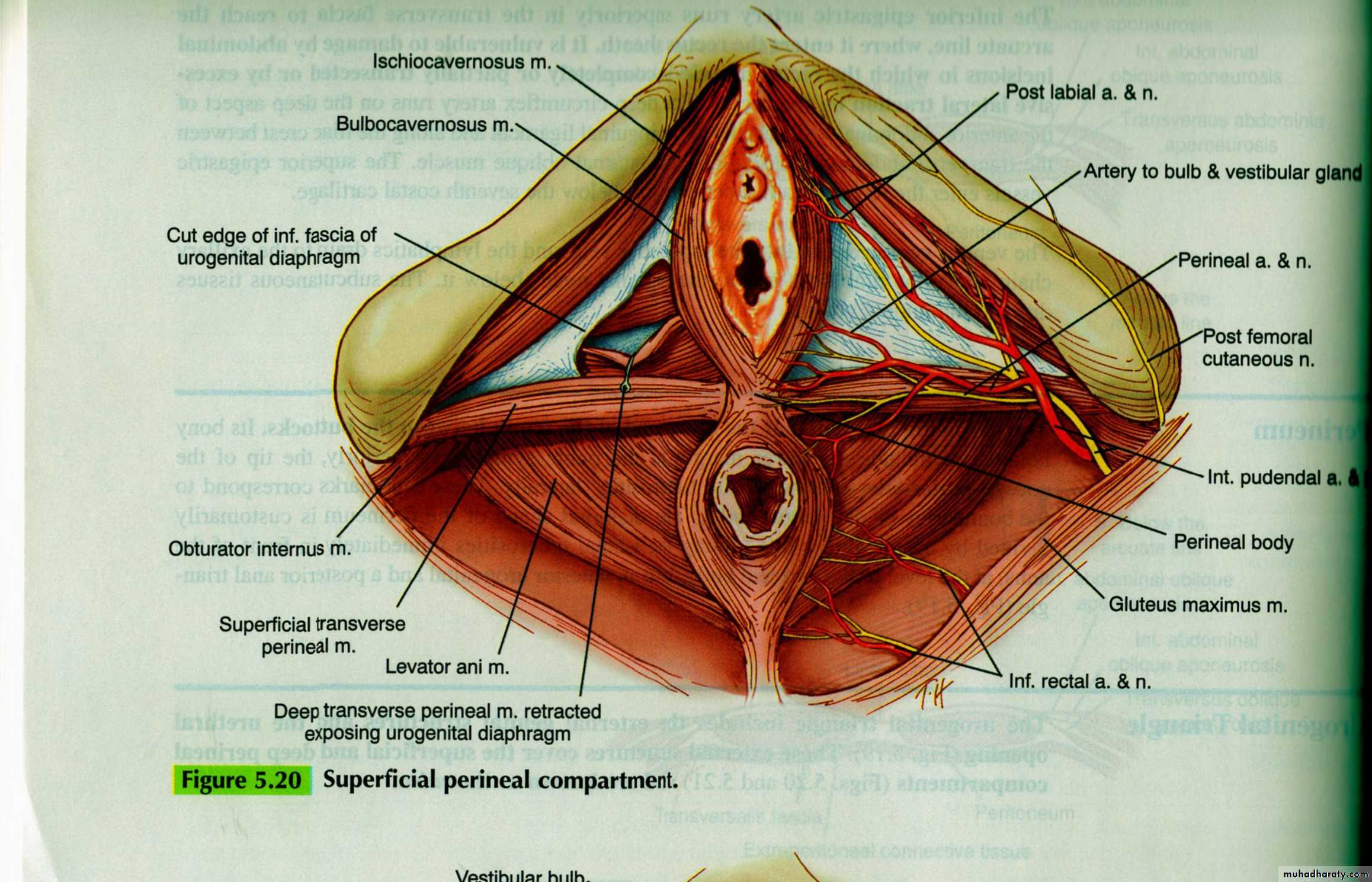
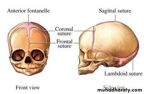
Fetal skull
Fetal skullIs made up of the vault, the face & the base.
The sutures are the lines formed where the individual bony plates of the skull meet one another.
At the time of labour, the sutures joining the bones of the vault are soft , unossified ,whereas that of the face and base are firmly united.
The fontanelles are the junctions of the various sutures
Fetal Skull
LANDMARKS
The bones form the vault are the parietal ,parts of the occipital,frontal &temporal bones.3sutures are of obstetric importance:
Sagittal suture:between the superior borders of parietal bones
Frontal suture:is the forward continuation of sagittal suture lies between the2 parts of frontal bones.
Coronal suture:between the anterior border of parietal &posterior border of frontal bones
LAMBDOIDAL SUTURE between the occipital bone behind and the parietal and temporal bones in front
TEMPORAL SUTURE between the temporal and the parietal bones
ANTERIOR FONTANELLE or BREGMA is the large diamond-shape depression at the anterior end of the cranium where frontal ,coronal &sagittal suturesmeet
POSTERIOR FONTANELL is the smaller triangular space at the posterior end of the cranium where sagital& lambdoid sutures meet.
Areas of skull
Vertex:-top of the skull,between the anterior &posterior fontanelles and the2 prietal eminenceSinciput:-the part of the head in front of the anterior fontanell .It is subdevided into the brow and the face
Occiput:-the back of he head,lies behind the posterior fontanelle.