Giardiasis
Giardia lamblia- LambliasisGiardia intestinalis
Beaver fever
Giardiasis
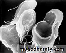
•Is a flagellated protozoan that infect the duodenum and small intestine. Range from asymptomatic colonization to acute or chronic diarrhea and malabsorption. • Inhabit duodenum, jejunum & upper ileum.G. intestinalis exists in 2 stages – trophozoite & cyst.
Most common causative agent of epidemic & endemic diarrhea throughout the world.
In endemic area giardiasis has been associated with growth stunting and with repeated Giardia infections would decreased cognitive function in children.
Incubation period 1-2 week
• Prevalence –
Population affected
Children
Travelers
Swimmers
2-5% in ideveloped countries (2% of adult , 6-8% children)
20-30% in developing countries• morbidity rate up to 20%
• Reservoir
• Man is the main reservoir• Infective form –mature cyst passed in feces of man
• Routes of transmission
– Feco-oral
• ingestion of contaminated water – most important
• Ingestion of contaminated food
– Person to person – day care, nursing homes,mental asylums (poor hygiene)
– Sexual – sexually active homosexual males
Pathology
• Do not invade tissues• Feed on mucous secretions
• May localize in biliary tract to avoid the acidity of duodenum
• Cause inflammation of duodenum & jejunum
• Cause malabsorption as the parasite coats the mucosa & damage epithelial brush border
• Stool contains large amounts of mucous & fat but no blood
Clinical manifestation of Giardiasis:
Asymptomatic : largest group
Acute : self-limiting infection, acute watery diarrhoea
, abdominal cramps, bloating, flatulence
*Stool is profuse & watery in earlier disease
*Voluminous, foul smelling & greasy (steatorrhoea) later
Chronic : chronic diarrhoea with malabsorption syndrome, steatorrhoea
Laboratory Parasitic Diagnosis
*Samples
• Stool
• Duodenal contents
– Duodenal fluid( Entero test )
– Duodenal/ jejunal biopsy
**Entero test
– gelain capsule containing a nylon string with a weight is swallowed by the patient. Free end of the string is fixed to the mouth. Capsule dissolves & the string is released in the duodenum. After overnight string is removed &bile stained mucus collected.
Microscopy
1-Direct Wet Mount
• Trophozoite with falling leaf motility in saline mount
• Cyst in iodine mount
2-Stained stool smears
3-serodiagnosis •ELISA
4-Culture • Not done routinely
Prevention
• Avoid food & water that might be contaminated
– filtration of water (be sure filter is fine enough to trap
the cysts)
– boiling water for at least 1 min.
– addition of a tincture of iodine are effective in killing cysts (chlorination of water does not affect the cysts)
-Travelers to endemic areas are advised to avoid uncooked food that might have been grown , washed ,or prepared with water that was potentially contaminated.
• Practice good hygiene
– Wash hands thoroughly with soap and water• after using the toilet
• before handling or eating food
Treatment
• Nitroimidazole derivatives, – Metronidazole,– Tinidazole
drugs of choice
• Acridine dye
– Quinacrine
• Nitrofurans
– Furazolidone