بسم الله الرحمن الرحيم
Urology Congenital anomalies of the upper urinary tract
د.أشرف إبراهيم العدول
دكتوراه بورد عربي جراحه الكلى
مدرس ـ فرع الجراحة
M.B.Ch.B., CABMS(Uro).
Congenital anomalies of the upper urinary tract
Anomalies of number -Agenesis: UnilateralBilateral -Supernumerary kidney Anomalies of volume and structure Hypoplasia Multicystic kidney Polycystic kidney
• Infantile
• AdultOther cystic disease
• Medullary cystic disease
Anomalies of ascent Simple ectopia Cephalad ectopia Thoracic kidney
Anomalies of form and fusion
Crossed ectopia with and without fusion
Unilateral fused kidney (inferior ectopia)
Sigmoid or S-shaped kidney
Lump kidney
L-shaped kidney
Disc kidney
Unilateral fused kidney (superior ectopia)
Horseshoe kidney
Anomalies of rotation Incomplete rotation Excessive rotation Reverse rotation
Anomalies of the collecting system Calyx and infundibulumCalyceal diverticulum
Hydrocalyx
Megacalycosis
Unipapillary kidney
Extrarenal calyces
Anomalous calyx (pseudotumor of the kidney)
Infundibulopelvic dysgenesis
Pelvis
Extrarenal pelvis
Bifid pelvis
Anomalies of renal vasculature Aberrant, accessory, or multiple vessels Renal artery aneurysm Arteriovenous fistula
Congenital anomalies of the upper urinary tract
In summery comprise a diversity of abnormalities, ranging from:-complete absent kidney, supernumerary Kidney
-aberrant location
-orientation
-shape of the kidney
-aberrations of the collecting system
-blood supply.
Unilateral Renal Agenesis (URA)
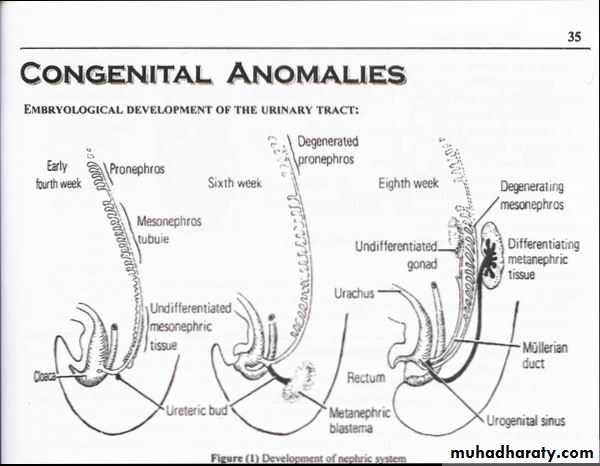
Incidence : 1: 1400 births Found accidentally, more frequently on the left side.Embryology :Complete absence of a ureteric bud or aborted ureteral development prevents maturation of the metanephric blastema into adult kidney tissue.
*Ipsilateral adrenal agenesis is rarely encountered with URA
*Other Genital anomalies are much more frequently observed
clinically: Asymptomatic
Diagnosis : U/S or IVU,CT scan: absent kidney on that side + compensatory hypertrophy of the contralateral kidney
Treatment: no specific treatment
Prognosis: no evidence that they have an increased susceptibility to other diseases
Bilateral agenesis: rare, incompatible with life
Supernumerary Kidney truly an accessory organ
Incidence very rare
Symptoms It may not produce symptoms until early adulthood, if at all.
Diagnosis accidentally by IVU or abdominal U/S
Treatment: no treatment
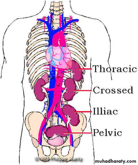
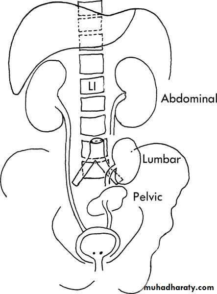
ANOMALIES OF ASCENT
Simple Renal EctopiaWhen the mature kidney fails to reach its normal location in the "renal fossa “
Incidence The incidence is 1 in 1000
Associated Anomalies
• The incidence of contralateral agenesis appears to be rather high• Clinical features Most ectopic kidneys are asymptomatic
• Diagnosis : U/S, IVU, CT scan
• Prognosis: The ectopic kidney is no more susceptible to disease than the normally positioned kidney except for the development of hydronephrosis or urinary calculus formation
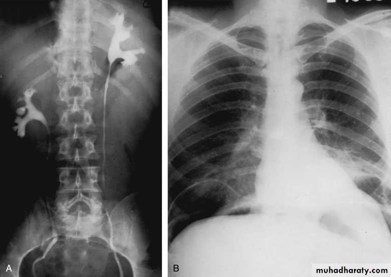
• Cephalad Renal Ectopia
• Thoracic Kidney
ANOMALIES OF FORM AND FUSION
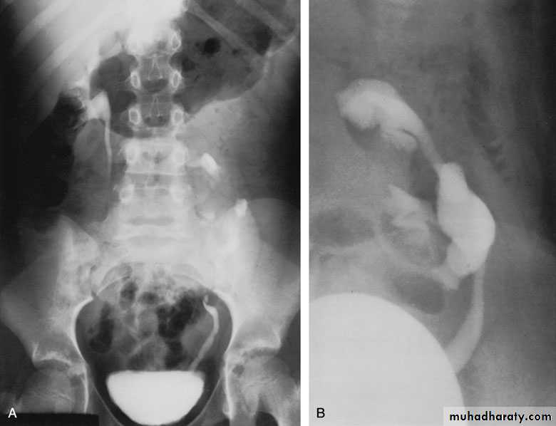
Crossed Renal Ectopia With and Without Fusion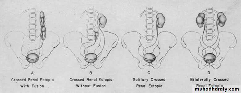
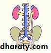
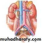
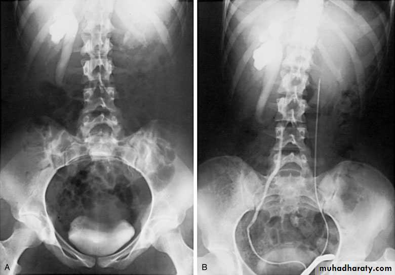
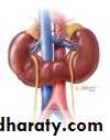
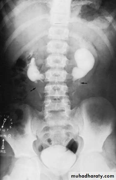
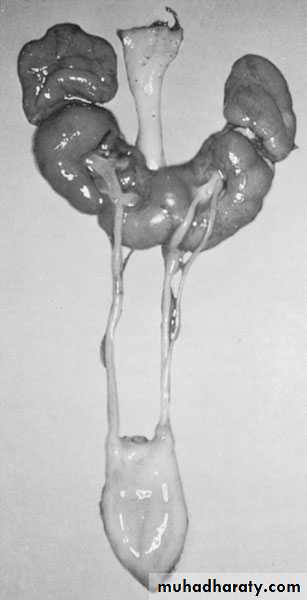
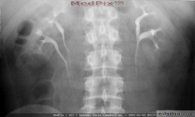
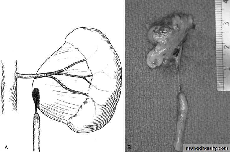
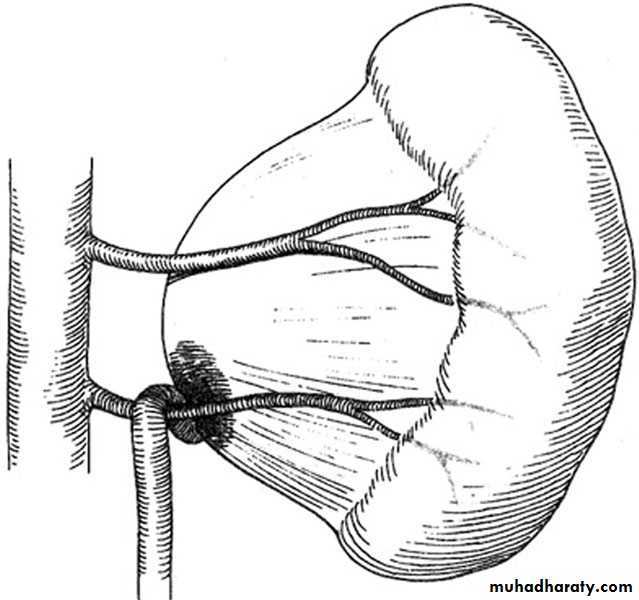
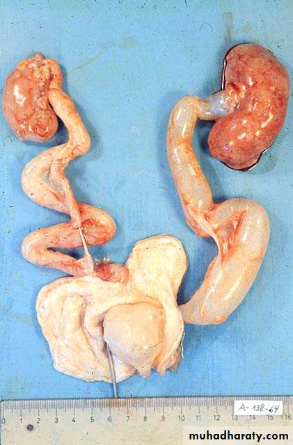
Horseshoe Kidney
Found in 1:1000 necropsies and is more common in men.probably the most common of all renal fusion anomalies
The anomaly consists of two distinct renal masses lying vertically on either side of the midline and connected at their respective lower poles by a parenchymatous or fibrous isthmus that crosses the midplane of the body.
Fusion of the renal masses occurs early in embryonic life, so its ascent will be impeded by inferior mesenteric artery.
The kidneys are low located, mal rotated and pelves lie anteriorly
Symptoms
When present, they are related to complications like hydronephrosis, infection, or calculus formation
Diagnosis ultrasound, IVU, CT scan
Treatment:
Medical: pain relief and to control infectionSurgical: stone removal, PUJ stenosis correction and isthmus division in cases of operations on the aorta
Prognosis: usually they have normal life.
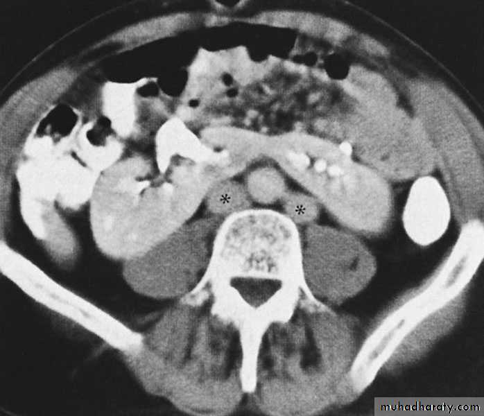
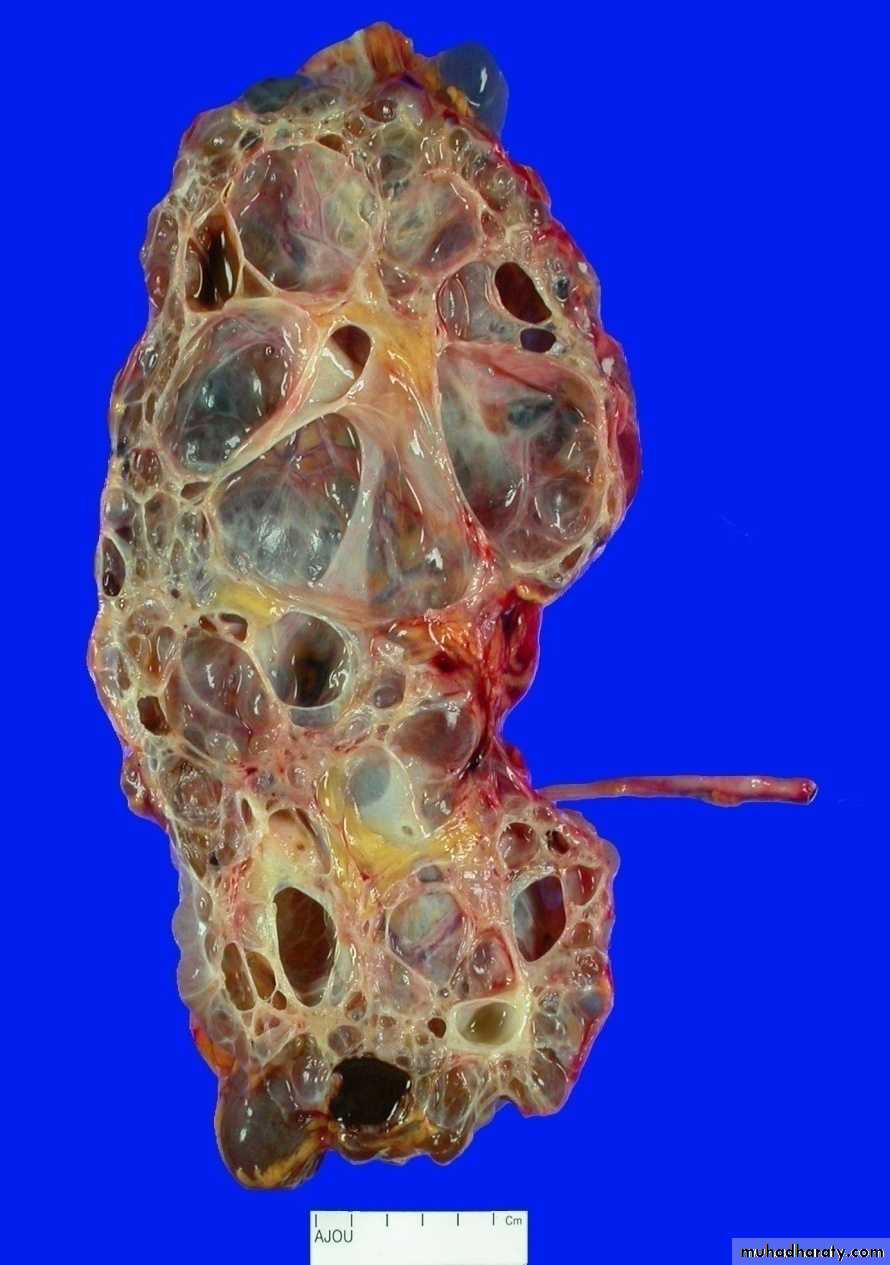
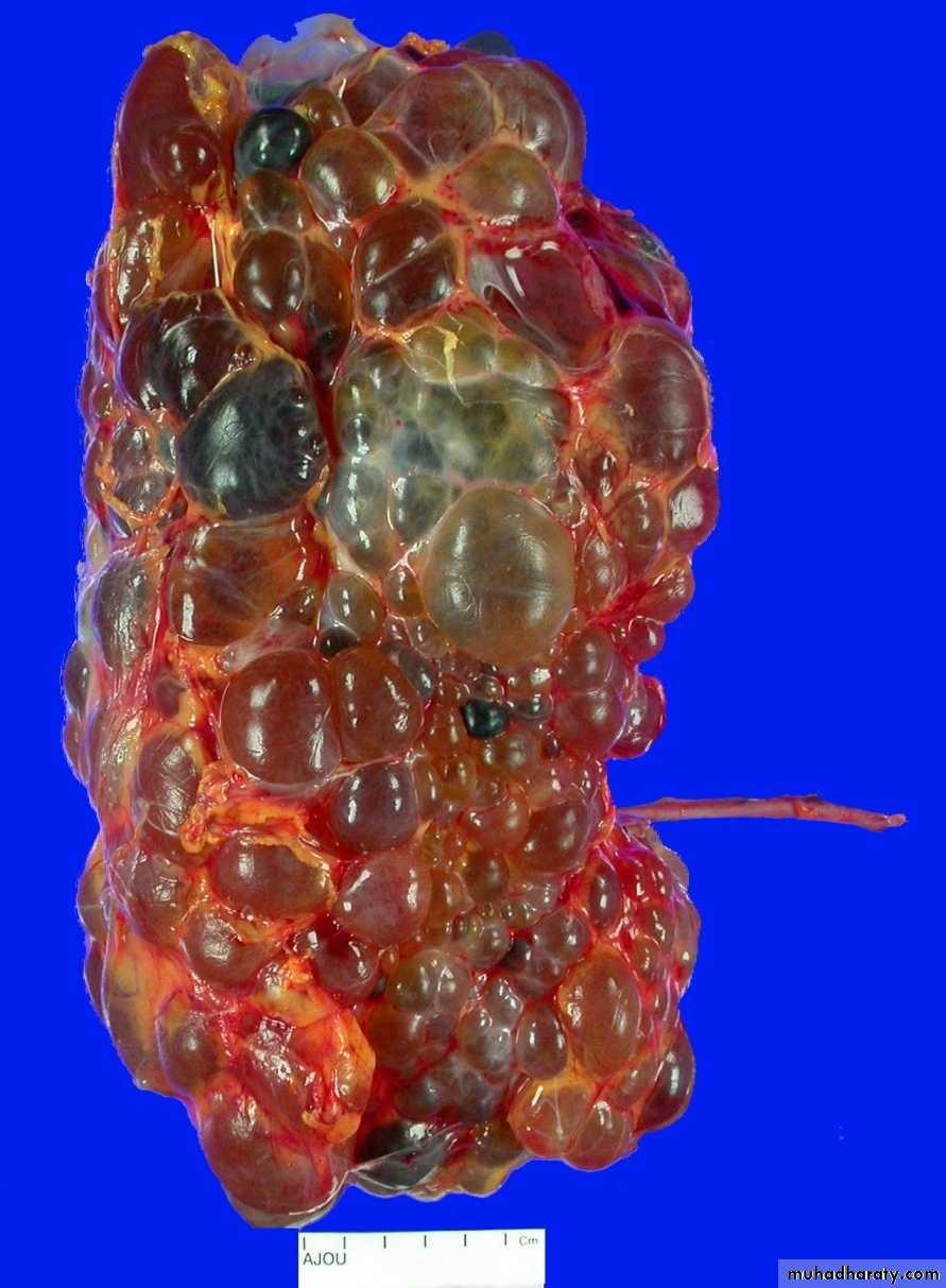
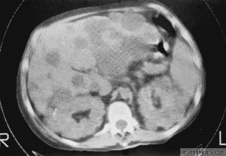
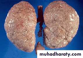
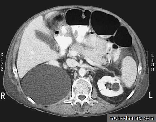
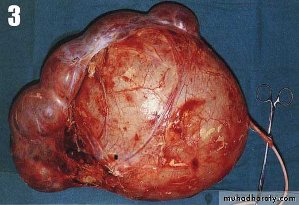
Cystic disease of the kidneys
• Polycystic kidney disease :• The kidney is one of the most common sites in the body for cysts
• Two types:
• AUTOSOMAL RECESSIVE ("INFANTILE") POLYCYSTIC KIDNEY DISEASE
• AUTOSOMAL DOMINANT ("ADULT") POLYCYSTIC KIDNEY DISEASE
Congenital cystic kidney (polycystic kidney) (Adult cystic renal disease)
Autosomal dominant, transmitted by either parents, 50% of offspring's affected.
Both kidneys replaced by large number of cysts of variable size which make the kidney of large size.
The cysts contain clear fluid but sometimes blood.
The cysts progressively increase in size causing pressure atrophy of the renal parenchyma and pressing the ureter.
15% associated with cystic disease of liver, lung, pancreas or spleen.
Etiology & Pathogenesis
The cysts occur because of defects in the development of the collecting and uriniferous tubules and in the mechanism of their joining. Blind secretory tubules that are connected to functioning glomeruli become cystic.Adult polycystic renal disease
Clinical pictures:
Rarely gives clinical manifestation before 4o yearsAsymptomatic: diagnosed accidentally.
Pain: due to pedicle stretching, stone, ureteric obstruction, bleeding inside cyst or infection.Hematuria: cyst distention and rupture to the collecting system.
Infection: renal or cyst infection causes fever, rigor and loin pain.
Hypertension: in 70%, unknown cause.
Renal impairment: anorexia, headache, nausea, vomiting, drowsiness and coma.
Renal enlargement: large knobby palpable kidney
Diagnosis: Family history of polycystic disease.
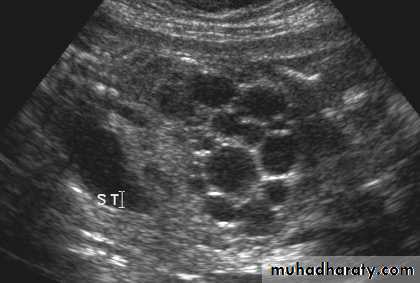
U/S, IVU, CT scan, MRI
Treatment:Medical: (Expectant)
To control infection, hypertension, pain and anemia.
Renal impairment: by low protein diet and dialysis.
Surgical:
Rovsing’s operation (deroofing) for large cysts causing symptoms or obstruction.
Stone removal.
Renal failure: Renal transplantation.
Infantile polycystic disease of the kidney
Rare autosomal recessive, incompatible with life.Both kidneys are large in size and replaced by large number of cysts which may obstruct labor.
The condition is due to failure of ureteric bud to fuse with metanephrose.
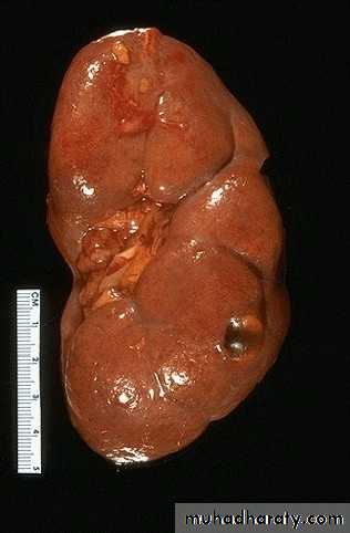
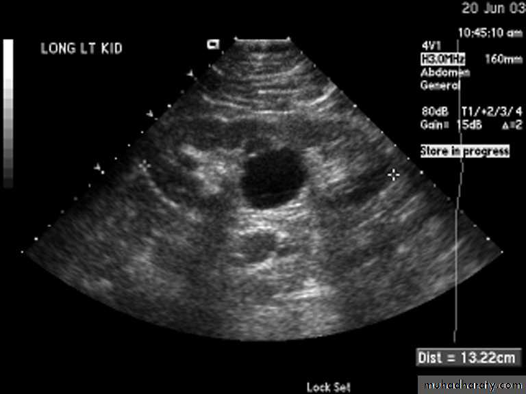
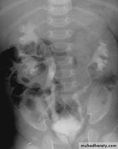
Simple (solitary) renal cyst
Common condition.Single or multiple.
Uni or bilateral.
Congenital or acquired.
Usually asymptomatic. In 10% symptomatic: pain, heaviness, infection, bleeding inside the cyst or pressure effect on the ureter causing hydronephrosis.
Diagnosis
Examination: usually –ve, big cyst cause painless loin mass, & painful if complicated by bleeding or infectionU/S: echo free area (cystic lesion).
KUB: soft tissue shadow.
IVU: stretched calyx, filling defect or hydronephrosis.
CT scan &MRI: are diagnostic.
Treatment: usually no treatment needed
Symptomatic cases:Aspiration and injection of sclerosing agent.
Rovsing’s operation (deroofing).
Partial or total nephrectomy in destructed kidney.
N.B. Malignant cyst: radical nephrectomy.
N.B. Hydatid cyst aspiration is contraindicated because of anaphylaxis and dissemination.
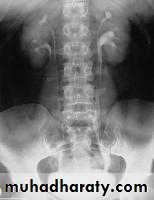
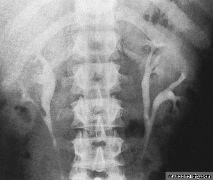
Congenital Anomalies of Renal pelvis & Ureter
Duplication of Renal PelvisIncidence: 4 %
More common on left side Renorenal reflux may occur from one pelvis to the other
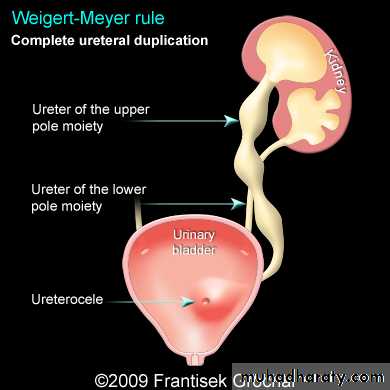
Duplication of the ureter
Incidence : 3 %Usually the ureters fuse & have common orifice in the bladder although they may open independently in which case the ureters cross each other so that the ureter that drain the upper pelvis open below (more distally) in the bladder & vise versa.
Clinical features : usually asymptomatic
More prone to infections, calculus disease & hydronephrosis
Ureteral duplication: partial and complete
Partial duplication: is more common. Two ureters draining single kidney for variable length, then unite together before entering the bladder in one ureteric orifice. Rarely the lower part is duplicated as inverted Y ureter.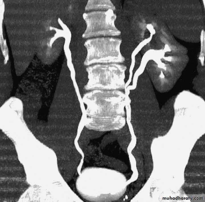
Complete duplication:
Less frequent, the whole ureter is duplicated, and each one opens in separate orifice in the bladder. The ureter draining the upper partopens more distally
in the bladder.
Treatment :expectant
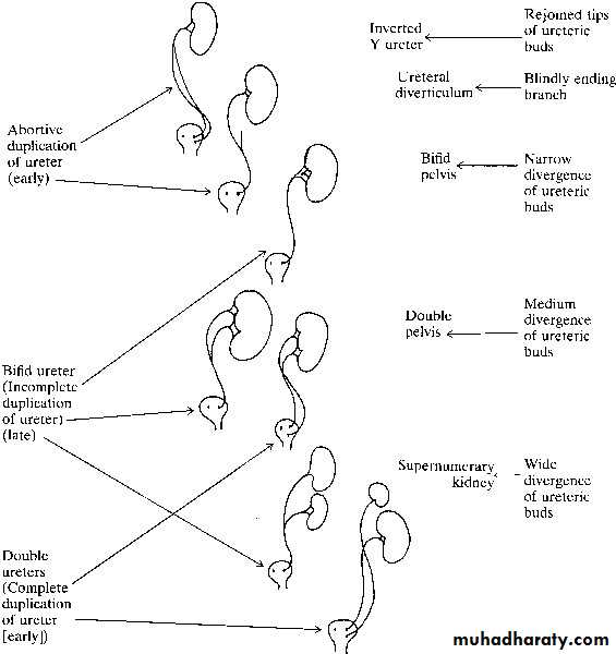
Bifid renal pelvis
i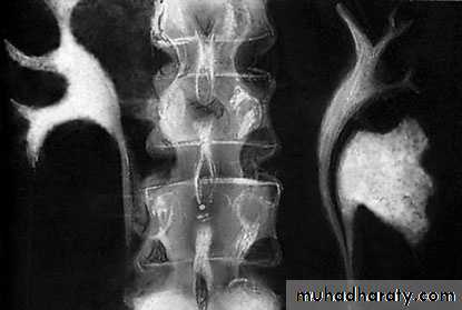
Complete ureteral duplication and ectopic ureteric orifice.
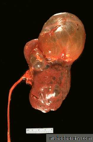
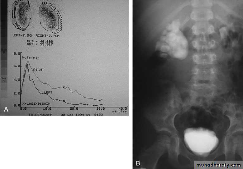
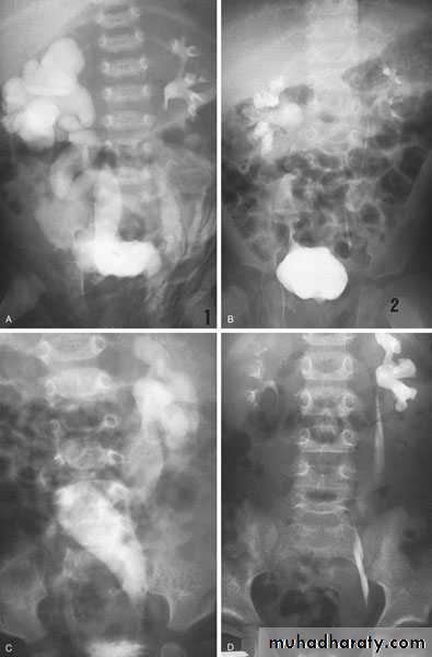
Ureteropelvic Junction (UPJ)(PUJ) Obstruction (stenosis)
The most common cause of significant dilation of the collecting system in the fetal kidneyBoys > Girls
Left-sided lesions predominate
15% bilateral
ETIOLOGY
Intraluminal : mucosal fold that causes valve like effect.
Intrinsic (intramural) interruption in the development of the circular musculature of the UPJ
Extrinsic An aberrant, accessory, or early-branching lower-pole renal artery
PUJ Obstruction – gross pathology
SYMPTOMS/PRESENTATION
Most infants are asymptomaticMost children are discovered because of their symptoms
Episodic flank or upper abdominal pain, sometimes associated with nausea and vomiting
DIAGNOSIS
U/S: hydronephrosisIVU: diagnostic , hydronephrosis with fixed stenotic segment or complete obstruction
CT scan: hydronephrosis that ends abruptly
Magnetic Resonance Imaging
Radionuclide Renography: to see the split function of each kidneyPressure-Flow Studies : Whitaker test
Treatment:
Medical: control infection and pain.Surgical:
Indications for surgery:
1-progressive hydronephrosis.
2- UTI, and symptomatic patients.
3- Severe hydronephrotic non functioning kidney.
4- Stone formation
Treatment
SURGICAL REPAIR including open surgical techniques, laparoscopic, & endoscopic approaches
Open & laparoscopic surgical techniques Anderson-Hynes dismembered pyeloplasty: excision of the pathologic UPJ & appropriate reanastamosis or flap technique or flap operation
Endoscopic Approaches
balloon dilatationAntegrade endopyelotomy
Nephrectomy for non functioning kidney
Bilateral PUJO
Ectopic Ureters
80% are associated with a duplicated collecting systemIn the male, the posterior urethra is the most common site of termination, also to semenal vesicle
In the female, the urethra and vestibule are the most common sites
Clinical features: According to the site of orifice
In females: continuous dribbling
In males: urinary tract infection
Diagnosis IVU, U/S, CT scan, cystoscopy
Treatment: Ureteric reimplantation to urinary bladder or implantation of one ureter to the other ureter is used
Ectopic ureters may drain renal moieties (either an upper pole or a single-system kidney) that have minimal function. Therefore, upper pole partial nephrectomy (or nephrectomy of single system) is sometimes recommended
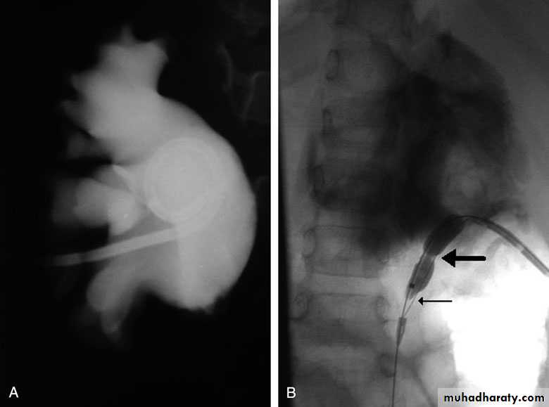
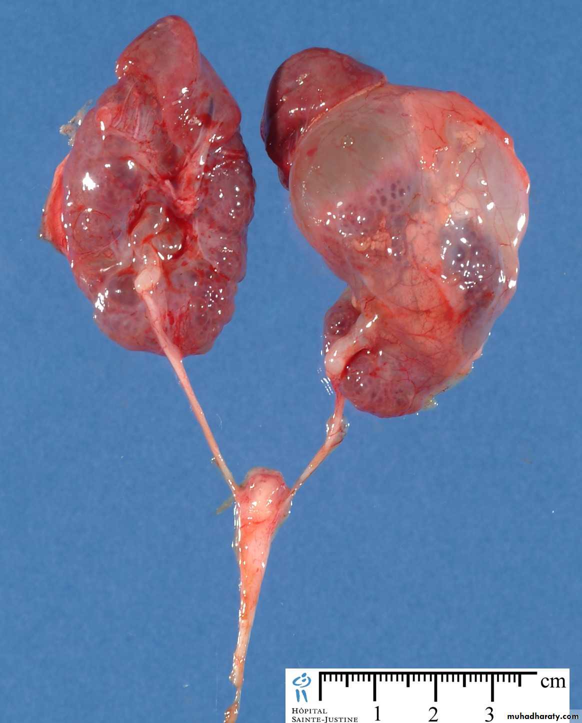
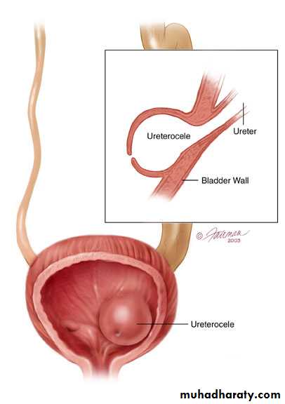
Ureteroceles
Is due to congenital atresia of the ureteric orifice which causes a cystic dilatation of the intramural portion of the ureter
Women > men
Sometimes involves with ectopic ureter
More prone to stone disease & UTIsClinical Features : asymptomatic
Repeated UTIs, Hematuria
Diagnosis
IVU, cystoscopy, cystogram
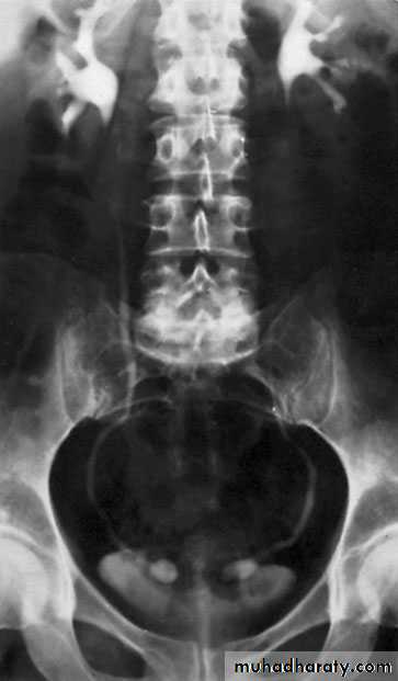
The ‘adder head’ on excretory urography
is typical.
Treatment
Asymptomatic : no treatment
Cystoscopy with diathermy cauterization of the hole
Nephrectomy in non functioning kidney
In complicated cases, ureteral reimplantation and vesical reconstruction
Cobra (Adder) head appearance of ureterocele
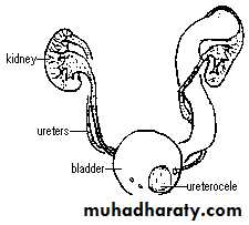
Ureterocele involving single system Ureterocele involving duplicated ureter
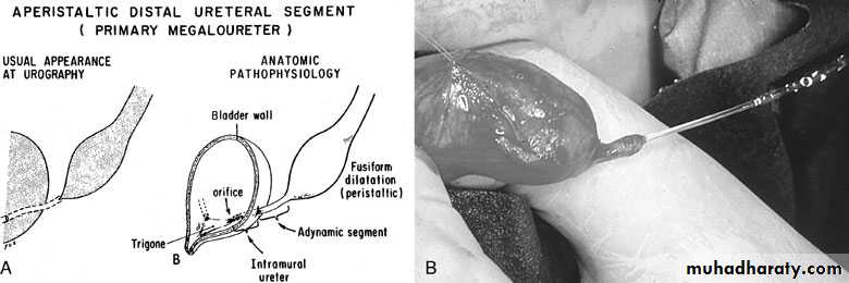
Congenital Megaureter
Grossly dilated ureterUnilateral or bilateral
More common in male
Clinical features:
Asymptomatic, pain, repeated UTIs
lower ureter might be obstructed
sometimes associated with vesicoureteral reflux
Diagnosis : IVU
Treatment
Infection should be controlled
Excision of the lower stenotic segment (if present)Ureteric tapering & reimplantation in
to the bladder
Nephroureterectomy for non functioning kidney
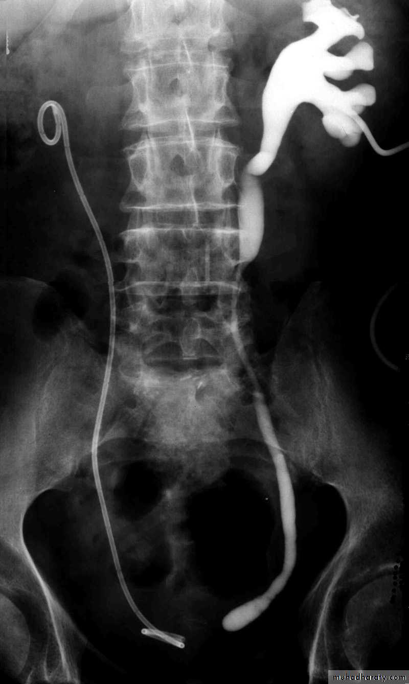
Postcaval (Retrocaval) ureter (Preureteral Vena Cava )
The right ureter pass behind the inferior vena cavaThis might causes obstruction
It is a vascular abnormality
Incidence: about 1 in 1500
Although it is congenital, most patients present at 3rd or 4th decade.
Diagnosis: IVU
Treatment
surgical correction involves ureteral division, with relocation and ureteroureteral or ureteropelvic reanastomosis, usually with excision or bypass of the retrocaval segment, which can be aperistaltic