بسم الله الرحمن الرحيم
Urology Congenital anomalies of the upper urinary tract
د.أشرف إبراهيم العدول
دكتوراه بورد عربي جراحه الكلى
مدرس ـ فرع الجراحة
M.B.Ch.B., CABMS(Uro).
Ureteropelvic Junction (UPJ)(PUJ) Obstruction (stenosis)
The most common cause of significant dilation of the collecting system in the fetal kidneyBoys > Girls
Left-sided lesions predominate
15% bilateral
ETIOLOGY
Intraluminal : mucosal fold that causes valve like effect.Intrinsic (intramural) interruption in the development of the circular musculature of the UPJ
Extrinsic An aberrant, accessory, or early-branching lower-pole renal artery
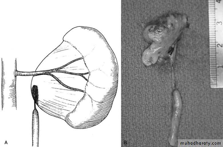
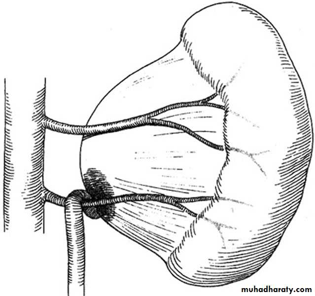
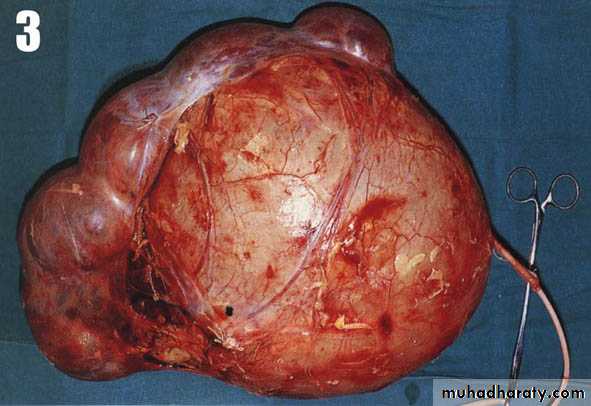
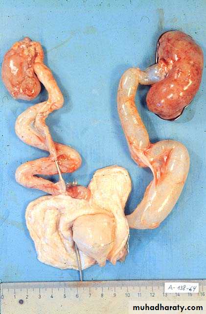
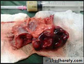
PUJ Obstruction – gross pathology
SYMPTOMS/PRESENTATION
Most infants are asymptomaticMost children are discovered because of their symptoms
Episodic flank or upper abdominal pain, sometimes associated with nausea and vomiting
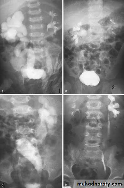
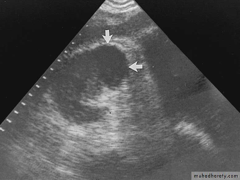
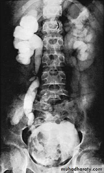
DIAGNOSIS
U/S: hydronephrosisIVU: diagnostic , hydronephrosis with fixed stenotic segment or complete obstruction
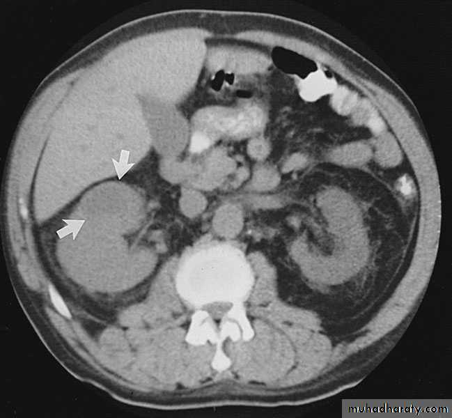
CT scan: hydronephrosis that ends abruptly
Magnetic Resonance Imaging
Radionuclide Renography: to see the split function of each kidney
Pressure-Flow Studies : Whitaker test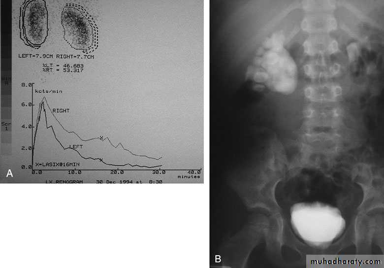
Treatment:
Medical: control infection and pain.Surgical:
Indications for surgery:
1-progressive hydronephrosis.
2- UTI, and symptomatic patients.
3- Severe hydronephrotic non functioning kidney.
4- Stone formation
Treatment
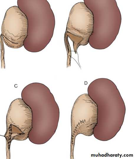
SURGICAL REPAIR including open surgical techniques, laparoscopic, & endoscopic approachesOpen & laparoscopic surgical techniques Anderson-Hynes dismembered pyeloplasty: excision of the pathologic UPJ & appropriate reanastamosis or flap technique or flap operation
Endoscopic Approaches
balloon dilatation
Antegrade endopyelotomy
Nephrectomy for non functioning kidney

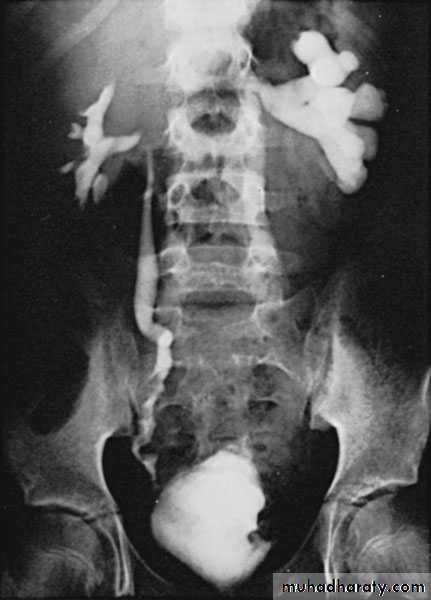
Bilateral PUJO

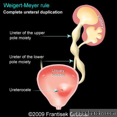
Ectopic Ureters
80% are associated with a duplicated collecting systemIn the male, the posterior urethra is the most common site of termination, also to semenal vesicle
In the female, the urethra and vestibule are the most common sites
Clinical features: According to the site of orifice
In females: continuous dribbling
In males: urinary tract infection
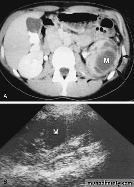
Diagnosis IVU, U/S, CT scan, cystoscopy
Treatment: Ureteric reimplantation to urinary bladder or implantation of one ureter to the other ureter is used
Ectopic ureters may drain renal moieties (either an upper pole or a single-system kidney) that have minimal function. Therefore, upper pole partial nephrectomy (or nephrectomy of single system) is sometimes recommended
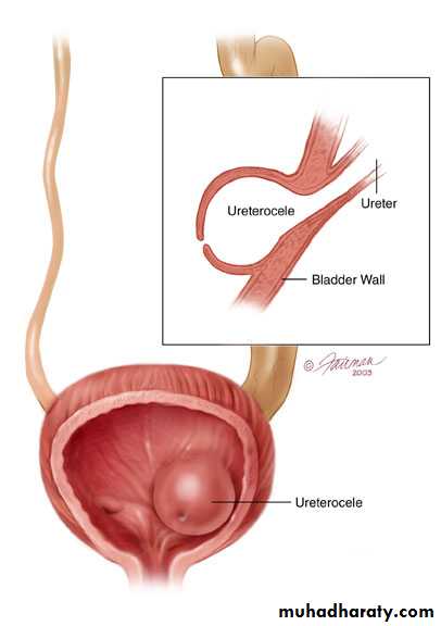
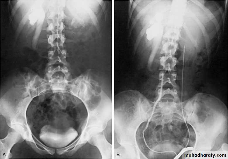
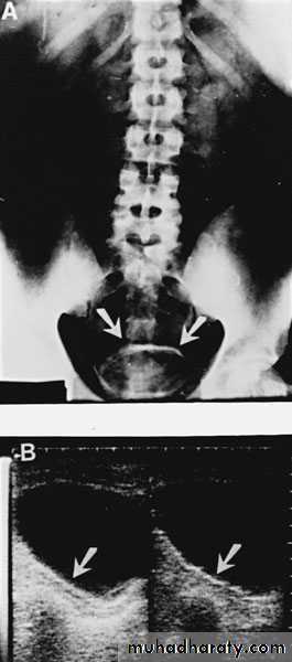
Ureteroceles
Is due to congenital atresia of the ureteric orifice which causes a cystic dilatation of the intramural portion of the ureter
Women > men
Sometimes involves with ectopic ureter
More prone to stone disease & UTIsClinical Features : asymptomatic
Repeated UTIs, Hematuria
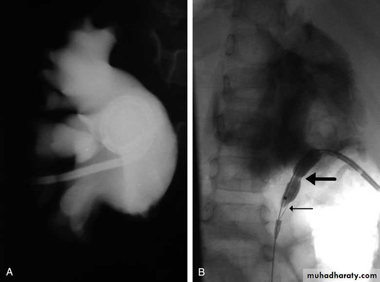
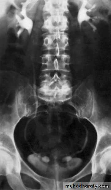
Diagnosis
IVU, cystoscopy, cystogram
The ‘adder head’ on excretory urography
is typical.
Treatment
Asymptomatic : no treatment
Cystoscopy with diathermy cauterization of the hole
Nephrectomy in non functioning kidney
In complicated cases, ureteral reimplantation and vesical reconstruction
Cobra (Adder) head appearance of ureterocele

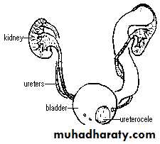
Ureterocele involving single system Ureterocele involving duplicated ureter
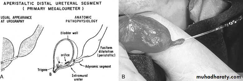
Congenital Megaureter
Grossly dilated ureterUnilateral or bilateral
More common in male
Clinical features:
Asymptomatic, pain, repeated UTIs
lower ureter might be obstructed
sometimes associated with vesicoureteral reflux
Diagnosis : IVU
Treatment
Infection should be controlled
Excision of the lower stenotic segment (if present)Ureteric tapering & reimplantation in
to the bladder
Nephroureterectomy for non functioning kidney
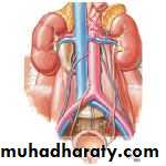
Postcaval (Retrocaval) ureter (Preureteral Vena Cava )
The right ureter pass behind the inferior vena cavaThis might causes obstruction
It is a vascular abnormality
Incidence: about 1 in 1500
Although it is congenital, most patients present at 3rd or 4th decade.
Diagnosis: IVU
Treatment
surgical correction involves ureteral division, with relocation and ureteroureteral or ureteropelvic reanastomosis, usually with excision or bypass of the retrocaval segment, which can be aperistaltic
Renal surgical infections
Renal surgical infectionsUrinary tract infection (UTI) is an inflammatory response of the urothelium to usually bacterial invasion that is usually associated with bacteriuria and pyuria.
Classification
Non specificSpecific ( T.B. & Bilharziasis )
Acute
Chronic
Bacteriology:
E.coli ( most common)
Proteous, Staph aurious, Klebsiella
Pathogenesis:
Ascending infection: most common routeHematogenic
Lymphatic
Direct extension
Introitus
• Urethra• Prepuce
Intrinsic factors: Bladder, ureteral & renal
Bacterial persistence
Urinary calculiObstructive uropathy
Renal pathology
Urethral infection
Foreign bodies
Urogenital & intestinal fistulae
Kidney Infections
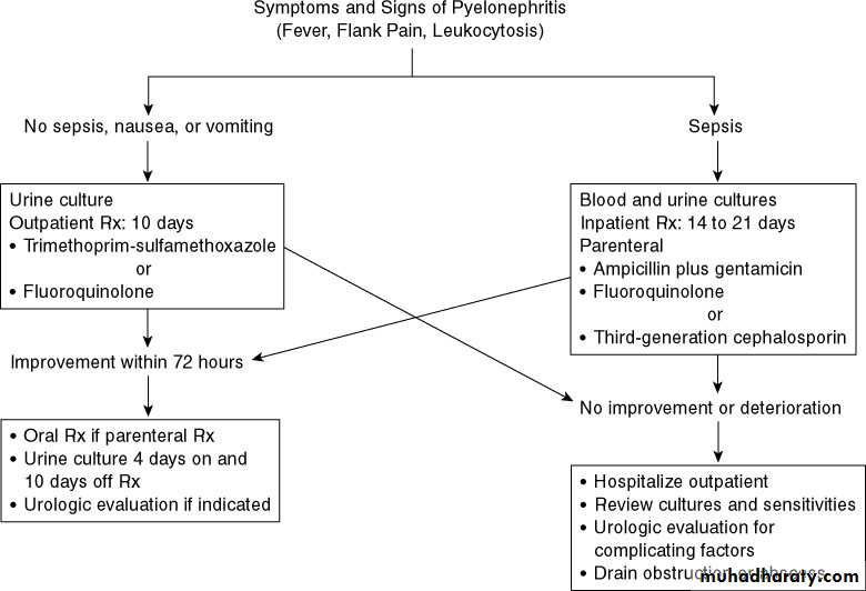
Acute pyelonephritis
Defined as inflammation of the kidney and renal pelvis
It is a clinical syndrome of chills, fever, and flank pain that is accompanied by bacteruria and pyuria, a combination that is reasonably specific for an acute bacterial infection of the kidney.
Female > male
Clinical features:
Constitutional symptoms
Flank & hypochondrial pain
Frequency, urgency, & dysuria
Investigations
GUEUrine +/- blood culture & sensitivity
U/S
KUB
IVU
Treatment
Depends on the severity ofthe infection
Admission to the hospital, Bed rest
Parenteral broad spectrum antibiotics until results of C&S
Analgesics
Encouraged copious fluid
intake otherwise give IVF
N.B. obstructive
Pyelonephritis needs
drainage
Pyonephrosis
Pyonephrosis refers to infected hydronephrosis where the kidney is converted into a sac containing pus associated with suppurative destruction of the parenchyma of the kidney, in which there is total or nearly total loss of renal function.It is usually unilateral
Causes
Infected hydronephrosis
Following acute pyelonephritis
Complication of renal calculus disease
CLINICAL FEATURES
The patient is usually very illFlank pain & Tenderness
High fever ,chills
Anaemia
Investigations
GUE + C&S + blood C&SCBC
KUB
U/S
IVU
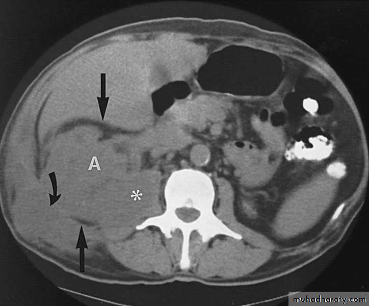
CT scan
Treatment
• It is Surgical Emergency that needs drainage
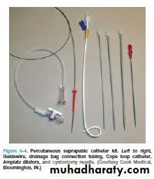
• ..nephrostomy: --percutaneous
• -- open
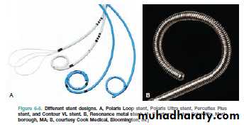
• .. JJ stint
The stone is removed
nephrectomy

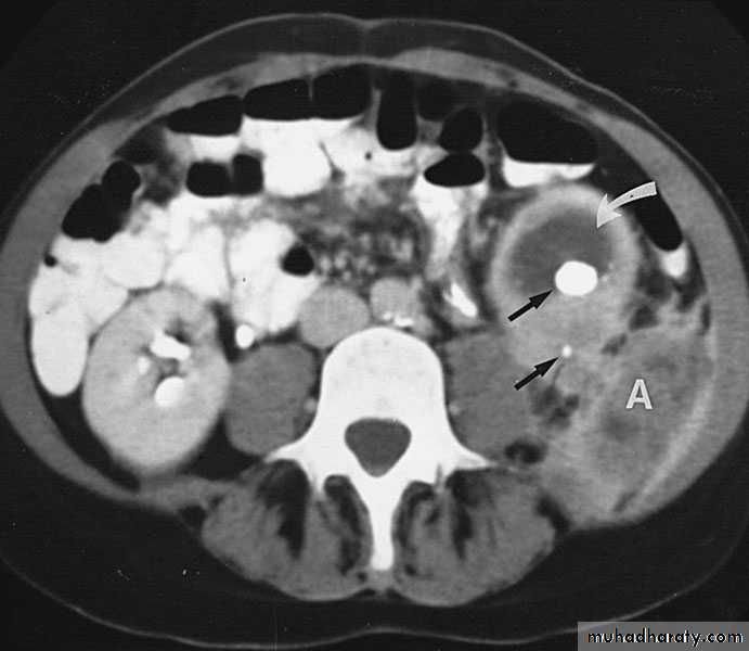
Renal Abscess or Renal Carbuncle
Renal abscess or carbuncle is a collection of purulent material confined to the renal parenchyma.The renal parenchyma contains an encapsulated necrotic mass
Insidious onset (may run > 2 weeks)
Obscure fever
Local pain
Symptoms of the primary cause
Tender renal angle
Tender mass : differentiate from malignant lesion
Bacteriology
Hematogenic infection
Commonly coliforms & staph aureous, proteous, klebsiella.
Predisposing factors
Diabetic patientsI.V drug therapy
Hemodialysis
Immunocompromized
Skin infection
Rarely ascending infection
Clinical picture
Usually underlying pathology:systemic bacterial infection, skin infections, urinary stones, vesicoureteric reflux, obstruction, DM
Infection—liquefaction—abscess formation
Male : female 3:1
Age : 20---30 year
Loin Pain
Fever
On exam.: renal angle tenderness
Investigations
GUE ????
Urine C&S ????
Blood culture ????
WBC:Leuckocytosis
U/S
KUB, IVU
CT scan
Treatment
Medical: RestAnalgesia
Antibiotics
Follow up examination
Surgical: Abscess drainage
Nephrectomy
Perinephric Abscess
Route of infection:Rupture of renal abscess
Infected perinephric hematoma or urinoma
Extension from nearby organs: Appendix, Gall Bladder, Pelvic organs.
Hematogenic: Tonsillitis, boils etc.
Bacteriology
EcoliStaph aureous
Proteous
Klebseilla
Pathology
Cortical abscess coallese, enlarge, rupture to the perinephric space, form a perinephric abscessFluid filled inflammatory mass
Thick wall, adhesions.
Clinical picture
Fever , rigor
Dysuria, frequency
Renal tenderness
Visible loin mass, tender, +ve fluctuation
Investigations
Leucocytosis, AnemiaPyurea, +ve bacterial culture
U/S
CT scan
KUB : soft tissue mass, stones.
IVU , Tomography
Chest x ray : ? Reactionary pleural effusion
Treatment
Bed restAntibiotics & analgesics
Always combined with drainage:
Under U/S or CT- scan guidance
Open drainage
Chronic non specific infectionXanthogranulomatous Pyelonephritis
Rare, severe, chronic renal infection typically resulting in diffuse renal destruction.
Commonly affect middle age
Mixed bacteria: E. coli, Proteous mirabilis
Predisposing factors:
DiabeticRenal stone disease
Neurogenic uropathy
Obstructive uropathy
Clinical picture
ChronicLoin pain
Low grade fever & malaise
Weight loss
Renal mass
Multiple fistulae
Macroscopic appearance: Excessive fatty infiltration, Xanthene deposit
Investigations
GUE
KFT
U/S
CT scan
KUB
IVU
Treatment
Always surgery… NephrectomyUnder antibiotic cover
prostatitis
Acute prostatitisBacteria: E. coli, staph aureus, S. faecalis, N. gonorrhoea
Route of infection: -Hematogenous
-2ry to UTI
Clinical features
Fever, shivering , rigorBackache, perineal pain
Irritative voiding symptoms: dysuria, frequency
Obstructive urinary symptoms
Pain on defecation
O/E: DRE : enlarged, extremely tender, hot, soft prostate
Treatment
Admission ?
Bed rest
Analgesics
Antipyretics
Parenteral antibiotics
If abscess: drainage
If retention: suprapubic catheterization.
Bacteria: Mycobacterium TB
Pathogenesis: HematogenicStart unilateral , late bilateral affection.
The 1st lesion starts usually in the pyramids
Chronic: Asymptomatic until late stage
TB granuloma, caseation, open to the calyces.
Renal destruction, calcification.
The ureteric upper & lower 1/3rd is affected
Ureteral & bladder involvement is commonly secondary to renal T.B.
Clinical picture
Always suspect if:Endemic area
Age : 20-30 year
Male : female 2:1
Chronic symptoms
Non responsive UTI to adequate therapy.
Unexplained hematuria.
loin pain
Night sweating, Wt loss
Fever when secondary bacterial infection
Chronic renal sinuses.
TB is the most common opportunistic infection in AIDS patients
InvestigationsGUE : RBC , Sterile acid pyuria.
-ve urine C&S
Three successive morning urine samples for AFB.
24 hours urine collection for AFB.
TB culture & sensitivity.
ESR
WBC total & differential.
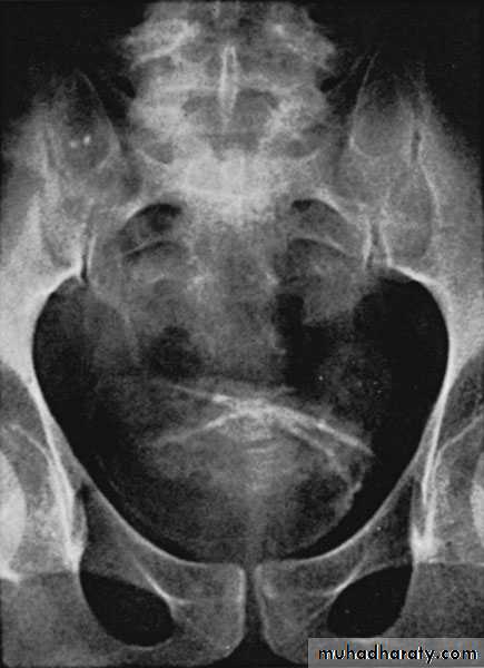
KUB: Renal calcification
IVU
CXR
Cystoscopy: for lower tract involvement.
Treatment
Medical:Surgical:
If complicated
No clinical control
Correct obstruction
Nephrectomy.
Complications
Perinephric abscessPyonephrosis
Renal stones
Ureteral strictures
Renal cutaneous sinuses
Chronic renal failure.
Autonephrectomy in ureteral obstruction
Bladder contracture (thimble bladder)
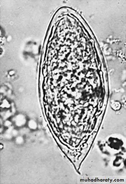
Bilharziasis
Trematode: schistosoma haematobiumMale: female 3:1
Endemic in Nile valley, Iraq, & middle east in general.
Marshes & slow running fresh water is the habitat of the fresh water snail ( bulinus truncatus ) which is the intermediate host.
Mode of infestation
The bifid tailed embryos (cercariae) penetrate the skin, enter the blood vessels, flourish in the liver, develop into male & female worms, they pass to the vesical venous plexusThe female pass to the submucous venule to lay its eggs with its terminal spine which penetrate the vessel wall & pass with urine & if reach fresh water it penetrates the intermediate host.
Clinical features
Urticaria ( swimming itch )
Fever , sweating
Hematuria: intermittent, terminal
Lymphadenopathy & splenomegaly
Investigations
GUE : early morning samples for several consecutive days – ovae with terminal spinesLeukocytosis – eosinophilia
Cystoscopy
Bilharzial pseudotubercles , nodules, sandy patches, ulceration, fibrosis, granulomas, papillomas, carcinoma (SCC).
Imaging study
KUBU/S
IVU
Treatment
Antimony e.g. praziquantel & metriphonatePapilloma : endoscopic removal
Carcinoma : radical cystectomy
Complications
2ry bacterial infectionVesical & ureteric calculus formation
Terminal ureteric stricture : needs dilatation or ureteric reimplantation
Prostatoseminal vesiculitis
Fibrosis of the bladder & bladder neck
Urethral stricture & fistula formation.