Anatomical landmarks of the maxilla & maxillary arch
Lecture 2Hasan khiraldeen mohialdeen
Maxillary anatomical land marks
Supporting structures• Mucous membrane
• Hard palate
• Rugae area
• Mid palatine raphae
• Residual ridge
• Incisive papillae
• Maxillary tuberosity
• Torus palatinus
Limiting and peripheral structures
• Labial frenum
• Labial vestibule
• Buccal frenum
• Buccal vestibule
• Hamular notch
• Vibrating line
• Fovea palatine
Maxilla & maxillary arch
Mucous membrane
A thin layer of tissue that lines a cavity, envelopes a vessel or separates a space or organ.It consists of:
• Mucosa
• Submucosa
• Masticatory mucosa
• Lining mucosa
• Specialized mucosa
Maxilla & maxillary arch
PalateThe palate extends from the roof of the mouth all the way back to the uvula: divided into
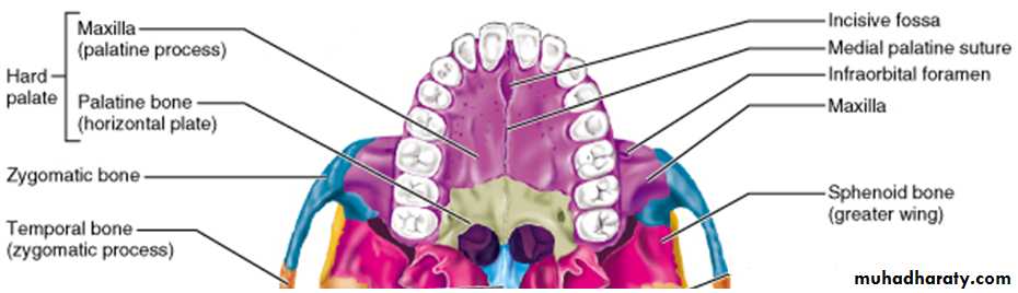
Hard Palate. The hard palate is made up of the anterior two-thirds of the palatal vault supported by bone (palatine processes of the maxillae and the horizontal plates of the palatine bones).
The horizontal portion of hard palate lateral to mid line provides primary support area for denture.
Soft Palate. The soft palate is made up of the posterior one-third of the palatal vault that is not supported by bone. The soft palate is a muscular extension from the posterior edge of the hard palate, and it is very movable, especially during speaking and swallowing.
Maxilla & maxillary arch
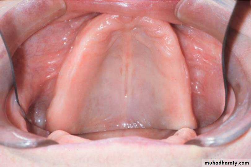
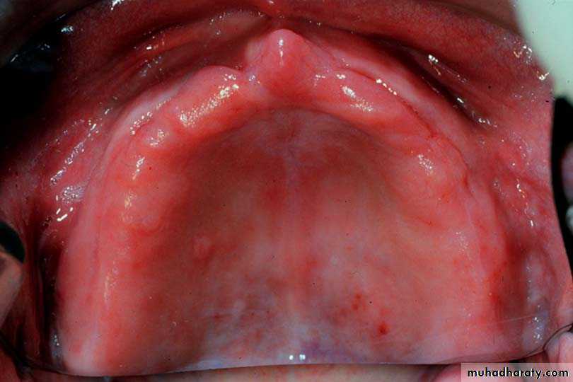
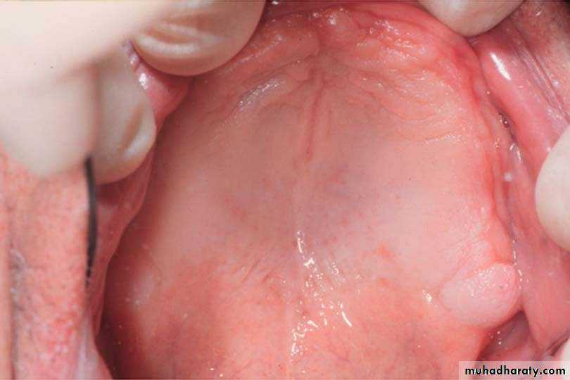
¨ Rugae
Rugae are irregular ridges of fibrous tissue found in the anterior one-third of the hard palate.
It plays important role in speech.
It is considered secondary stress bearing area.
Aids in stability and retention of denture.
Maxilla & maxillary arch
Median Palatine RapheThe medial palatine raphe is a slight tissue elevation which occurs in the midline of the hard palate, immediately over the median palatine suture.
May require relief when covered by a denture.
Submucosa is extremely thin.
Maxilla & maxillary arch
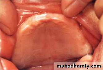
Torus palatinusIs defined as a smooth round anatomical protuberance.
It is a hard bony enlargement that occurs in the midline of hard palate and is found in 20% of the population.
Require relief, if sever should be surgically removed.
Maxilla & maxillary arch
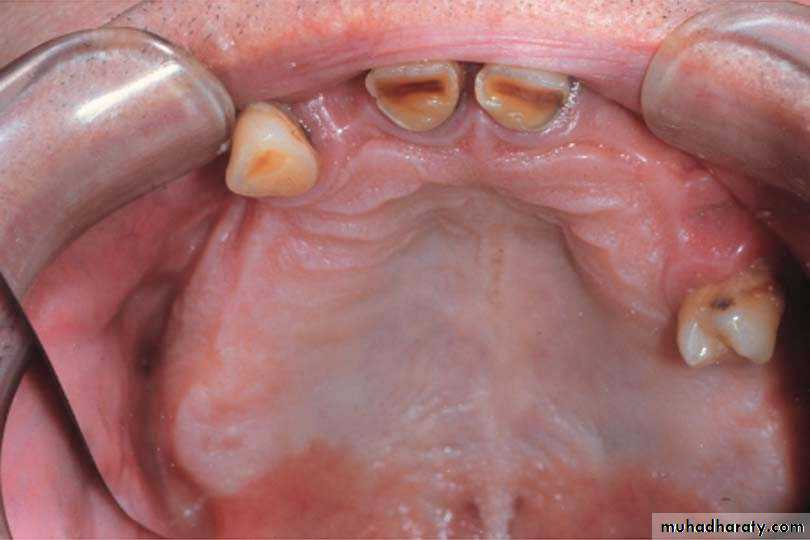
Alveolar ProcessThe alveolar process is a process of the maxilla that surrounds the roots of natural teeth. The right and left alveolar processes combine to form the maxillary arch.
Alveolar Ridge (Residual Ridge)
Defined as the portion of the residual bone and its soft tissue covering, that remains after the removal of teeth.
The residual ridge is the reminant of the alveolar process which originally contained sockets for natural teeth. After natural teeth are extracted, the alveolar ridge can be expected to get smaller (resorb). The rate of resorption varies considerably from person to person.
It is considered secondary support bearing area.
Maxilla & maxillary arch
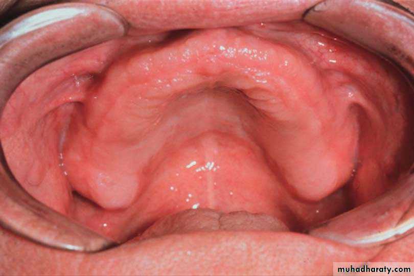
Maxillary TuberosityThe maxillary tuberosity is the most distal (posterior) portion of the maxillary alveolar ridge.
It is considered secondary stress bearing area.
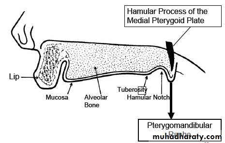
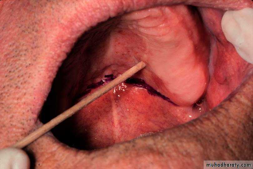
Hamular Notch
The hamular notch is a deep depression located posterior to the maxillary tuberosity. The depths of this depression is part of a series of guides used to determine the posterior border of a maxillary denture.
Maxilla & maxillary arch
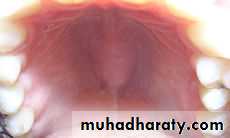
Incisive PapillaThe soft tissue elevation immediately over the incisive foramen.
The incisive foramen is located in the midline of the hard palate, immediately behind the central incisor teeth. The foramen is an exit hole for blood vessels and nerves.
Since the incisive papilla is visible in the exact midline of the hard palate, just behind the natural central incisors, the papilla is a reliable guide for determining the midline relationships of upper anterior denture teeth.
It may require releif.
Maxilla & maxillary arch
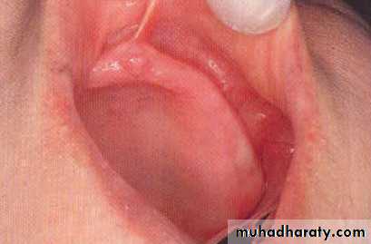
Labial FrenumThe labial frenum is a narrow fold of oral mucosa, which is found in the approximate midline. It extends from the inner surface of the lip to the labial surface of the alveolar ridge. The labial frenum is not a reliable guide for determining the midline of the face when natural teeth are absent.
Need relief.
Buccal Frenum
There are two buccal frena. These frena are located on each side of the arch, usually in the first bicuspid region. Each frenum extends from the mucosa of the cheek to the buccal aspect of the alveolar ridge.
Need relief.
Maxilla & maxillary arch
Sulci (VESTIBULES)The maxillary sulcus (vestibule) is a groove formed by the mucosa of the cheek or lip and the mucosa at the base of the alveolar ridge.
The portion of the sulcus which lies between the labial and buccal frena is called the labial sulcus (vestibule).
The part of the sulcus between the buccal frenum and the hamular notch is the buccal sulcus (vestibule). The muscles shaping the sulcus cause its depth to change with every facial expression a person makes.
Retrozygomal Fossae (Space)
Palpate zygomatic process in buccal vestibule just buccal to first maxillary molarVestibular space posterior to zygoma
Pterygo-Mandibular Raphe
Connects from the hamulus to the mylohyoid ridge,When prominent, can cause pain, or loosening,
Requires relief “groove ” if prominent.
Maxilla & maxillary arch
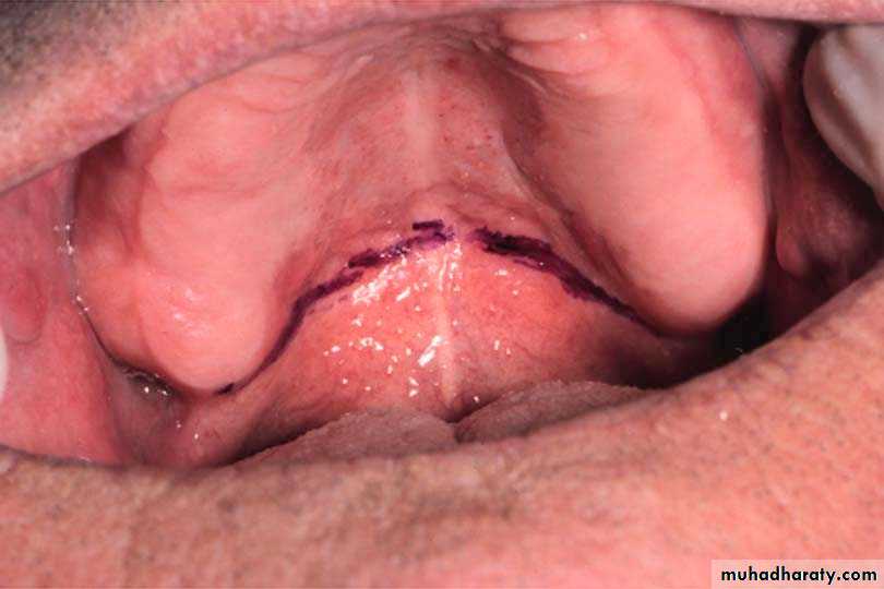
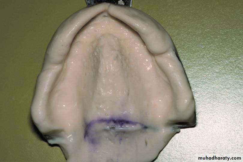
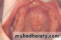
Vibrating LineThe vibrating line is an imaginary line drawn across the palate that marks the beginning of motion in the soft palate when an individual says “ah”.
It extends from one hamular notch to the other.
At the mid line it usually passes about 2 mm in front of the foveae palatine.
Fovea Palatine
The fovea palatine are indentations located on each side of the midline of the palate and slightly posterior to the junction of hard and soft palates formed by coalescence of several mucous gland ducts.
They are always in the soft tissue which makes them an ideal guide for the location of the posterior border of the denture.