Introducing cross-sectional imaging
Dr.Ahmed S. TawfeekFIBMS(Rad.)
Introducing cross sectional imaging
CAT principalCAT comparisons
MRI principle
MRI comparisons
MRI
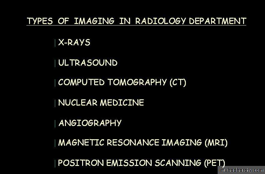
Computerized Axial Tomography (CAT)
Using CT scanners shows sections of the body resembling anatomical sections

Generally, images are obtained in the transverse plane
Computerized Axial Tomography (CAT)In this process a small beam of x-ray is passed through a plane of the body while the x-ray tube moves in an arc or a circle around the body
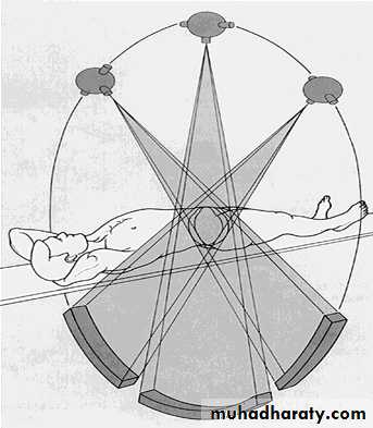
Computerized Axial Tomography (CAT)
The amount of radiation absorbed by different elements of the chosen plane variesComputerized Axial Tomography (CAT)
A computer stores a large amount of data from a selected region of the body, making it possible to determine the spatial relationship of the radiation-absorbing structures within it
Computerized Axial Tomography (CAT)
Important diagnostic information about tissues in the scanned regions of interest is thereby madeContrast enhancement may be used
Contrast enhancement of the bowel after oral administration of barium
Computerized Axial Tomography (CAT)Sections are visualized as if you were looking at cross sections from below with the right side to your left
right
leftliver
spleen
liver
Comparisons
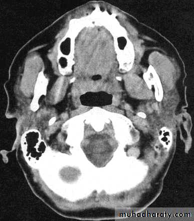
Cross sectionCAT image
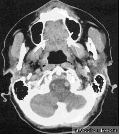
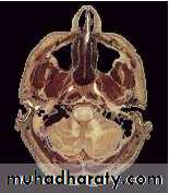
head
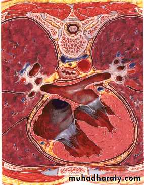
ComparisonsCross section
CAT image
thorax
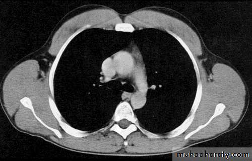
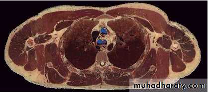
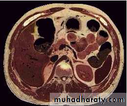
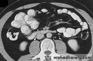
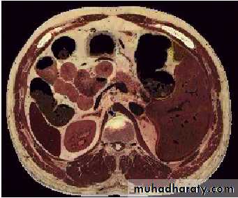
Comparisons
Cross sectionCAT image
abdomen
Cross-sectional imaging
Magnetic Resonance Imaging (MRI)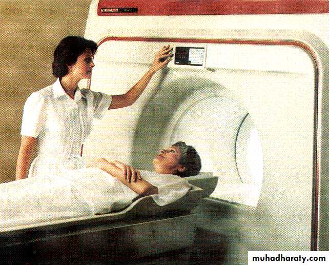
Magnetic Resonance Imaging (MRI)
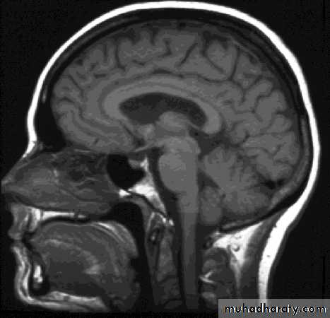
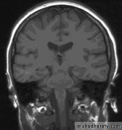
Uses non-ionizing radiation and has no demonstrated adverse biological effects.Magnetic resonance images can be obtained in any tissue plane
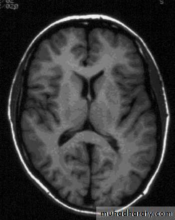
transverse
sagitttalcoronal


Magnetic Resonance Imaging (MRI)
The appearance of an MR image is a function of the chemical composition of the various types of tissue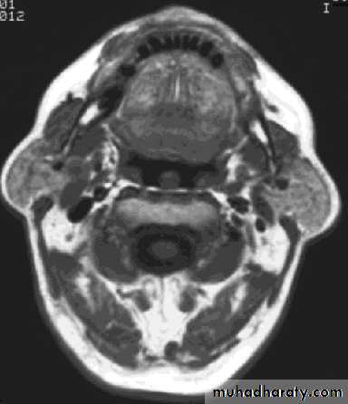
bone
fatmuscle
Magnetic Resonance Imaging (MRI)
At the atomic level, water and adipose are composed of hydrogen, oxygen, carbon, and phosphorus atoms. The hydrogen atom contains a proton and an orbiting electron.A spinning charged particle (the proton) produces a local magnetic field
Magnetic Resonance Imaging (MRI)
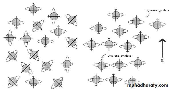
In the absence of any external forces, the magnetic moments of protons in tissue are oriented randomlyMagnetic Resonance Imaging (MRI)
If the protons are placed in a strong magnetic field, their magnetic dipoles align with and against the strong magnetMagnetic Resonance Imaging (MRI)
Slightly more than half of the magnetic moments align parallel to the field, because it takes less energy for the small magnetic moments to align with the stronger main magnetic fieldMagnetic Resonance Imaging (MRI)
This slight excess of protons in the lower energy state, whose individual magnetic moments add up, creates the net magnetization and causes the patient to acquire a slight magnetismMagnetic Resonance Imaging (MRI)
Magnetic resonance imaging (MRI) combines a strong magnetic field and radiofrequency (RF) energy to study the distribution and behaviour of hydrogen protons in fat and water
RF energy is used to generate a second magnetic field, perpendicular to the static magnetic field of the machine.
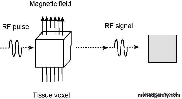
The result of this second magnetic field is to rotate or flip the protons away from the static magnetic
Once the RF field is switched off, the protons experience only the effects of the static magnetic field and flip back to their original position
During this return to equilibrium, a process which is called relaxation, protons emit the RF energy which they had acquired
This energy is detected by the antenna in the MRI machine, digitised, amplified, and, finally, spatially encoded by the array processor
The resulting images are displayed on the operators console and can be recorded
ComparisonsMRI image
CAT image
thorax
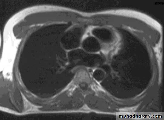
Compare bone and soft tissue density
ComparisonsMRI image
CAT image
abdomen
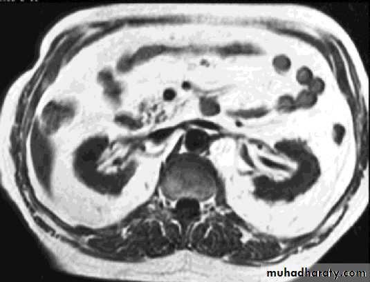
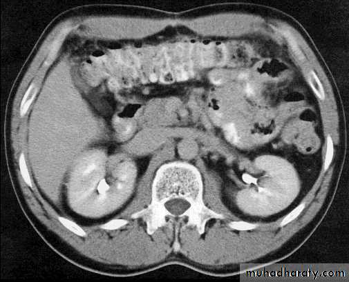
Compare bone and soft tissue density
ComparisonsMRI image
CAT image
head
Compare bone and soft tissue density