Oral Histology
Lec.3 د.سحرغانمEnamel structures:
Enamel forms a protective covering of variable thickness over the entire surface of the crown.
On the cusps of human molars and premolars the enamel attains a maximum thickness
of about 2 to 2.5 mm, thinning down to almost a knife edge at the neck of the tooth.
The enamel was found to be thicker in the lingual surfaces of maxillary molars and in the buccal surfaces of mandibular molars.
As these are supporting cusps, it is suggested that the increased thickness in these areas may be viewed as an adaptation to functional demands.
Because of its high content of mineral and their crystalline arrangement, enamel is the hardest calcified tissue in the human body.
The function of the enamel is to form a resistant covering of the teeth, rendering them suitable for mastication..
Enamel is an active chemical system that participates in a variety of reactions,
including solute and ion transport from saliva to dentin and back, ion-exchange reactions with saliva and demineralization-remineralization processes.
Chemical composition of Enamel:
Highly mineralized crystalline structure, 96% inorganic materials by weight; hydroxyapatite (HA), 4% by weight organic content and water.The organic matrix of enamel is made from non-collagenous proteins and enzymes.
Of the enamel proteins 90% are amelogenins and 10% are non-amelogenins.
The different types of non amelogenins associated with formation of enamel are meloblastin, enamelin and tuftelin. The primary function of the organic material is to direct the growth of enamel crystals .
The inorganic component of enamel is comprised almost entirely of HA crystals.
Enamel HA crystals are the largest HA crystals of all the calcified tissues in the body.
In addition to HA crystals enamel also contains carbonates and trace elements.
These crystals are susceptible to dissolution by acids and hence provides the basis for dental caries.
Physical Properties of E:
Very hard (like ceramics) hardest substance of human body.
Very brittle and low tensile strength (like ceramics), therefore enamel requires base of dentin to withstand forces (dentin acts as a support for enamel).
Enamel is translucent and varies in color from light yellow to whitish.
It varies in thickness, with maximum over cusps (2.5 mm) to a feather edge at the cervical line. Thickness of enamel in primary teeth is nearly half than that in permanent teeth
Although enamel is an extremely hard tissue it is partially permeable to some fluids, bacteria and other products of the oral cavity. The permeability of enamel is due to the presence of cracks and microscopic spaces on the surface of enamel which allows penetration of fluids.
Cracks are often seen in the enamel of teeth.
Unlike other calcified structures in the body enamel is unique as it is totally acellular .
Unsupported enamel is subject to easily fracture or cleave along rod boundaries (organic sheath). This is an important concept in dental preparations which has to do specifically with tooth microstructure.
Structure and Organization of E.:
Enamel is made up of 3 structures:
1-E. rods or prisms
2- E.rod sheaths3-Cementing inter-rod substance
Each Rod (Prism) is made up of millions of crystallites, and each rod is formed by four ameloblasts.
Rods run from DEJ to the external surface of the tooth.
Rods are formed nearly perpendicular to DEJ and curve slightly towards the cusp tip. The follow a wavy course as the traverse from the DEJ to the surface of the crown. The length of most rods is much longer than the thickness of enamel.
The diameter of the rod at the outer surface is double the diameter at DEJ. Crystals that surround each rod are called interrod enamel. Rod and inter-rod enamel is formed from the Tomes process of Ameloblasts.
The crystals making up the rod and interrod enamel have same composition but are oriented in different direction .
The boundary between rod and interrod enamel is marked by a narrow space filled with organic materials known as rod sheath.
In cross section, the E. rods have a rounded head or body and a tail (look like keyholes) forming a repetitive series of interlocking prisms; rounded head of each prism (rod) lies between the narrow tail portions of 2 adjacent prisms; usually the rounded head is oriented incisally or occlusally, and the tail cervically. Crystals are long hexagonal needle-like in shape.
Histological features of enamel:
Gnarled enamel:
Most enamel rods follow an undulating pathway from DEJ to the tooth surface. But in the cusps tips of molars groups of enamel rods twist about one another. This twisting pattern of enamel rod is known as Gnarled enamel. Gnarled enamel makes the enamel strong and more resistant to fracture.
Hunter-Schreger bands:
Hunter-Schreger bands are an optical phenomena and are seen in reflected light. They can be seen in ground longitudinal sections as alternating dark and light bands . The dark bands correspond to the cross sectional enamel rods and the light bands represent the longitudnally sectioned interrod enamel.Surface structures of E.
1-Perikymata:
They are transverse, wave like grooves, believed to be the external manifestations of the striae of retzius. They are continuous around a tooth & usually lie parallel to each other & to the cementoenamel junction.
Their course is usually fairly regular, but in the cervical region it may be quite irregular.
2-E. cuticle
Primary enamel cuticle covers the entire crown of the newly erupted tooth , has wavy course and it of no major clinical significance . Is secreted by the ameloblasts when enamel formation is completed. probably soon removed by mastication and its remnants called Nasmyth’s membrane .
3-E.Pellicle
Formed after the tooth erupted in the oral cavity, acquired from saliva and the oral flora. May contain factors which hinder the attachment of bacteria to tooth surfaces.
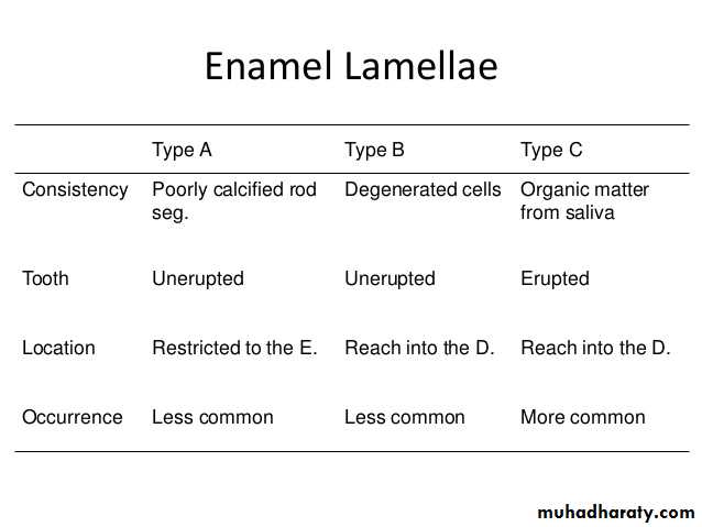
4-E. Lamllae
Thin leaf like structures that extend from the enamel surface toward the dentinoenamel junction and may sometimes extend to dentin. Consist of organic material, with but little mineral content. E. lamellae usually developed in planes of tension. E. lamellae type of hypomineralized structure in teeth that extend either from the dentinoenamel junction (DEJ) to the surface of the enamel.
E.Structures near DEJ:
1- Enamel spindles:Enamel spindles originate from odontoblastic process which cross the DEJ. Before enamel forms, some developing odontoblastic process extend into the ameloblast layer, and when enamel formation begins become trapped to form enamel spindles (which represent the only ectomesenchymal structure present in the E.
2- Enamel tufts:
Enamel tufts also originate from the DEJ, run a short distance in the enamel or sometimes to one half of the E. thickness. They represent protein (enamelin) rich areas in the enamel matrix that fail to mature. They are formed during the formative stages of enamel.
Incremental lines of E:
1-Cross striations:
Cross striations are periodic bands that appear along the full length of enamel rod . Because of this the enamel rod appears like a ladder with cross striations being the rungs of the ladder. They appear at regular intervals that is in agreement with the rate of enamel deposition (which is approximately 4 μ m per day).
2-Striae of Retzuis:
Striae of Retzuis also represent incremental growth. In ground cross sections they appear like concentric growth rings similar to those found in trees. In ground longitudinal sections they appear to be dark line extending from the DEJ to the tooth surface . Along the Retzuis striae fewer enamel crystals are found and this is related to physiologic disturbances in the body. Striae of Retzuis often extend from the DEJ to the outer surface of the enamel, where they end in shallow furrows know as perikymata (or imbrication lines).3- Neonatal line :
Neonatal line is a Striae of Retzuis that forms at birth, because it reflects the great physiologic changes occur at birth. So these lines demarcating the boundary between E. formed before and after birth.
Age changes in enamel:
1- With age enamel becomes worn out because of masticatory attrition.
2- Age also causes a decrease in the permeability of enamel.
3- Other characteristics of aging of enamel are discoloration and a change in the surface layer