• ENAMEL
Dr asmaa s

• PHYSICAL CHARACTERISTICS
• Hard, brittle, totally acellular , highly mineralized• Secretory product of stratified squamous epithelium
• Calcified tissue
• Hydroxyapatite crystal arrange in prism or rods Density:-
• Decreases from the surface of enamel to the dentino-enamel junction.
• Thickness:-
• Thickness over the cusps of the molars where it measures 2.5 mm & incisal edges of incisors where it is
• 2.0 mm.

• ENAMEL
• Forms a protective covering (2 mm – knife edge).• Forms a resistant covering (suitable for mastication).
• The hardest calcified tissue in human body.
• enamel is very brittle but the underlying dentin provides some resilience
• Acts as semipermeable membrane (selectively permeable).
• Color: yellowish white to grayish white depends on translucency.
• Enamel gains mechanical strength by interweaving HAP crystals
• Enamel rod – 5-12 million/tooth• Appatite crystal is hexagonal
• Enamel initially starts with a high protein content, but these are removed and the voids backfilled with HAP as the tooth matures


• CHEMICAL PROPERTIES
• 96% inorganic - by weight• inorganic crystalline calcium phosphate – hydroxyapatite
• various ions like strontium, magnesium, lead and fluoride are present at some point during enamel formation

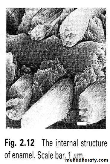
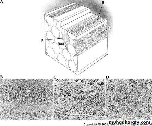
• ENAMEL STRUCTURE
• 1)• Enamel (rods(prisms)
• Rod sheaths
• Inter-rod substance.

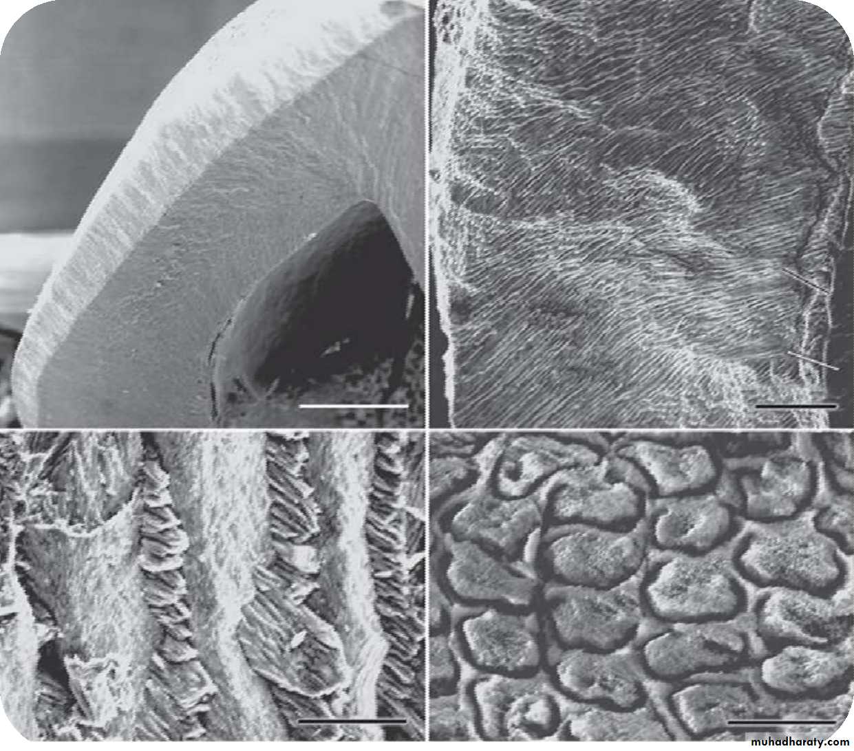

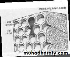
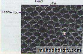
• CROSS SECTION
• Cross section of enamel rod shows the key hole pattern• Head represents the rod and key shows the inter rod region
• Head is directed towards the occlusal aspect and tail towards the cervical region of the tooth

• CROSS SECTION OF ENAMEL

• ENAMEL


• CHARACTERISTICS - ENAMEL ROD/PRISM
• Number: 5 – 12 millions.• Direction: Run in oblique direction and wavy course.
• Length: greater than the thickness.
• Diameter average: 4 µm.
• Appearance: Have a clear crystalline appearance.
• Cross-section: hexagonal, round, oval, or fish scales.
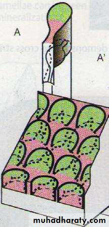
• Enamel Rod: Basic Structural Unit
• Cross section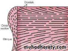
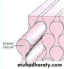
• Head of enamel rod is formed by one ameloblast and tail is formed by three ameloblasts
• Thus each rod is formed by four ameloblasts


• SUBMICROSCOPIC STRUCTURE OF ENAMEL RODS
• Keyhole or paddle-shaped.• Separated by interrod substance.
• About 5 µm in breadth and 9 µm in length.
• The bodies are near the occlusal or incisal surface.
• The tails point cervically.
• The crystals; parallel to the long axis of the prism heads.
• Deviate about 65° from the tails.

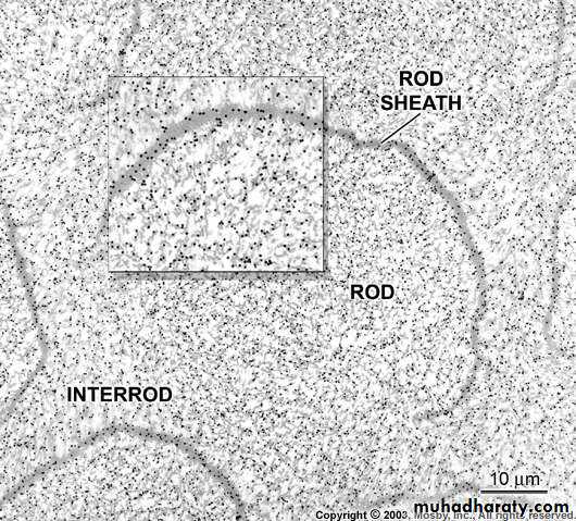
• ROD SHEATH
• the boundary between rod and interrod is delimited by a narrow space containing organic material – rod sheath• A thin peripheral layer.
• Darker than the rod.
• Relatively acid-resistant.
• Less calcified and contains more organic matter than the rod itself.
• Electron Microscope : often incomplete.

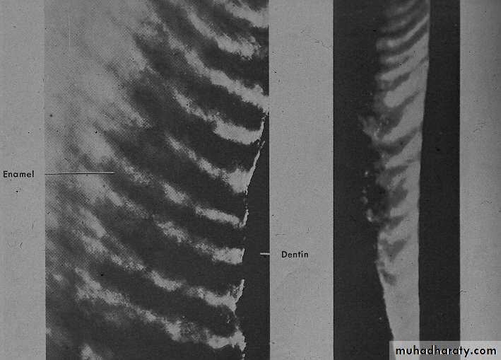
• ALTERNATING ROD DIRECTIONALITY
• Hunter Schreger bands are alternating light and dark bands seen in a section of enamel when cut longitudinally and illuminated in a special way.• The bands are produced by the orientation of groups of rods.
• If the light passes through rods cut in cross-section, the band appears light.
• If the light passes through rods cut in longitudinally, the band appears dark.

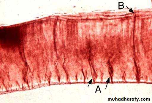
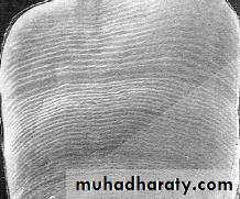
• STRIATIONS
• E. rod is built-up of segments (dark lines).• Best seen in insufficient calcified E.
• In a longitudinal section dark lines are seen that shows the daily deposition of enamel (rhythmic manner of E. matrix formation).
• These lines are known as cross striation
• Segment length: about 4 µm.


• CROSS-STRIATIONS
• Cross striations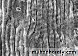

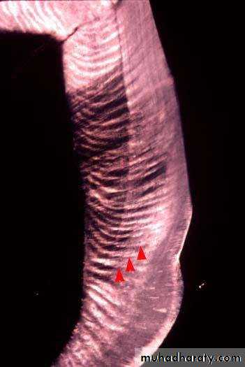
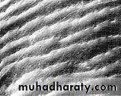
• DIRECTION OF RODS
• Near the edge or cusp tip they are oblique• At the cusp tip they are almost vertical
• Run from DEJ to surface of enamel
• Usually at right angles to the Dentin surface.
• Follow a wavy course in clockwise and anticlockwise deviation full thickness of enamel
• At the cusps or incisal edges: gnarled enamel.
• At pits and fissures: rods converge in their outward course.


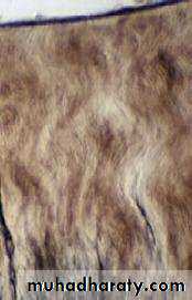
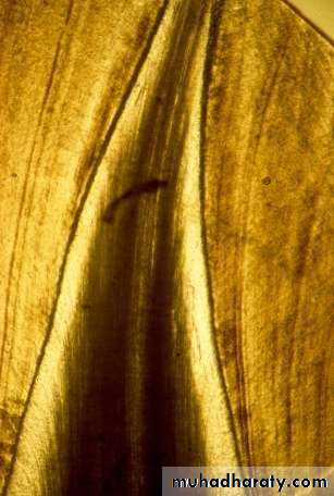
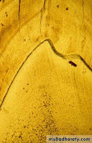
• STRAIGHT ENAMEL RODS -LONGITUDINAL LABIOLINGUALSECTION
• The enamel rods project in the direction of the arrow.• Can you see the striaof Retzius?
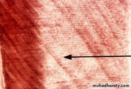
• Wavy course of enamel rod
• A more spiral course is noted at cusps & incisal areas Gnarled enamel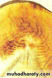

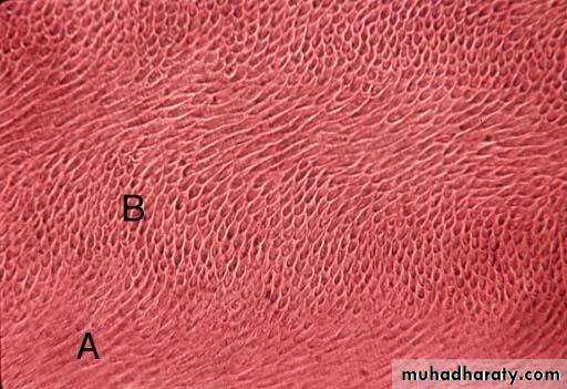
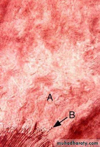
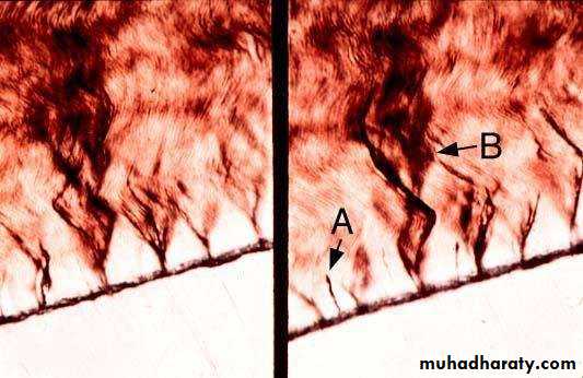
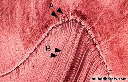
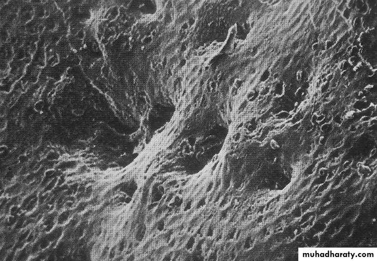
• GNARLED ENAMEL
• Enamel rods are general not straight throughout their length.• In the cuspal region, the rods
• are very wavy.
• This is referred to as gnarled enamel.
• In this section, you can see the end of an odontoblasticprocess penetrating the enamel just past the DEJ.
• This structure is called an
• enamel spindle.
• Legend
• Legend: A, Gnarled enamel; B, Enamel spindle
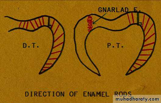

• DIRECTION OF ENAMEL RODS


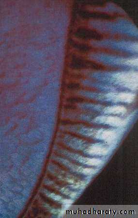
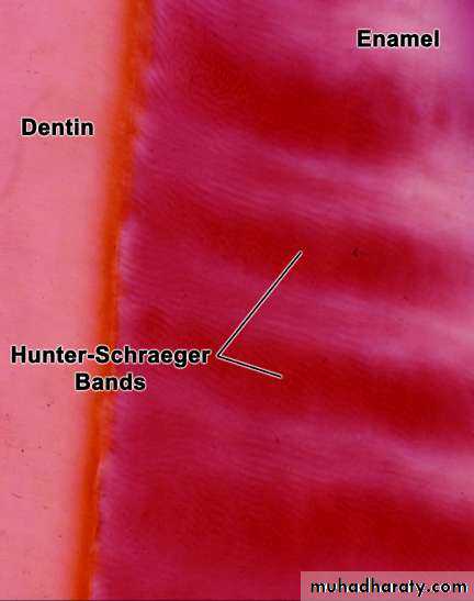
• HUNTERSCHRAGER BANDS
• Optical phenomenon seen in reflected light• Alternate light and dark bands
• Seen in ground longitudinal section
• Due to abrupt change in the direction of enamel rod
• Originate from the DEJ.


• HUNTER-SCHREGER BANDS
• HUNTER-SCHREGER BANDS


• ENAMEL -TRANSVERSE GROUND SECTION
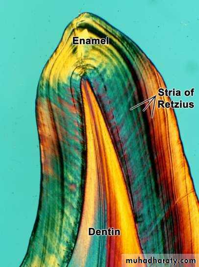
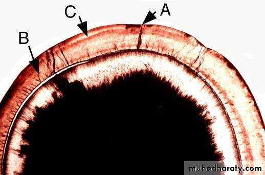
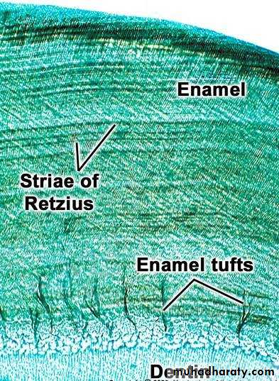
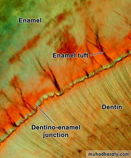
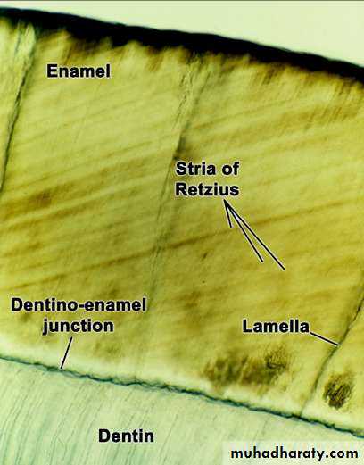
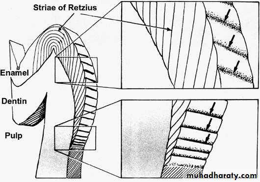
• In a transverse section of tooth, the stria of Retzius appear as concentric bands parallel to the dentino-enamel junction (DEJ). In addition to the "hypo-mineralized" dark striaof Retzius, there also exist hypo-mineralized areas perpendicular to the DEJ. These are enamel lamellae(that traverse the entire thickness of enamel) and enamel tufts(that traverse the inner third of enamel adjacent to the DEJ• sectionLegend: A, Striaof Retzius; B, Enamel tuft; C, Enamel lamella; D, DEJ
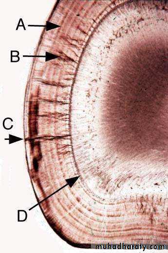


• STRAE OF RETZIUS
• Incremental lines of growth• Eccentric growth rings
• DEJ to outer surface of enamel
• Where they end as shallow furrows known as perikymata


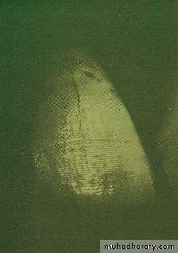
• INCREMENTAL LINES OF RETZIUS:


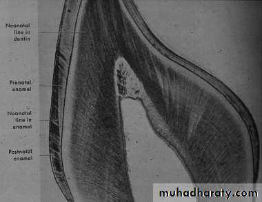
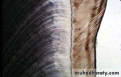
• NEONATAL LINE
• The E. of the deciduous teeth and the 1st permanent molar (It is incremental line that is the boundary between the enamel forms before and after the birth)• The neonatal line is usually the darkest and thickest striaof Retzius.
• Etiology
• Due to sudden change in the environment and nutrition.
• The antenatal E. is better calcified than the postnatal E.


• NEONATAL LINE

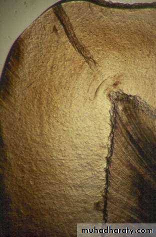
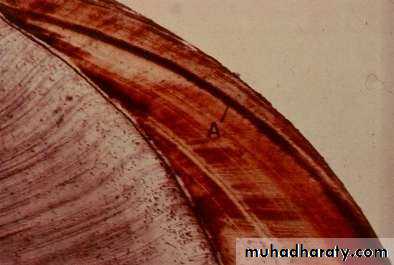
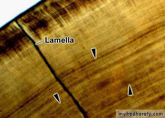
• ENAMEL LAMELLAE
• Are thin, leaf like structures,• Develop in planes of tension.
• Extends from E. surface towards the DEJ.
• Confused with cracks caused by grinding (decalcification).
• Extend in longitudinal and radial direction.
• Represent site of weakness in the tooth and three types; A, B, and C.

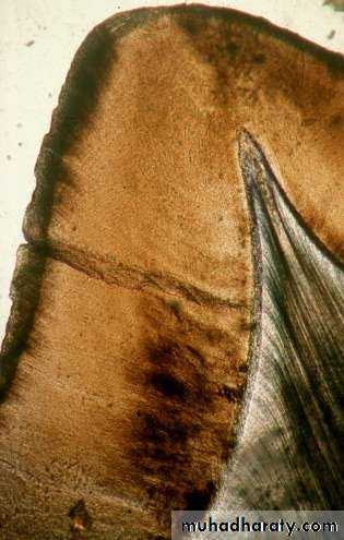
• ENAMEL LAMELLAE
• In this ground cross- section of tooth, you can see enamel lamellae and enamel tufts You can also see the neonatal line.• •What do all three of these structures have in common?
• Answer: They are all hypocalcified.
• Legend: A, Enamel lamella; B, Enamel tuft; C, Neonatal line

• ENAMEL LAMELLAE
• ENAMEL LAMELLAE
• Enamel tufts are less mineralized areas of enamel in the inner third of enamel adjacent to the DEJ. They resemble tufts of grass.
• •They are wavy due to the waviness of the adjacent rods.
• •Structures rich in organic matter (i.e. less mineralized) that project to the surface of the enamel are enamel lamellae.
• Legend: A, Enamel tufts; B, Enamel lamella


• ENAMEL TUFTS -TWO PLANES OF FOCUS
• Enamel tufts consist of several unconnected "leaves" of hypo- calcified enamel.• •They display a wavy twisted appearance.
• •Enamel spindles are the processes of odontoblastsprojecting into the enamel.
• Legend: A, Enamel spindle; B, Enamel tuft

• ENAMEL TUFTS
• ENAMEL TUFTS


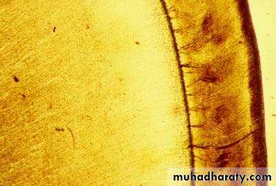
• DENTINO-ENAMEL JUNCTION
• Scalloped junction – the convexities towards D.• At this junction, the pitted D. surface fit rounded projections of the enamel.
• The outline of the junction is performed by the arrangement of the ameloblasts and the
• B. M.
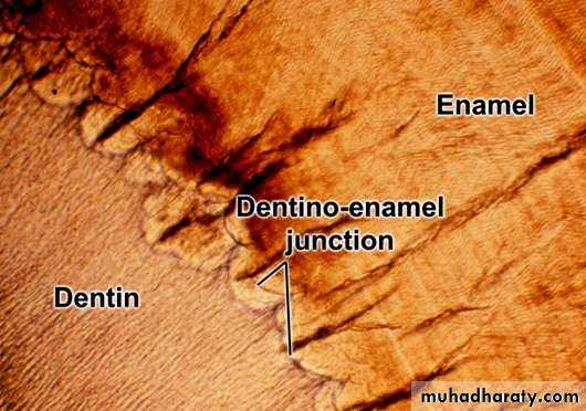
• DENTINO-ENAMEL JUNCTION

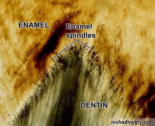
• ENAMEL SPINDLES
• Odontoblast processes usually end at the DEJ. However, sometimes the ends of the process become embedded in the enamel as it forms.• These very small, usually straight structures that you can see adjacent to the DEJ are enamel spindles.
• They are only about one tenth the length of an enamel tuft. Legend: A, Enamel
• spindle;
• B:Odontoblastprocesses in dentin
• Legend: A, Enamel spindle; B, Odontoblastprocess; C,
Enamel rod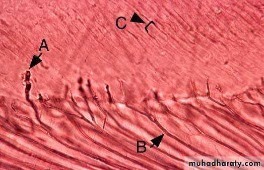


• ODONTOBLASTIC PROCESSES AND ENAMEL SPINDLES


• THE RELATIONSHIP BETWEEN THE STRIAE OF RETZIUZ AND SURFACE PERIKYMATA
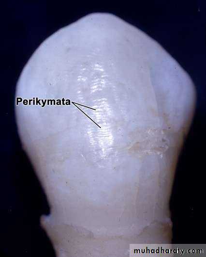


• PERIKYMATA (IMBRICATION LINES) ARE EXTERNAL MANIFESTATIONS OF RETZIUS STRIAE

• C. ROD ENDS
• Are concave and vary in depth and shape.• Are shallow in the cervical regions.
• Deep near the incisal or occlusal edges.

• ROD ENDS

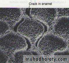
• D. CRACKS
• Narrow fissure like structure.• Seen on almost all surfaces.
• They are the outer edges of lamellae.
• Extend for varying distance along the surface.
• At right angles to CEJ.
• Long cracks are thicker than the short one.
• May reach the occlusal or incisal edge.

• CRACKS


• LIFE CYCLES OF THE AMELOBLASTS
• According to their function, can be divided into six stages:• Morphogenic stage.
• Organizing stage.
• Formative stage.
• Maturative stage.
• Protective stage.
• Desmolytic stage.


• Morphogenic stage.
• React by differential growth• Produce shape of the crown
• Terminal bar appears
• Basal lamina separates the inner enamel epithelium and cells of the dental papilla
• Pulpal layer adjacent to the basal lamina is a cell free zone
• At cervical region – cell is relatively undifferentiated
• Organizing stage.
• Inner enamel epithelium interact with the cells of dental papilla which differentiate into odontoblast• Cells become elongated
• Proximal part contain nuclei
• Distal end is nucleus free zone
• Dentin formation begins
• Cell free zone disappear
• As dentine is formed nutrition supply of the inner enamel epithelium changes from dental papilla to the capillaries that surround the outer enamel epithelium
• Reduction and gradual disappearance of the stellate reticulum
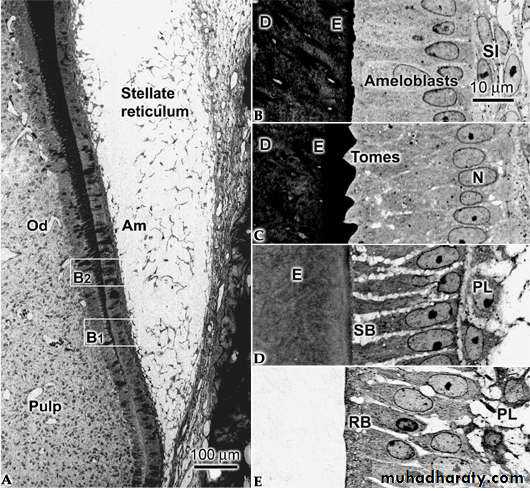
• Formative stage.
• Formatve stage starts After the dentine formation• Enamel matrix formation starts
• Development of blunt cell process on the ameloblast surface which penetrate the basal lamina and enter the predentin
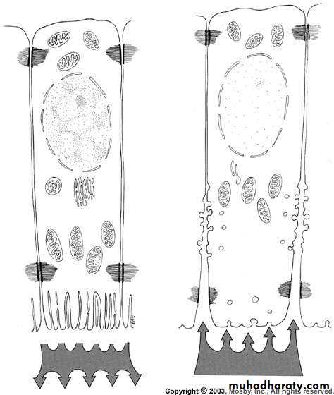
• Maturative stage.
• Maturation starts after most thickness of enamel matrix formation in occlusal and incisal area. In cervical area matrix formation is still in progress• Ameloblast reduce in length
• Cells of stratum intermedium takes spindle shape
• Protective stage.
• After enamel calcification cells on ameloblast can no longer be differentiated from stratum intermedium and outer enamel epithelium• These layer forms reduced enamel epithelium
• Protect the enamel from connective tissue until the tooth erupts, if it contacts then anomalies develop enamel may be resorbed or cementum cover may form (afibrillar cementum)
• Desmolytic stage.
• Reduced enamel epithelium induces atrophy of connective tissue separating it with oral epithelium thus fusion of the two epithelia can occur• Premature degeneration of the reduced enamel epithelium may prevent the eruption of he tooth

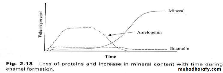
• AMELOGENESIS
• Organic matrix formation (follows incremental pattern – brown striae of Retzius).• Mineralization.
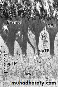
• dpTP=distal portion of Tome’s process
• ppTP=proximal portion of Tome’s process• Sg=secretory granules(E. protein)

• ORGANIC MATRIX FORMATION

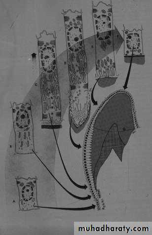
• AMELOGENESIS
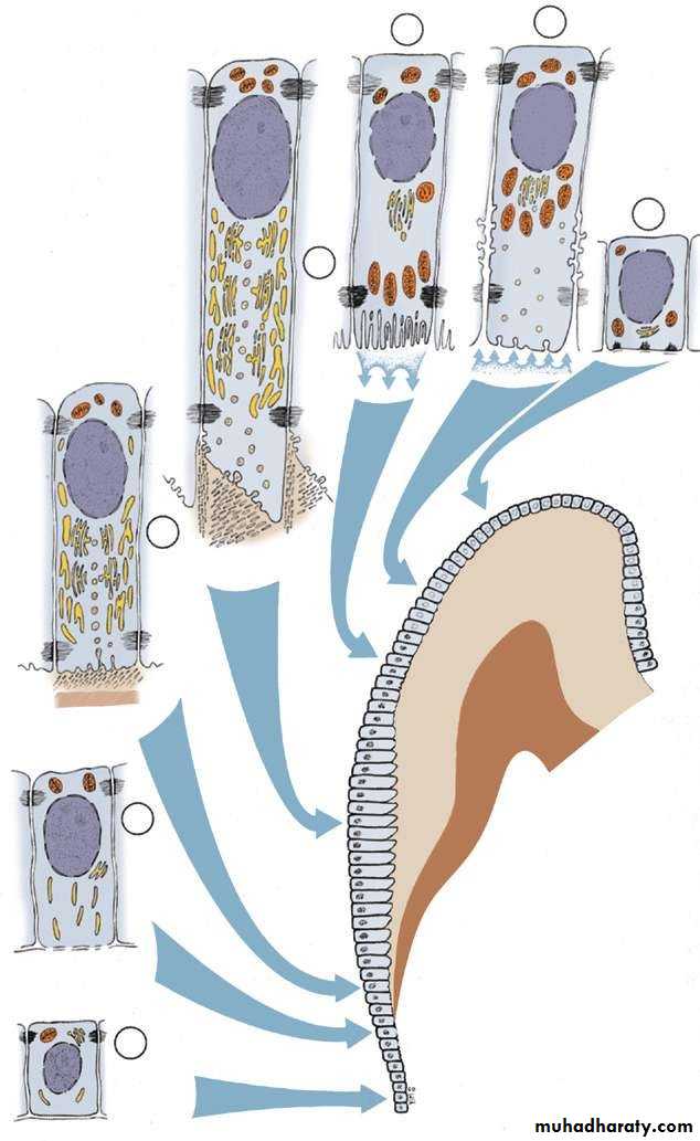
• Schematic representation of the various functional stages in the life cycle of ameloblasts as would occur in a human tooth.
• Morphogenetic stage;
• histodifferentiation stage;
• initial secretory stage (no Tomes’ process);
• secretory stage (Tomes’ process);
• ruffle-ended ameloblast of the
• maturative stage;
• 6. smooth-ended ameloblast of the maturative stage;
• 7.protective stage.
• AMELOGENESIS

• RUFFLED AND SMOOTH AMELOBLASTS