Anatomy of Thoraxد.رندعبداللطيف
The thoracic wall is formed posteriorly by the vertebral column; anteriorly by the sternum and costal cartilages; laterally by the ribs and intercostal spaces; superiorly by the suprapleural membrane; and inferiorly by the diaphragm, which separates the thoracic cavity from the abdominal cavity.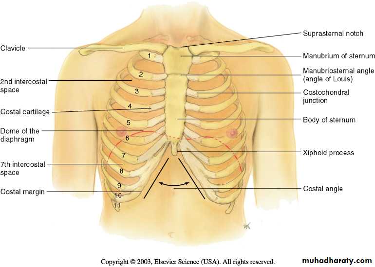
Sternum
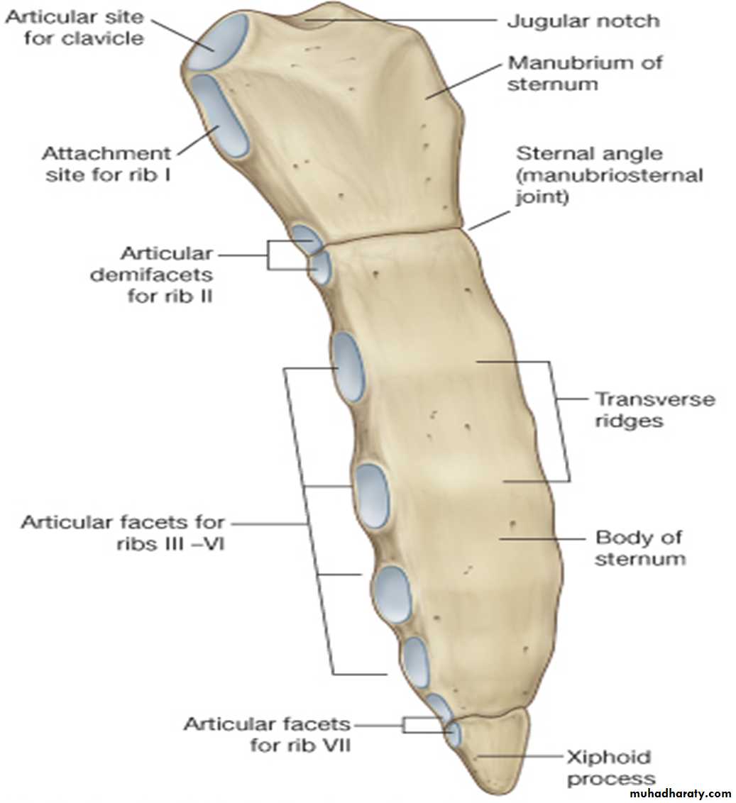
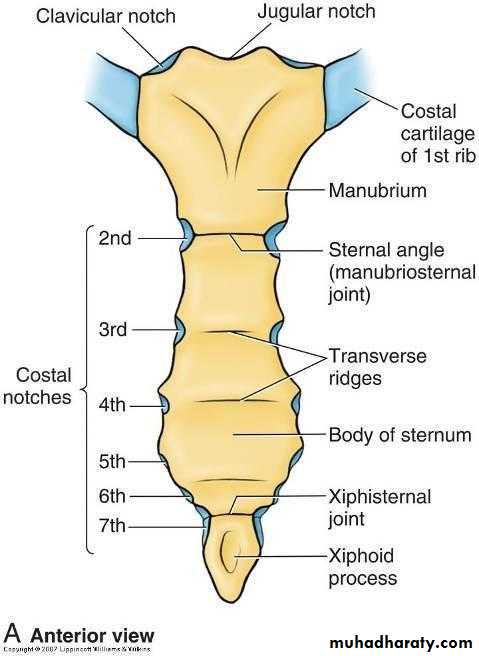
The sternum lies in the midline of the anterior chest wall. It is a flat bone that can be divided into three parts:Manubrium sterni.
Body of the sternum.
Xiphoid process.
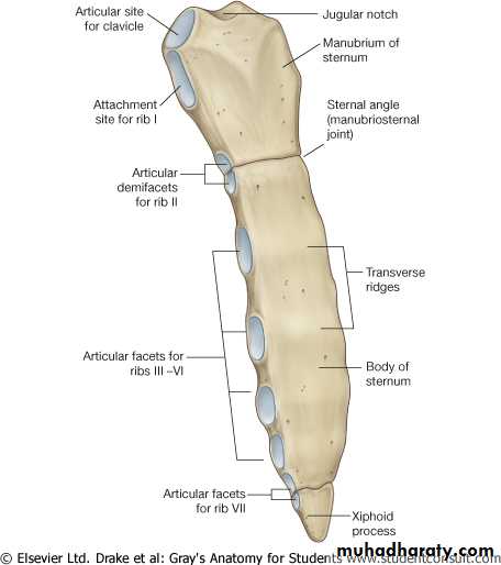
The manubrium is the upper part of the sternum. It articulates with the body of the sternum at the manubriosternal joint, and it also articulates with the clavicles and with the1st costal cartilage and the upper part of the 2nd costal cartilages on each side. It lies opposite T3&T4
.
The body of the sternum articulates above with the manubrium at the manubriosternal joint and below with the xiphoid process at the xiphisternal joint.
it articulates with the 2nd to the 7th costal cartilages
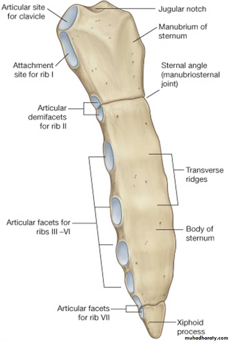
The xiphoid process is a thin plate of cartilage that becomes ossified at its proximal end during adult life. No ribs or costal cartilages are attached to it.
The sternal angle (angle of Louis)
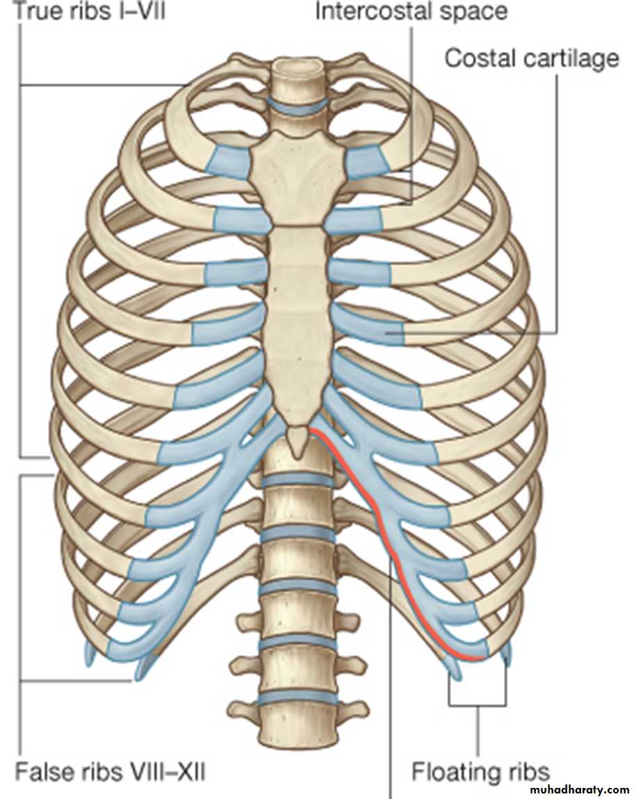
Ribs
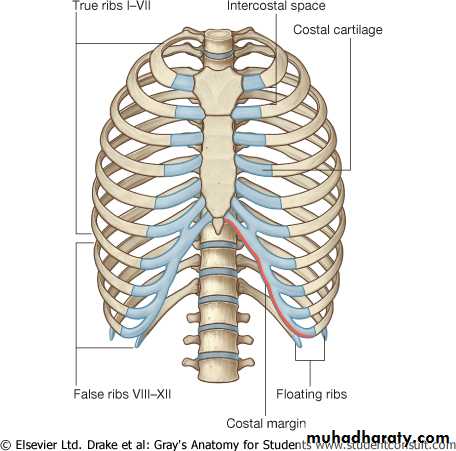
There are 12 pairs of ribs, all of which are attached posteriorly to the thoracic vertebrae. The ribs are divided into three categories:True ribs: The upper seven pairs are attached anteriorly to the sternum by their costal cartilages.
False ribs: The 8th, 9th, and 10th pairs of ribs are attached anteriorly to each other and to the 7th rib by means of their costal cartilages and small synovial joints.
Floating ribs: The 11th and 12th pairs have no anterior attachment.
Typical Rib
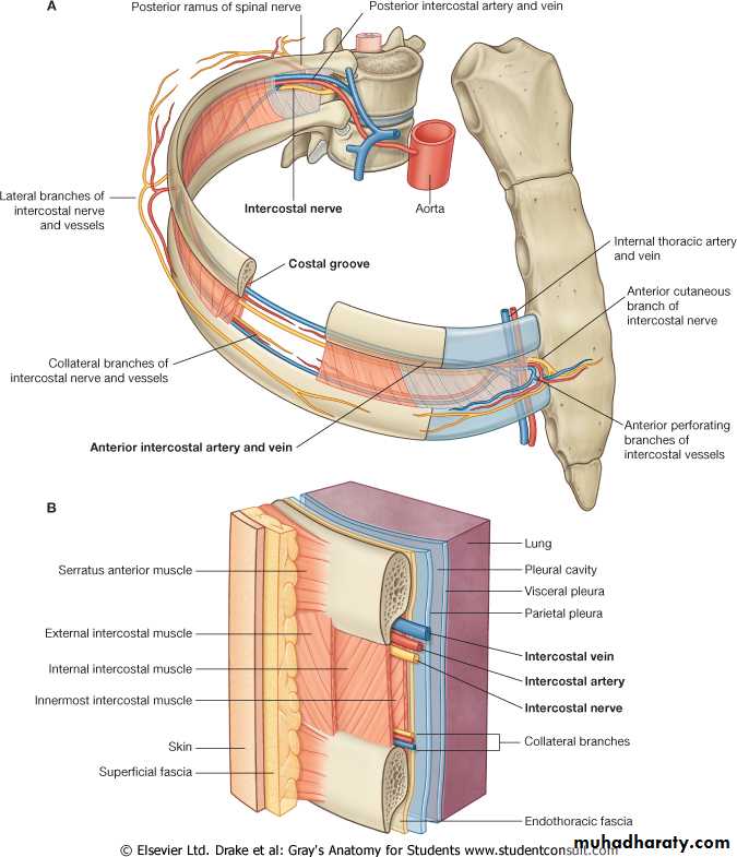
A typical rib is a long, twisted, flat bone having a rounded, smooth superior border and a sharp, thin inferior border. The inferior border overhangs and forms the costal groove, which accommodates the intercostal vessels and nerve.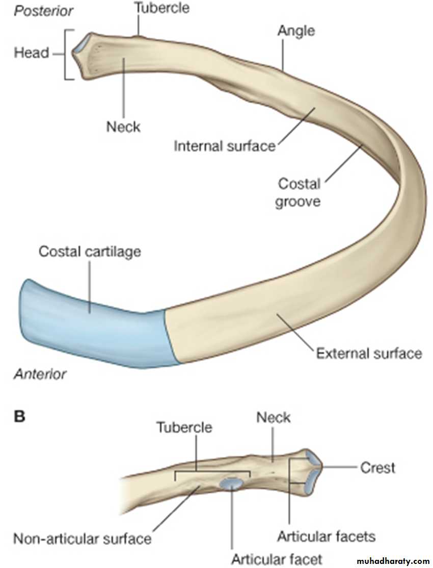
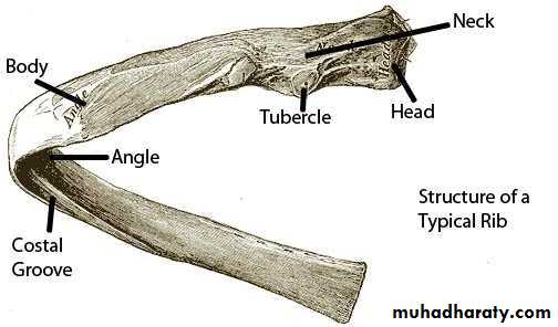
A rib has a head, neck, tubercle, shaft, and angle. The head has two facets for articulation with the numerically corresponding vertebral body and that of the vertebra above.
The neck is a constricted portion situated between the head and the tubercle. The tubercle is a prominence on the outer surface of the rib at the junction of the neck with the shaft. It has a facet for articulation with the transverse process of the corresponding vertebra
The shaft is thin and flattened and twisted .Its inferior border has the costal groove. The angle is where the shaft of the rib bends sharply forward
Atypical ribs:
The first rib:- Short, acutely curved, has superior & inferior surfaces .
The head has single facet for T1
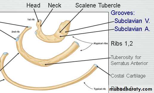
The superior surface is rough & characterized by the scalene tubercle for insertion of Scalenus anterior muscle which separates the area of subclavian vein in front from that of subclavian artery behind
- No costal groove
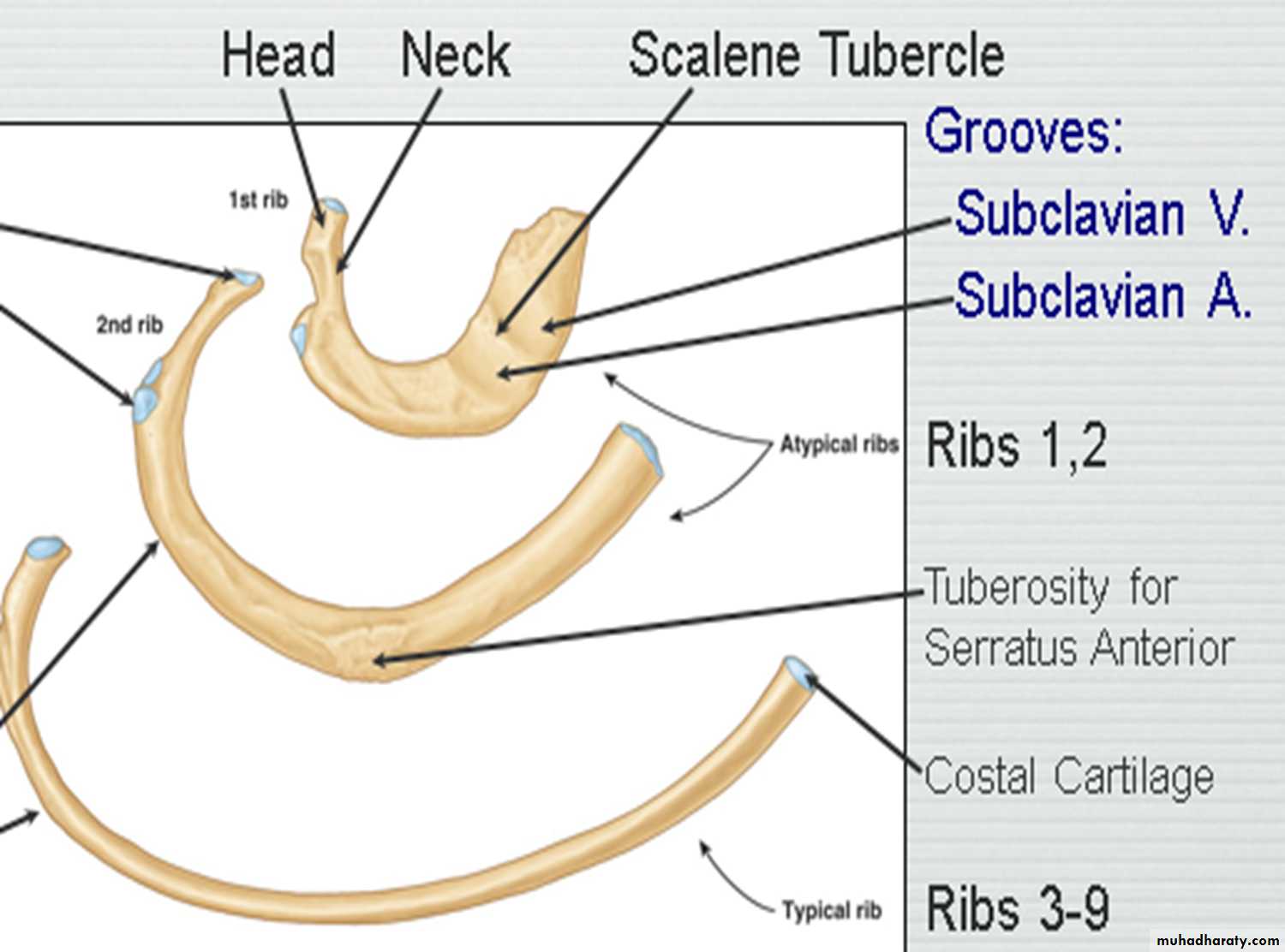
The second rib:
- Is midway in position between the 1st & typical ribsThe 10 rib:.
The 11 rib:
The 12 rib
all are atypical ribs
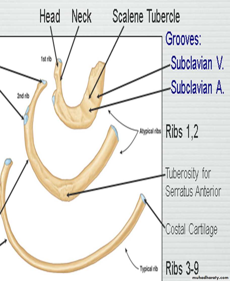
General Characteristics of a Vertebra
The typical vertebra consists of a rounded body anteriorly and a vertebral arch posteriorly. These enclose a space called the vertebral foramen. The vertebral arch gives rise to seven processes: one spinous, two transverse, and four articular processes are vertically arranged and consist of two superior and two inferior processes.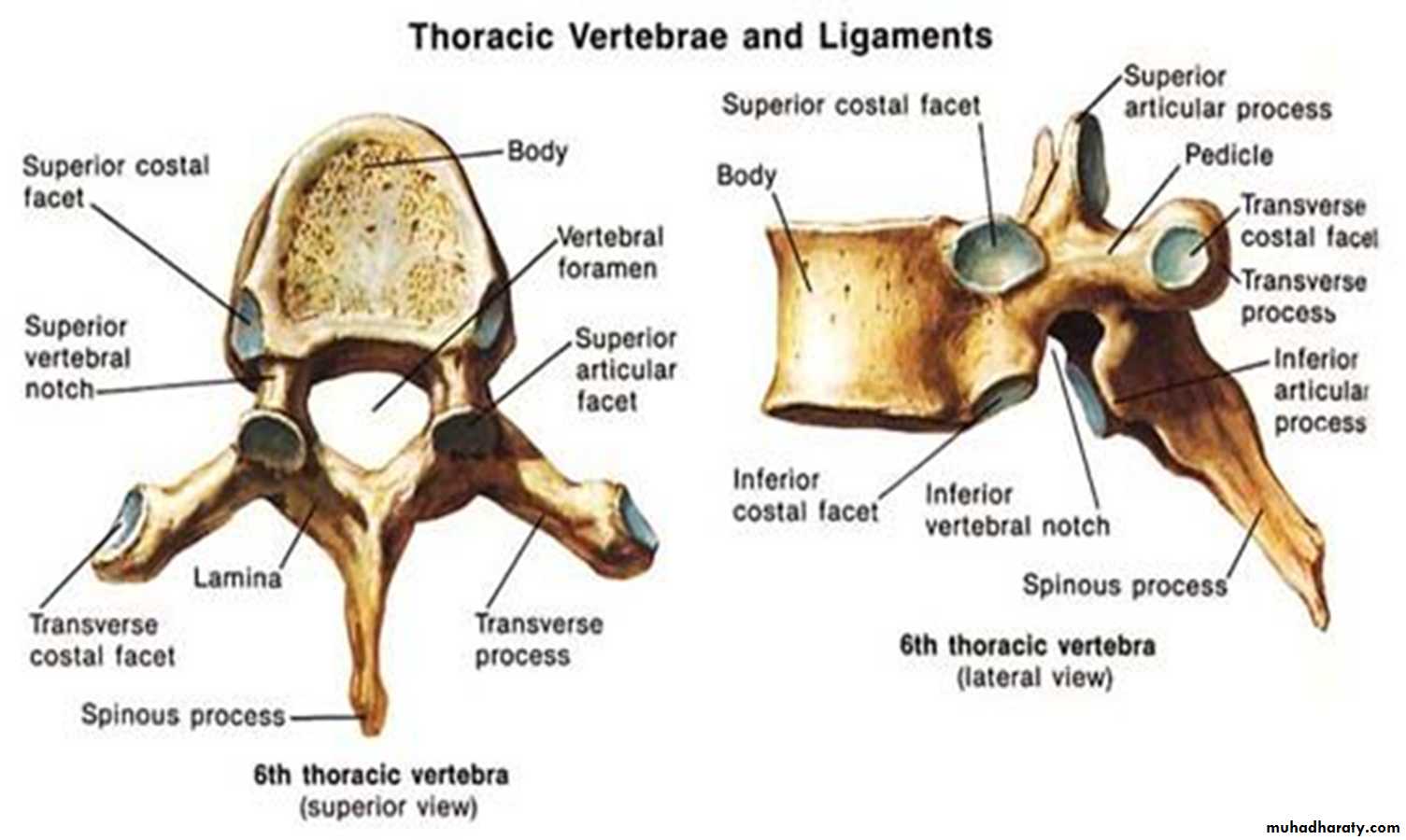
The spinous process, or spine, is directed posteriorly.
The thoracic spines are long and inclined downward. The body is medium size and heart shaped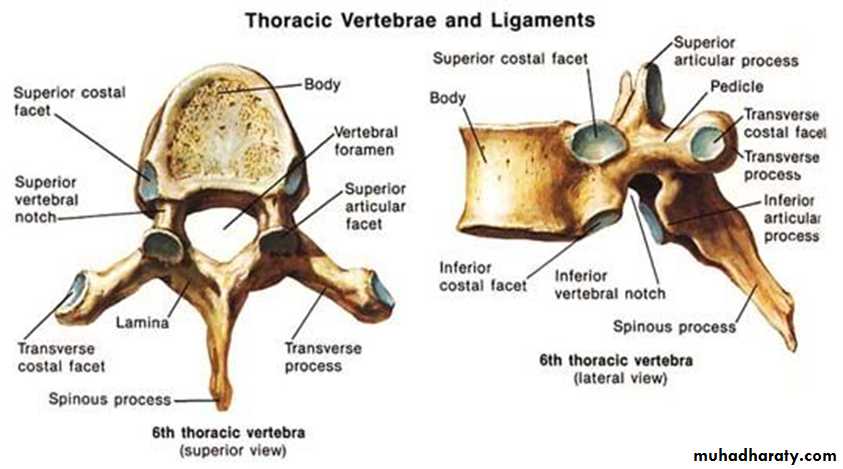
Openings of the Thorax
Thoracic outletthe outlet is closed by a dense fascial layer called the
suprapleural membrane
The opening is attached at its apex to the tip of the transverse process of the \C7, laterally by the medial borders of the 1st ribs and their costal cartilages, and anteriorly by the superior border of the manubrium sterni
The thoracic cavity communicates with the abdomen through a large opening which is bounded posteriorly by the T12, laterally by the curving costal margin, and anteriorly by the xiphisternal joint.
Intercostal Spaces
The spaces between the ribs contain three muscles of respiration:The external intercostal m.
The internal intercostal m.
The innermost intercostal m
lined internally by endothoracic fascia
The external intercostal muscle
It forms the most superficial layer.Its fibers are directed downward and forward from the inferior border of the rib above to the superior border of the rib below.
The muscle extends forward to the costal cartilage where it is replaced by an aponeurosis, which is named as the anterior (external) intercostal membrane
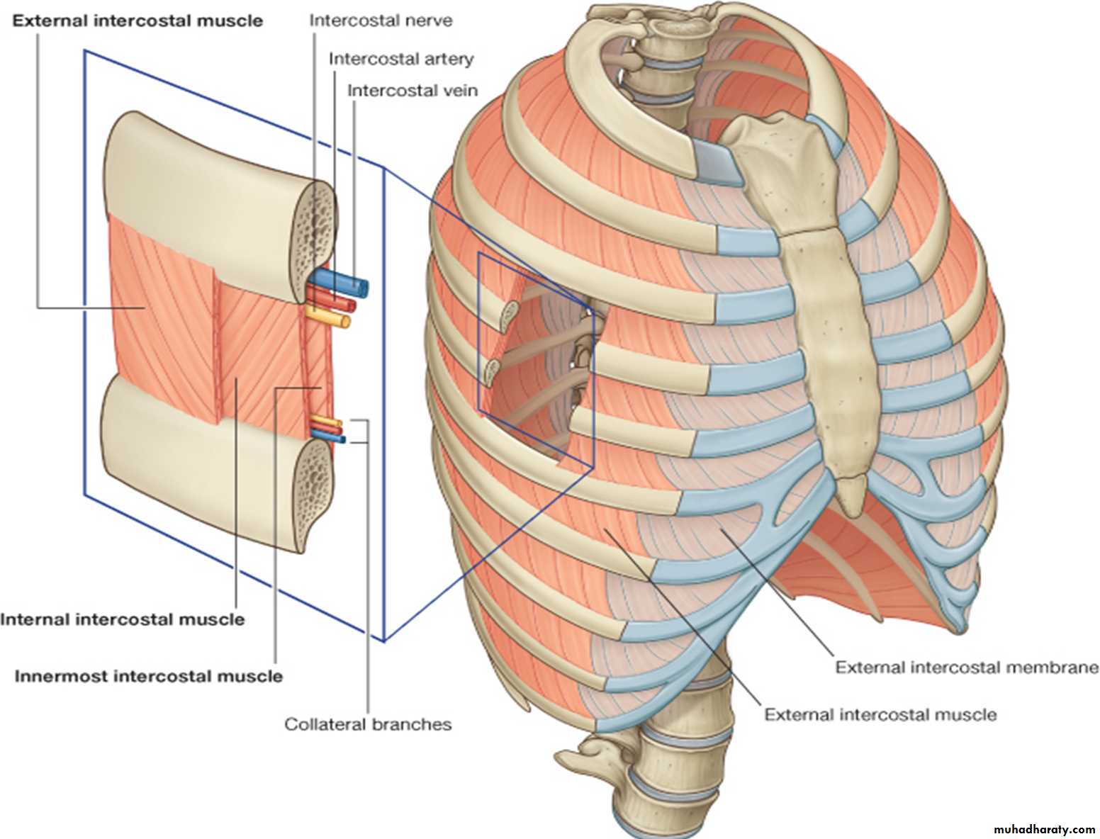
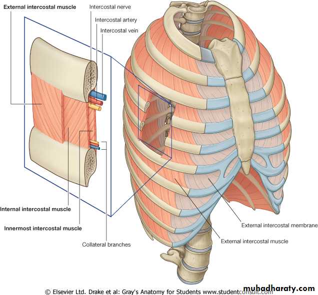
The internal intercostal muscle
Its fibers are directed downward and backward from the subcostal groove of the rib above to the upper border of the rib below.The muscle extends backward from the sternum in front to the angles of the ribs behind, where the muscle is replaced by an aponeurosis, which is named as the posterior (internal) intercostal membrane
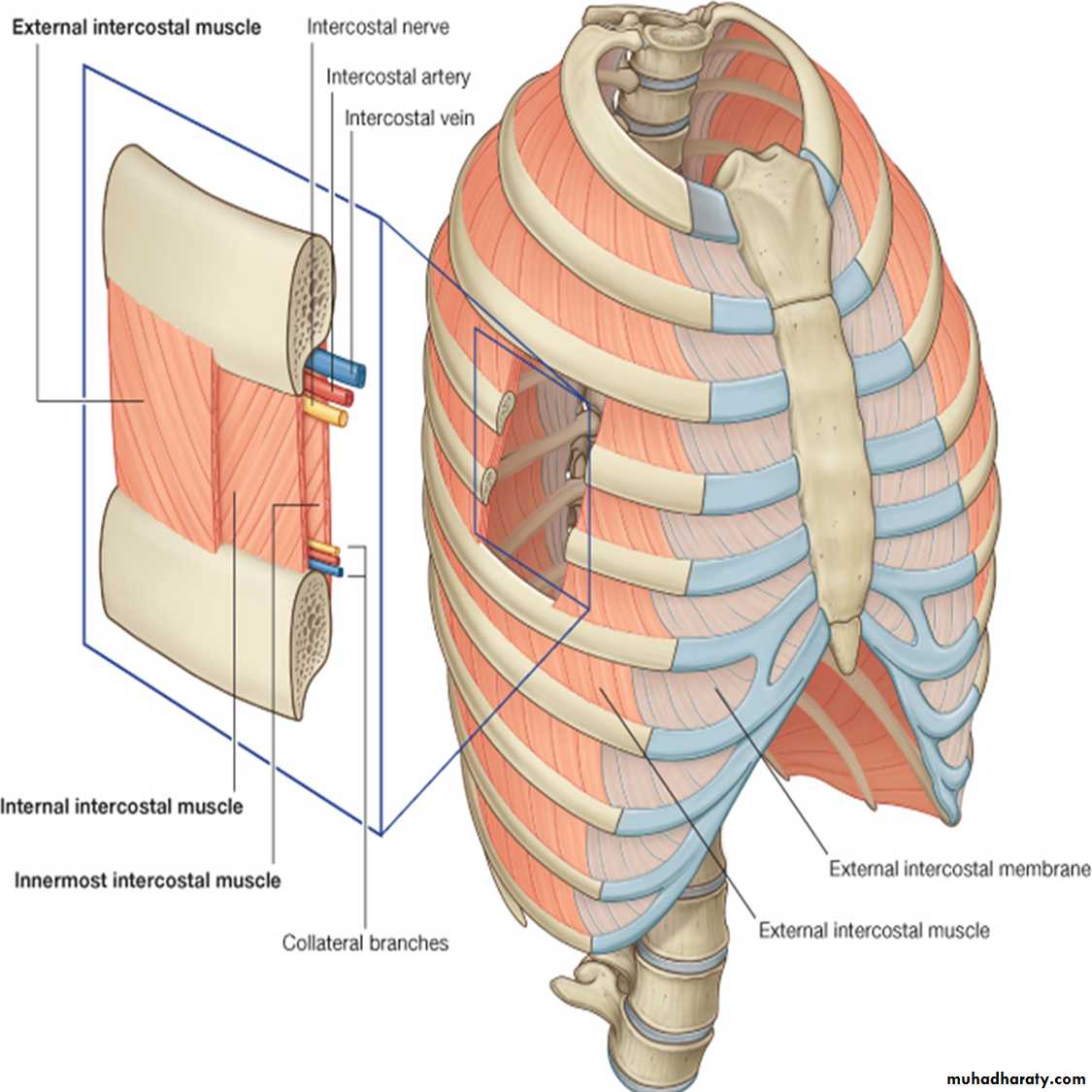
The innermost intercostal muscle
It forms the deepest layer. It is an incomplete muscle layer and crosses more than one intercostal space within the ribs.It is related internally to endothoracic fascia and parietal pleura and externally to the intercostal nerves and vessels. The innermost intercostal muscle can be divided into three portions, which are more or less separate from one another.
Nerve Supply of I.C.M
The intercostal nerves• Collateral branches;. passes below & parallel to the main nerve contain most of the motor fibers
• Lateral cutaneous branches; arise at the midaxillary line from the remaining part of the nerve, each divides into anterior & posterior branches
Anterior cutaneous branches
pierce the muscles lateral to the sternum & ends into lateral (large) & medial (small) branches
Intercostal Arteries
Each intercostal space contains a large single posterior intercostal artery and two small anterior intercostal arteriesThe anterior intercostal arteries of the first six spaces are branches of the internal thoracic artery, which arises from the first part of the subclavian artery. The anterior intercostal arteries of the lower spaces are branches of the musculophrenic artery, one of the terminal branches of the internal thoracic artery.
Those of the 2nd, 3rd & 4th spaces supply the mammary gland in female.
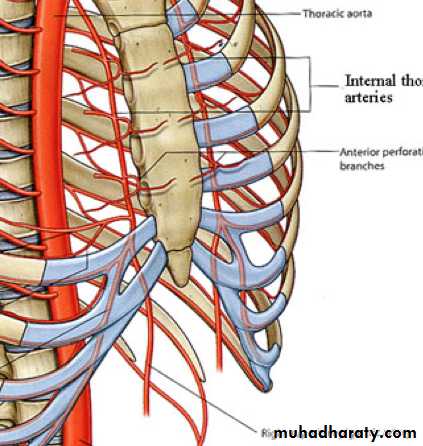
The posterior intercostal arteries of the first two spaces are branches from the superior intercostal artery, a branch of the costocervical trunk of the subclavian artery. The posterior intercostal arteries of the lower nine spaces are branches of the descending thoracic aorta.
Intercostal veins:
The anterior intercostal veinsThe posterior intercostal veins
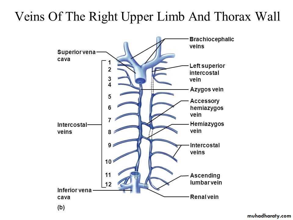
The highest intercostal vein (1st post. I. V) drains on each side to the brachiocephalic v.
• The 2nd & 3rd and sometimes the 4 th post. I. V unite to form the superior I.V. which drains on the left in the brachiocephalic v & on the right in the azygos arch.
The eight lower right IVS drain to azygos v. The fourth to the eighth left IVS will unite to form the Accessory Hemiazygos Vein which joins the azygos vein at the level of T7
The 9th to 11 th post. left I.VS drain into the Hemiazygos V which join the azygos v that empty to superior vena cava.