Organs of special senses
Histology
د.رندعبداللطيف
The EYE
Is a complex and highly specialized organ for perception of form, light, and color.
The eyes are located in the protective cavities within the skull called orbits.
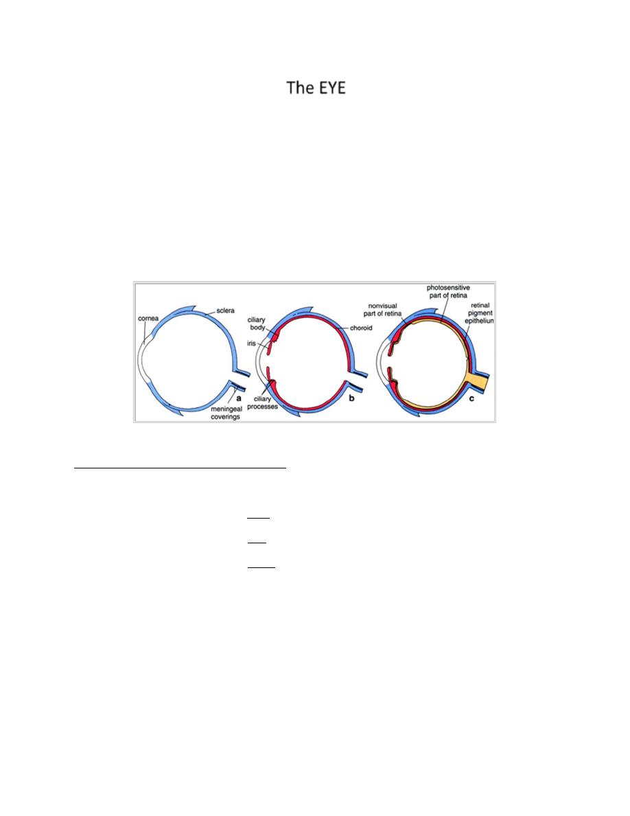
Each eye consists of 3 concentric layers:
1. external layer consist of sclera posteriorly and cornea anteriorly
2. middle layer consist of choroids, ciliary body & ciliary processes
3. inner layer of nerve tissues called retina.
Each eye consist of 3 compartments:
1. ant. Chamber: space between the cornea ant. & iris post.
2. post. Chamber: limited ant. by iris
lat. by ciliary processes
post. by lens & zonal fibers of the lens
3. vitreous space: lies behind the lens & its zonal fibers and it is surrounded by
the retina.
Both ant. & post. chambers are filled with a protein-rich fluid called aquous humor
while the vitreous space is filled with gelatinous substance called vitreous body.

Organs of special senses
Histology
د.رندعبداللطيف
I.
External layer of the eye (Tunica fibrosa ):
Consist of two parts:
Sclera & Cornea ant. 1/6
th
it is colorless & transparent.
Sclera:
post. 5/6
th
it is opaque white layer. Consist of tough dense C.T. formed mainly of
flat collagen bundles oriented in different directions with a moderate amount of
ground substance. The cells of the sclera are few fibroblasts which contain pigment
near the corneo-scleral junction and near the exit of the optic nerve. The mu7scles
of the eye are attached to the sclera & control its movement.

Organs of special senses
Histology
د.رندعبداللطيف
The sclera is composed of 3 layers:
1) Episclera: is the external layer of loose C.T. running adjacent to the
periorbital fat. The episclera is connected to a dense layer of C.T. (Tenon’s
capsule) by a thin bundles of collagen fibers, between the tenon’s capsule &
the sclera is a space called tenon’s space & because of the presence of this
space the eyeball can make rotatory movements.
2) Middle layer (stroma): consist of bundles of collagen fibers
3) Inner layer of the sclera which is adjacent to the choroid and called
suprachoroidal lamina it is thin layer rich in melanocytes, fibroblasts &
elastic fibers.
Lamina fibrosa :
The part of the sclera at the exit of the fibers of optic nerve appear to be
reduced in thickness and show perforations through which fibers of the optic
nerve pass outwards, it constitute a weak spot so any rise in the intra ocular
pressure such as in glaucoma cause bulging of the sclera out words.
The sclera is relatively avascular & ant. it blends with the cornea in a
transition zone called limbus.
Cornea
Is the ant. 1/6
th
of the tunica fibrosa it is colorless & transparent, a transverse
section of the cornea shows that it consist of five layers:
1) Corneal epithelium: non-keratinized stratified squamous epithelium
consist of 5-6 layers with numerous mitotic figures at the basal layer
indicate its regenerative capacity. Numerous free nerve endings terminate
in this epithelium which is part of the blink reflex.
2) Bowman’s membrane: thick homogenous layer lies beneath the corneal
epithelium, it consist of collagen fibers with condensation of intercellular

Organs of special senses
Histology
د.رندعبداللطيف
substance and no cells. This membrane contribute greatly to the stability
& strength of the cornea.
3) Corneal stroma: is the main layer formed of many layers of parallel
collagen bundles, between these fibers few fibroblast cells (keratocytes)
are found, the cornea is avascular & this feature together with the paucity
of cells render the cornea transparent to transmit light.
4) Decemet’s membrane: it is hyaline homogenous layer on the post. aspect
of corneal stroma composed of thin collagen fibers.
5) Corneal endothelium: single layer of simple squamous cells which line
the inner surface of the cornea, these cells have organelles characteristic
of cells engaged in the process of active transport & protein synthesis so
these cells are responsible for:
a. Synthesis & maintenance of Decemet’s membrane.
b. Maintaining the transparency of the cornea (together with the
corneal epithelium) both layers pumps fluid from the corneal
stroma to the outside keeping the stroma in a dehydrated state
which is very important factor in the transparency of the cornea.
Excessive hydration of the stroma will result in a state of
opacification of the cornea.
The cornea is avascular and receive its nutrition by diffusion from nearby vessles
& from fluid in the ant. chamber of the eye.
Corneo-scleral junction: (limbus)
Is the area of transition from the transparent collagen bundles of the cornea to the
white opaque fibers of the sclera this area is highly vascularised so it consist of
highly communicating channels lined with endothelial cells, these channels unite to
form the Canal of sclemm which will drain the fluid in the ant. chamber of the eye
(aquous humor). Sclemm’s canal communicate externally with the venous system.

Organs of special senses
Histology
د.رندعبداللطيف
Any block in this drainage pathway will lead to increase intraocular pressure
leading to glaucoma. Normal intraocular pressure is 24 mmHg (similar to CSF).
Middle layer (vascular layer, uvea):
Uvea is the intermediate layer in the eye between the sclera & retina, it contains
blood vessels, nerves, supportive cells, contractile cells & melanocytes. It is
devided into 3 areas: choroids, ciliary body & iris (uveal tract).
1- Choroid
extends from the ora serrata to the optic nerve. It is highly vascularized
coat, between its blood vessels there is loose C.T. which is rich in
fibroblasts, macrophages & melanocytes.
The inner layer of the choroids is richer in blood vessels & called
choriocapillary layer it has an important function in nutrition of the retina
Choriocapillary layer is separated from the retina by a thin hyaline
membrane called Bruch’s membrane. The choroids is bound to the sclera by
the suprachoroidal lamina which is a loose C.T. rich in melanocytes.
2- Ciliary body & Ciliary processes:
Is the ant. expansion of the choroids at the level of the lens, it extend from the base
of the iris to the ora serrata where it is continuous with the choroid. In transverse
section the ciliary body is triangular in shape, composed of vascular stroma,
smooth muscles & melanocytes. Its inner surface is lined by a double layer of
columnar epithelium, the basal cells are pigmented while the superficial cells are
not (this epithelium is regarded as a continuation of the non-nervous part of the
retina).
Ciliary processes are ridge like extensions of the ciliary body with a core of
loose C.T. & numerous fenestrated capillaries. from these processes emerge
fibers called zonule fibers that will insert into the lens. Ciliary processes are

Organs of special senses
Histology
د.رندعبداللطيف
covered by the same cells covering the ciliary body and they are responsible
for formation of aquous humor.
3-
Iris:
Is the ant. continuation of the choroids it lies infront of the lens leaving a
hole in the center called pupil,
the iris consist of a vascular core with pigmented cells and iridial muscles.
It is lined by double layer of columnar pigmented cells posteriorly, while
ant. it is covered by simple squamous epithelium which represent the
continuation of the post. epithelial layer of the cornea.
The iridial muscles
1-Constrictor pupilli muscle: lies internally, circularly arranged, supplied by
parasympathetic fibers. It causes constriction of the pupil.
2-Dilator pupilli muscle: radially arranged, lies externally supplied by
sympathetic fibers and their contraction lead to dilatation of the pupil.
Inner layer ( Retina)
Is the inner most layer of the eye & considered as a photosensitive nervous layer. It
extend from the optic disc to the ora serrata which limits the beginning of the non-
nervous part. When the retina is examined by ophthalmoscope it appear red in
color due to reflection of light from the vascular choroids layer. At the center of
the retina lies the yellowish spot (macula) which contain a central depression called
fovea centralis this is the area of higher visual acuity in the eye. Medial to the
macula we have the optic disc (blind spot) where the fibers of optic nerve leave the
eye.

Organs of special senses
Histology
د.رندعبداللطيف
The retina consist of many types of cells:
1) Pigment cells: large hexagonal cells contain pigments, send processes (large
microvilli) that extend between the rods & cones, each pigment cell cover 9-
10 rodes & cone, these cells lies on the bruch’s membrane.
2) Photoreceptor cells of rods & cones: rods are more numerous then cones
(rods are 110-115 million while cones are 6-7 millions) nuclei of rods lie in
the inner part of the outer nuclear layer, cones are concerned with color
vision and bright light vision while rods are concerned with low light vision.
3) Supportive cells (muller’s cells & astrocytes): the muller cells are tall
supporting cells extend from the base of the photoreceptor cells up to the
retinal surface, they link to each others by processes forming the outer
limiting membrane. Astrocytes found throughout the retina and there
prosesses form a scaffold for other nerve cells.
4) Nerve cells: are bipolar cells, horizontal cells, amacrine cells. They
interconnect with each others and with the photoreceptor cells to finally
transmit signals to the optic nerve.
The retina is formed of 10 layers from out side to inside:
1)
Pigment cell layer
2)
Photoreceptor cell layer
3)
External limiting membrane
4)
Outer nuclear layer
5)
Outer plexiform layer
6)
Inner nuclear layer: nuclei of bipolar, horizontal, amacrine & muller
cells
7)
Inner plexiform layer
8)
Ganglion cell layer: a row of ganglion cells which have large nuclei
and prominent nucleoli

Organs of special senses
Histology
د.رندعبداللطيف
9)
Nerve fiber layer: the axons of ganglion cells in their way to the optic
nerve.
10)
Inner limiting membrane:a barely visible membrane between the
vitreous and retina.