Emberyology
Dr.Muna ZuhairLec. 2
Respiratory System

Emberyology
Dr.Muna ZuhairLec. 4
Genital System
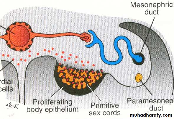
Development of testis:
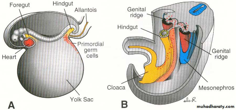
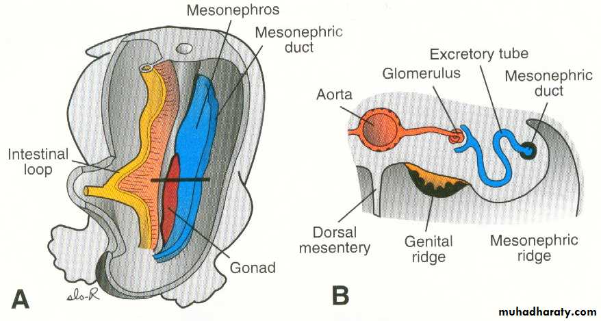
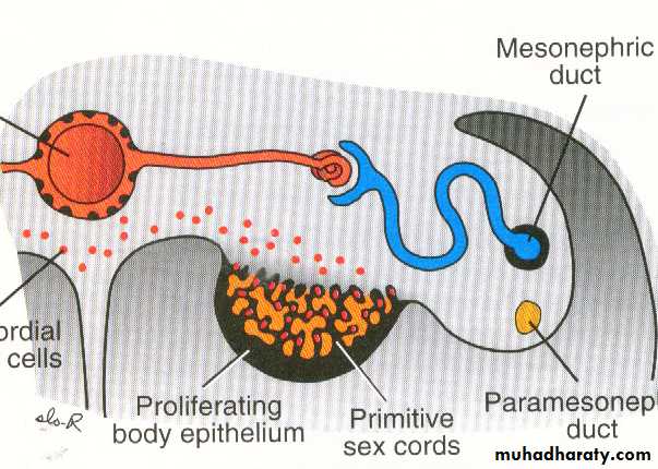
• The gonads appear as a pair of longitudinal ridge called genital ridge.• The primordial germ cells appear in the wall of yolk sac and migrate to reach the gonads at 5th week and invade the epithelium of the genital ridge which proliferate to form primitive sex cords.
The testis develops from 3 sources:
Coelomic epithelium: give rise to Sertoli cells.
Primordial germ cells : give rise to spermatogonia.
Intermediate cell mass of mesoderm: forms the connective tissue of testis and its covering tunica albuginea.
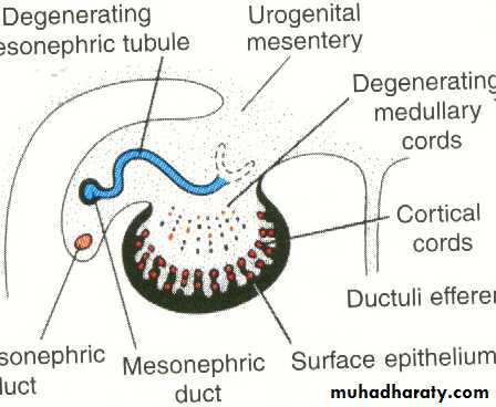
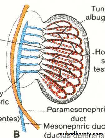
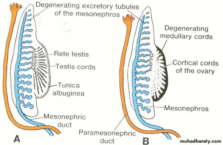
The primitive sex cords continue to proliferate and enter the medulla forming medullary cords (testis cords) formed from germ cells and Sertoli cells then become separated from the surface by tunica albuginea forming horse-shoe shaped testis cords, then undergo canalization to form the seminiferous tubules.
Descent of testis:
• The testis descends from its higher position on the posterior abdominal wall to enter the scrotom.Its descend is guided and facilitated by a fibrous cord called gubernaculum which extends from the lower pole of testis to the floor of the scrotal pouch.• The gubernaculum traverses the muscles of the anterior abdominal wall (in the inguinal canal) to reach the scrotom where it is accompanied by an evagination of peritoneal sac called Processus vaginalis which follow the course of gubernaculum to the scrotal swelling.
• Shortening of the gubernaculum together with lengthening of the embryo will help in the descent of testis.It descend retroperitoneally to enter:The inguinal canal : at 7th month.Inside the scrotom: at birth (or in the 9th month).
• The canal connecting processus vaginalis with the peritoneal cavity obliterated .
Congenital anomalies:
• Indirect inguinal hernia: due to patent processus vaginalis or it become filled with peritoneal fluid forming (congenital hydrocele).• Undescended testis : due to failure of descent of testis which may retained in the abdomen or may stop at : superficial inguinal ring, Root of penis, perineum.
• Ectopic testis: the testis may take a position away from the normal pathway of descent as to be found in the lower part of anterior abdominal wall or upper part of thigh.
• Gonadal dysgenesis: failure of formation of genital duct and external genital organ.
• Hermaphroditism (intersex):
• True hermaphroditism- gonads +external genetalia of both sexes
• Pseudo hermaphroditism-gonads of one sex+external genetalia of the opposite sex.
Anomalies of the processus vaginalis:
It may remain patent in the whole or in part leading to the following:
• Congenital hydrocele.
• Congenital inguinal hernia.
• Cyst in the spermatic cord.
Development of the ovary:
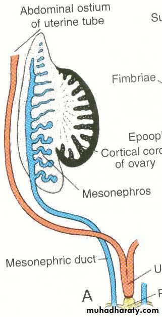
• The primitive sex cords dissociate forming degenerating medullary cords which disappear and replaced by vascular stroma (ovarian medulla).• The germinal epithelium proliferate together with the primordial germ cells form cortical cords which become divided into isolated cell clusters called (primordial follicles). Each follicle is formed from oogonia surrounded by a single layer of follicular cells.
Thus the ovary develops from 3 sources:
Coelomic epithelium (germinal epithlium): forms the follicular cells.Primordial germ cells : give rise to oogonia.
Intermediate cell mass of mesoderm: forms the stroma of the ovary and its covering tunica albuginea.
Descent of ovary:
The ovary is attached by gubenaculum which traverse the anterior abdominal wall in the inguinal canal to reach the labium majus and it get attachment to the side of the uterus close to the entrance of uterine tube and as a result it become divided into 2 parts:Cranial part: forms the ligament of the ovary (between the uterus and ovary).
Caudal part : forms the round ligament of uterus (between the uterus and labium majus).
Paramesonephric duct:
Development:Two longitudinal ducts one on each side and descend very close to Mesonephric (Wollfian duct).Each duct has 2 ends:
• Cranial end: open into the coelomic (peritoneal) cavity.
• Caudal end: blind and comes in contact with the posterior wall of the definitive urogenital sinus.
Its course divided into 3 patrs:
Cranial part: descend vertically lateral to the mesonephros.Intermediate part : crosses transversely (horizontally) ventral to the mesonephric duct.
Caudal part: descends vertically medial to the mesonephric duct.
The caudal vertical parts of both ducts descend in contact with each other inside a mesodermal partition called genital cord. This cord extends between the rectum posteriorly and urinary bladder anteriorly and its mesoderm differentiates into the fibromuscular wall of the uterus and vagina (not its epithelial lining).
Fate of Paramesonephric duct:
In the female :• The cranial vertical part : form uterine tube with its cranial end opening into the coelomic cavity
• The intermediate transverse part: form the fundus and upper part of the body of the uterus.
• The caudal fused vertical parts of both sides: forms the lower part of the body of uterus and cervix.
In the male:
The duct disappears almost completely except for 2small parts at its ends:
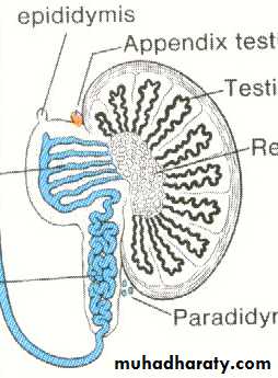
Cranial end : form the appendix of testis.
Caudal end: form the prostatic utricle.
In contrast to the paramesonephric duct, the mesonephric duct disappears completely in female but persist in the male.
Female genital organs:
Uterine tube:
Each tube is developed from the cranial vertical part of the paramesonephric duc. The cranial end of the duct will become the limbricated end of the uterine tube which persist communicating with the peritoneal cavity.
Uterus:
The fundus and upper part of the body of uterus: develop from the intermediate transverse parts of the paramesonephric ducts.The cervix and lower part of the body of uterus: develop from the fused caudal vertical parts of the 2 paramesonephric ducts which extend inside the genital cord.
Anomalies of uterus:
• Uterus didelphys (double uterus): the 2 paramesonephric ducts fail to fuse completely in the genital cord where each duct forms a separate uterus and each one has a separate vagina.• Uterus bicornis bicollis: double uterus with double cervix but with a single vagina.
• Bipartite uterus: The caudal part s of the 2paramesonephric duct fuse together but the septum in between persist and thus there is a single uterus with its cavity divided into 2 parts by a septum .
• Bicornuate uterus: The uterus is single but has 2 separate horns due to improper development of the fundus.
Development of the vagina:
The caudal vertical part of the 2 paramesonephric ducts come in contact with the dorsal wall of the definitive urogenital sinus to produce a bulge into the cavity of the sinus called Mullerian tubercle.
At the point where the Mullerian tubercle lies, the endodermal cells lining the urogenital sinus proliferate to form 2 masses called sino-vaginal bulbs which fuse together to form the vaginal plate.
The vaginal plate grows cranially then become canalized to form the lumen of the vagina. Expansion of the lumen forms:
• Vaginal fornices around the cervix at the cranial end of the vagina.
• The hymen at the caudal end of the vagina.
Anomalies:
• Absent vagina: due to failure of canalization of the vaginal plate.
• Imperforated hymen: when the continuity between the vaginal lumen and the cavity of urogenital sinus is not established.
Prostate gland:
It develops as multiple (15-20) outgrowths (buds) from the lining of the prostatic urethra (caudal part of the vesicourethral canal and pelvic part of the urogenital sinus). They arise mainly from the lateral aspect of the urethra, although few buds also arise from its dorsal and ventral aspect.The lining of the prostatic urethra is mainly endodermal except in its posterior wall which is mesodermal and therefore, the prostatic buds are mostly endodermal (except posteriorly). The cellular buds become canalized to form the alveoli and tubules of the prostate.
The fibromuscular capsule and the connective tissue of the gland are derived from the surrounding mesoderm in the genital cord.