
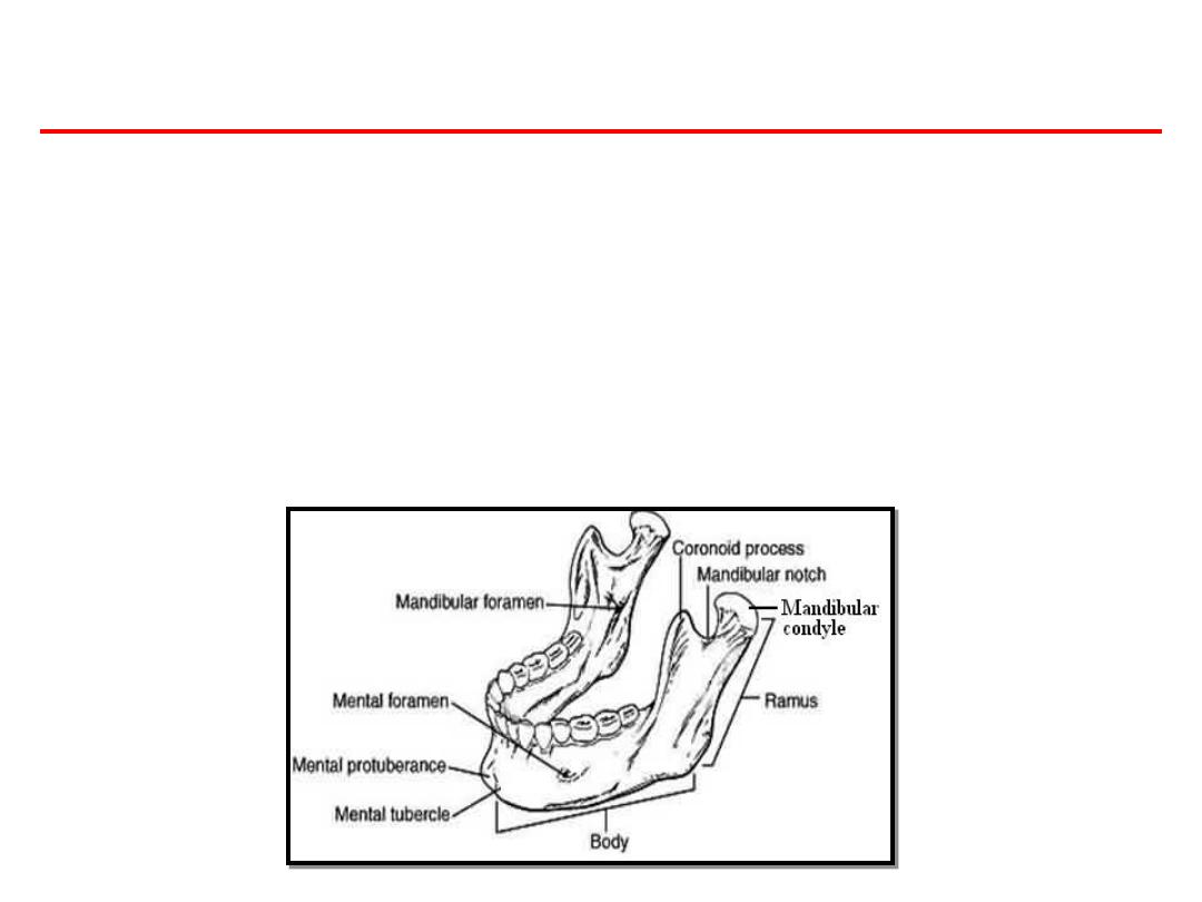
GROWTH OF MANDIBLE

• The mandible derives from the first pharyngeal arch
and ossifies intramembranously, beginning in the 6th
week i.u. It is the second bone to ossify after the
clavicle.
• It ossifies laterally to Meckel’s cartilage with the
ossification centres appearing bilaterally at the
bifurcation of the inferior alveolar nerve into the
mental and incisive branches.
• Ossification extends forwards, backwards and upwards
to form the body, alveolar processes and ramus.

• Secondary cartilages appear, including the
condylar cartilage during the 10th week i.u.
Endochondral bone appears in the condylar
cartilage by the 14th week i.u.

• The role of the condylar cartilage in the
growth of the mandible is not yet entirely
clear. It is not a primary growth centre in its
own right, but rather it grows in response to
some other controlling factors.
• However, normal growth at the condylar
cartilage is required for normal mandibular
growth to take place.

• Postnatal growth of the mandible follows a
pattern intermediate between a neural and
somatic pattern, although it follows the
somatic pattern more closely than does the
growth of the maxilla (Fig 4.8). Most
mandibular growth occurs as a result of
periosteal activity.
• While the mandible appears in the adult as single bone, it is
developmentally and functionally divisible into a several
skeletal sub-unit.
• Basal bone forms one unit, to which is attached the alveolar
process, coronoid process, condylar process, angular process,
the ramus and the chin.
POST-NATAL GROWTH OF MANDIBLE

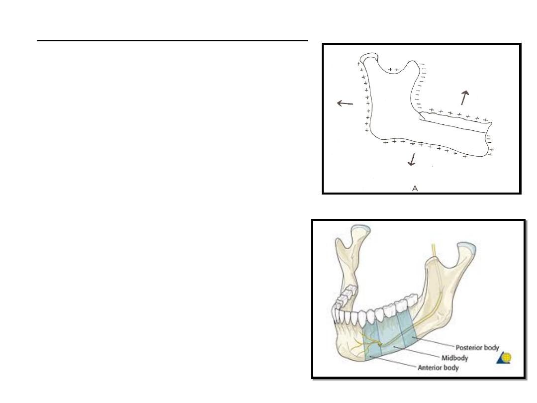
RAMUS:
• It moves progressively
posterior by a combination of
deposition and resorption.
Resorption occurs on anterior
part of ramus while bone
deposition occur on the
posterior region.
• This result in a “drift” in
posterior direction.
• Function of the remodelling
of ramus is to facilitate the
lengthening of the
mandibular body, which in
turn accommodates the
erupting molars.
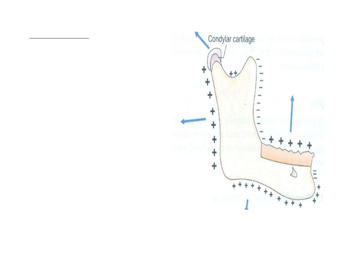
THE BODY OF THE MANDIBLE:
• Displacement of the ramus
results in the conversion of the
ramal bone into the posterior
part of the body of the
mandible.
• In this manner, it lengthens.
Thus additional space made
available by means of
resorption of the anterior
border of the ramus is made
use of to accommodate the
erupting molar.
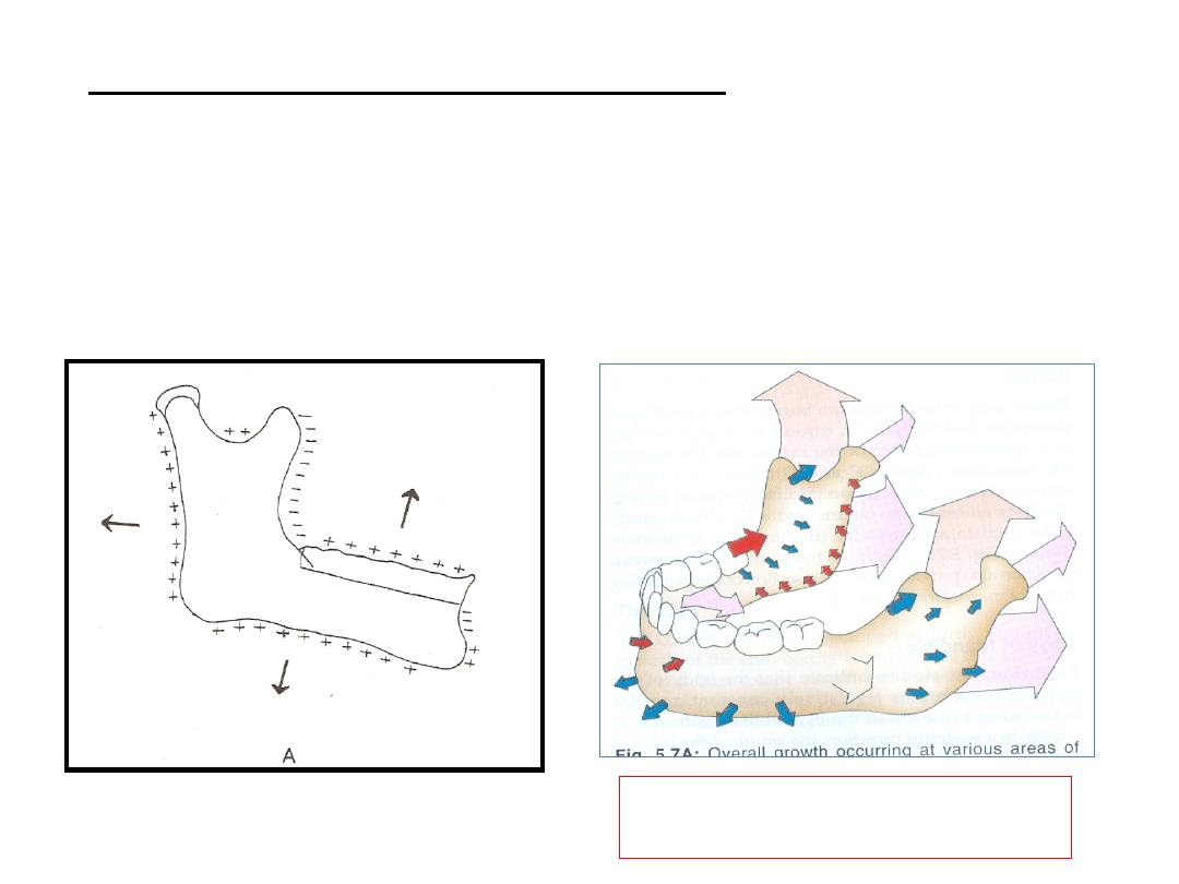
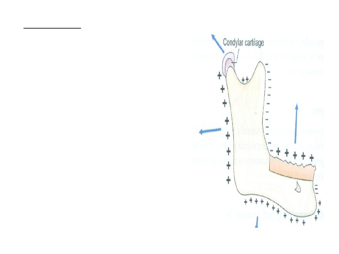
THE BODY OF THE MANDIBLE:
Appositional growth occurs along the lower
border of mandible and on its lateral surface.
Red arrows - bone resorption
Blue arrows - bone deposition
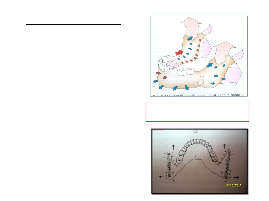
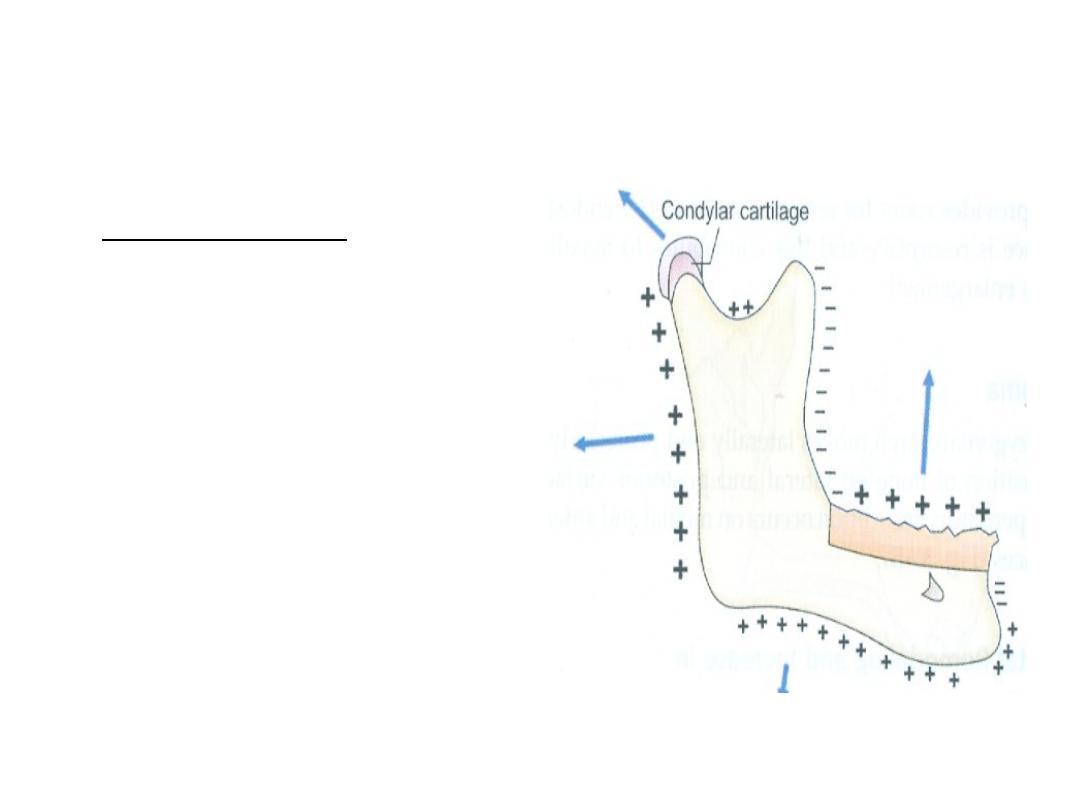
ANGLE OF THE MANDIBLE:
• On the lingual side of the
angle of the mandible,
resorption take place on the
posterio-inferior aspect
while deposition on the
anterio-superior aspect.
• On the buccal side,
resorption occur on the
anterio-superior part while
deposition takes place on
posterio-superior part.
• This results in flaring of the
angle of mandible as age
advances.
Red arrows - bone resorption
Blue arrows - bone deposition
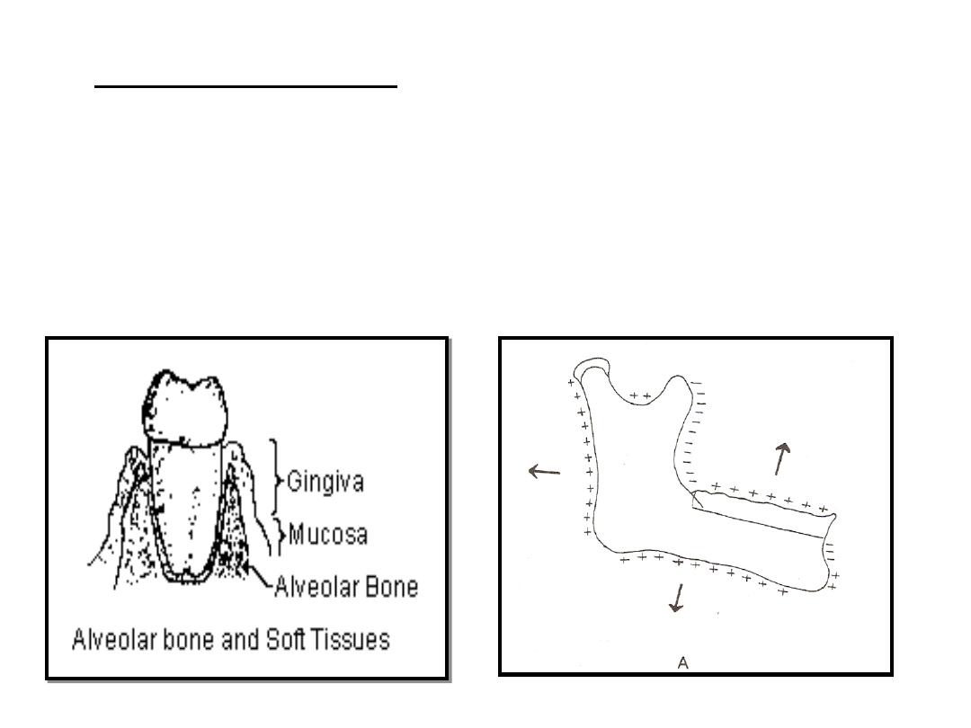
ALVEOLAR PROCESS:
• It develops in response to the presence of tooth
buds.
• As the teeth erupt, it develops and increases in
height by bone deposition at the margins.
THE CHIN:
• As the age advances the
growth of chin becomes
significant.
• Usually males are seen to have
prominent chin as compared
to females.
• Bone deposition on mental
protuberance.
• Bone resorption on alveolar
region above the prominence,
creating a concavity.
THE CONDYLE:
• The head of the condyle
is covered by the thin
layer of cartilage called
the condylar cartilage.
• The presence of conylar
cartilage is an
adaptation to withstand
the compression that
occurs at the joint.
THE CONDYLE:
• It is believed that the growth of
the soft tissues including the
muscles and the connective
tissues carries the mandible
forward away from cranial
base. Bone growth follows
secondary at the condyle to
maintain constant contact with
cranial base.
• The condylar growth rate
increases at puberty reaching a
peak between 12-14 years. The
growth ceases around 20 years
of age.

THE CORONOID PROCESS:
• The growth follows the enlarging ‘V’ principle.
• Viewing, the logitudinal section of coronoid
process from posterior aspect, it can be seen that
deposition occurs on the lingual (medial) surfaces
of the left and right coronoid process .
• Although additions take places on the lingual side,
the vertical dimension of the coronoid process
also increases. This follows the ‘V’ principle.
• Viewing from the occusal aspect, the deposition
on lingual of coronoid process brings about
posterior growth movement in ‘V’ pattern
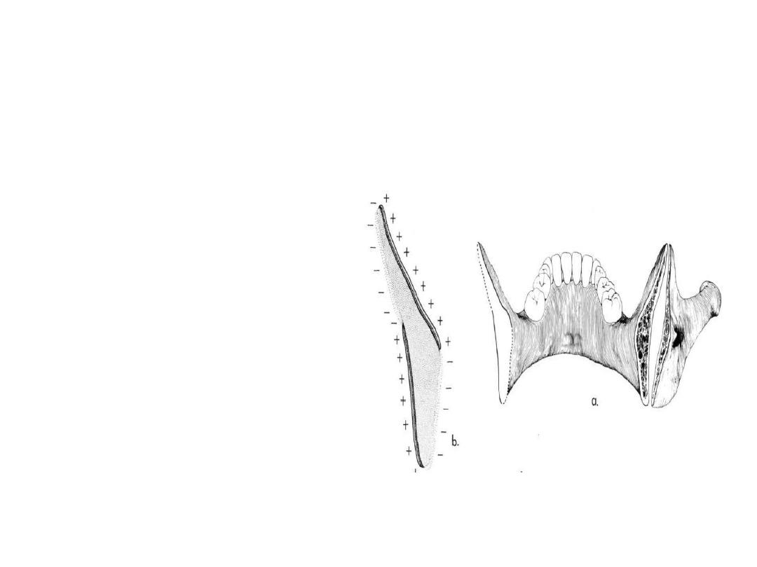
Vertical section of coronoid process
• BONE DEPOISITION -
lingual surface (+ +)
• BONE RESORPTION -
buccal surface (- -)
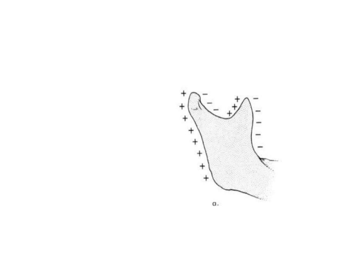
SIGMOID NOTCH
•
Bone deposition -
posterior border of
coronoid process
•
Bone resorption -
anterior Face of neck of
condyle.

Age changes in mandible
• INFANTS –
Mental foramen - near lower border
Mandibular canal - lower border of body of mandible
Angle of mandible - obtuse around 140* or more
• Mandibular canal - runs
parallel with mylohyoid line.
• Angle of mandible - 110* -
120*
• Mental foramen -
midway of upper & lower
border.
ADULTS-
• OLD AGE
Mandibular foramen - near alv. Bone
Mandibular canal - near alv. Bone
Angle of mandible - obtuse 140*


• Muscular processes develop at the angles of
the mandible and the coronoid processes, and
the alveolar processes develop vertically to
keep pace with the eruption of the teeth.

• As the mandible is displaced forwards growth
at the condylar cartilage fills in posteriorly
while at the same time periosteal remodelling
maintains its shape ( Fig. 4.12 ).
• Bone is laid down on the posterior margin of
the vertical ramus and resorbed on the
anterior margin, and this posterior drift of the
ramus allows lengthening of the dental arch
posteriorly.

• At the same time the vertical ramus becomes
taller to accommodate the increase in height
of the alveolar processes.

• Remodelling also brings about an increase in
the width of the mandible, particularly
posteriorly.

• Lengthening of the mandible and anterior
remodelling together cause the chin to
become more prominent, an obvious feature
of facial maturation especially in males.
• Indeed, just as in the maxilla, the whole
surface of the mandible undergoes many
complex patterns of remodelling as it grows in
order to maintain its proper anatomical form.

• Before puberty growth occurs at steady rate
with an increase of 1–2 mm per year in ramus
height and 2–3 mm per year in body length.
• However, growth rates can double during
puberty and the associated growth spurt.
• Mandibular growth slows to adult levels rather
later than maxillary growth, on average at
about 17 years in girls and 19 years in boys,
although it may continue for longer.
