Chapter 56 699Contributions of the Cerebellum and Basal Ganglia to Overall Motor Control
The cerebellum plays major roles in the:Timing of motor activities.
Rapid and smooth progression from one muscle movement to the next.
Control the intensity of muscle contraction when the muscle load changes.
Controlling necessary instantaneous interplay between agonist and antagonist muscle groups.
Cerebellum and its motor functions
The cerebellum has been called a silent area of the brain, because electrical excitation of the cerebellum does not cause any conscious sensation and rarely causes any motor movement.Removal of the cerebellum, cause body movements to become highly abnormal.
The cerebellum is especially vital during rapid muscular activities such as running, typing, playing the piano, and even talking.
Loss of this area of the brain can cause almost total incoordination of these activities even though its loss causes paralysis of no muscles.
The cerebellum receives continuously updated information about the desired sequence of muscle contractions from the brain motor control areas.
It also receives continuous sensory information from the peripheral parts of the body, giving sequential changes in the status of each part of the body—its position, rate of movement, forces acting on it, and so forth.
The cerebellum also aids the cerebral cortex in planning the next sequential movement a fraction of a second in advance while the current movement is still being executed, thus helping the person to progress smoothly from one movement to the next.
Also, it learns by its mistakes—that is, if a movement does not occur exactly as intended, the cerebellar circuit learns to make a stronger or weaker movement the next time.
Anatomical functional areas of the cerebellum:
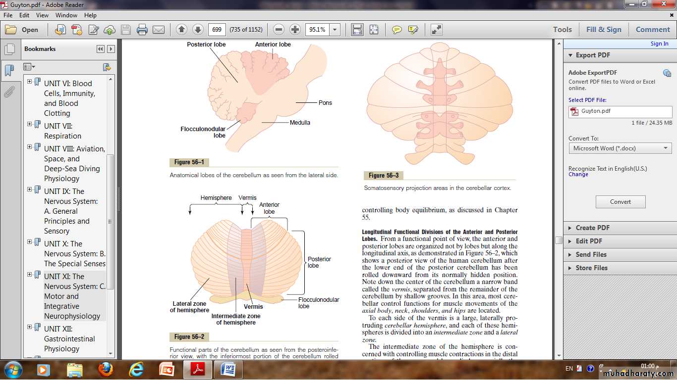
Anatomically, the cerebellum is divided into three lobes by two deep fissures:(1) The anterior lobe.
(2) The posterior lobe.
(3) The flocculonodular lobe (it developed along with and functions with the vestibular system in controlling body equilibrium).
Longitudinal functional divisions of the anterior and posterior lobes.
In the vermis , most of cerebellar control functions for muscle movements of the axial body, neck, shoulders, and hips are located.To each side of the vermis is a large, laterally protruding cerebellar hemisphere, and each of these hemispheres is divided into an intermediate zone and a lateral zone.
The intermediate zone of the hemisphere is concerned with controlling muscle contractions in the distal portions of the upper and lower limbs, especially the hands and fingers and feet and toes.
The lateral zone of the hemisphere operates at a much more remote level because this area joins with the cerebral cortex in the overall planning of sequential motor movements.
Without this lateral zone, most discrete motor activities of the body lose their appropriate timing and sequencing and therefore become in coordinate.
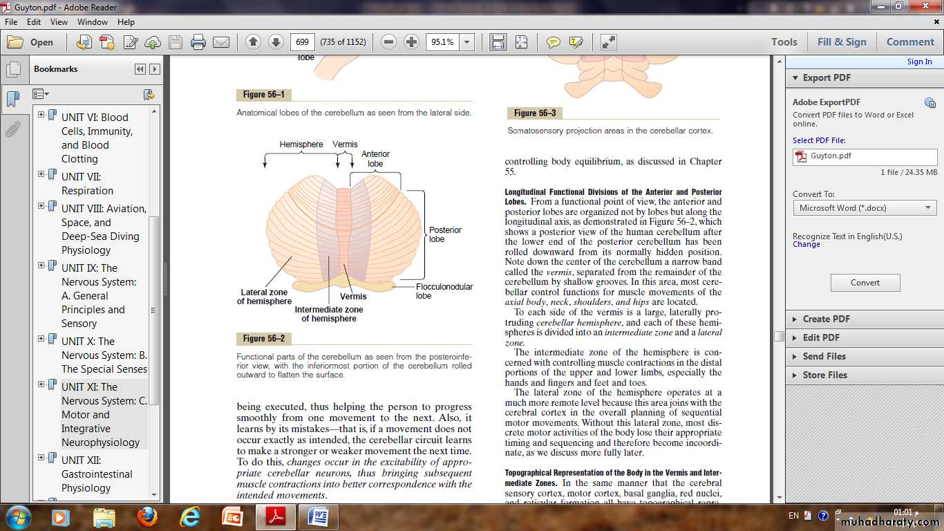
Topographical Representation of the Body in the Vermis and Intermediate Zones.
The axial portions of the body lie in the vermis part of the cerebellum, whereas the limbs and facial regions lie in the intermediate zones.These topographical representations receive afferent nerve signals from all the respective parts of the body as well as from corresponding topographical motor areas in the cerebral cortex and brain stem.
In turn, they send motor signals back to the same respective topographical areas of the cerebral motor cortex, the red nucleus and reticular formation in the brain stem.
The large lateral portions of the cerebellar hemispheres do not have topographical representations of the body.
These areas of the cerebellum receive their input signals almost exclusively from the cerebral cortex, especially from the premotor areas of the frontal cortex and from the somatosensory and other sensory association areas of the parietal cortex.
This connectivity with the cerebral cortex allows the lateral portions of the cerebellar hemispheres to play important roles in planning and coordinating the body’s rapid sequential muscular activities that occur one after another within fractions of a second.
Neuronal circuit of the cerebellum
The human cerebellar cortex is actually a large folded sheet, about 17 centimeters wide by 12 centimeters long, with the folds lying crosswise.Each fold is called a folium, lying deep beneath the folded mass of cerebellar cortex are deep cerebellar nuclei.
Afferent pathways from other parts of the brain:
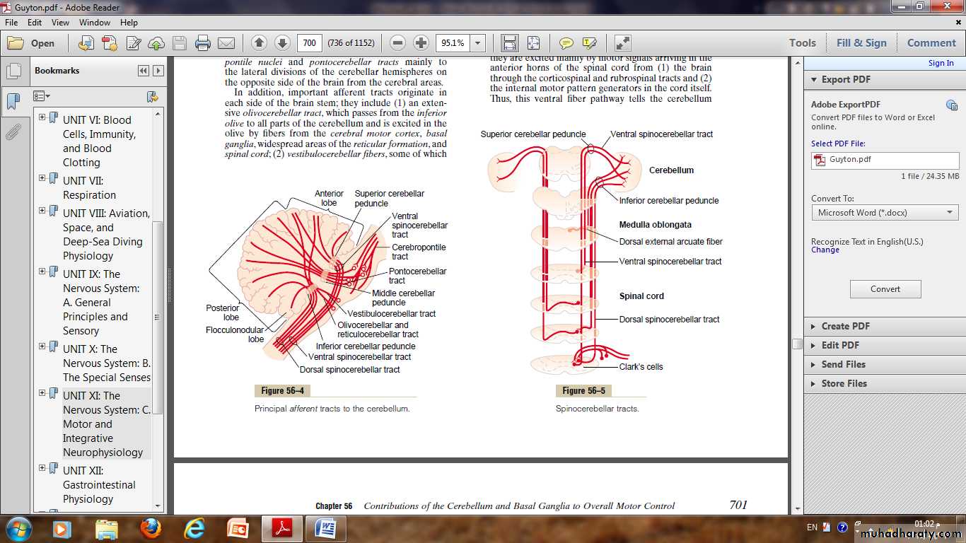
The basic input pathways to the cerebellum are: (1)Corticopontocerebellar pathway, which originates in the cerebral motor, premotor cortices and cerebral somatosensory cortex.(2) Olivocerebellar tract, which passes from the inferior olive to all parts of the cerebellum.
(3) Vestibulocerebellar fibers.
(4) Reticulocerebellar fibers.
Afferent pathways from the periphery.
The cerebellum receives sensory signals from the peripheral parts of the body:1) The dorsal spinocerebellar tract.
2) The ventral spinocerebellar tract.
The signals transmitted in the dorsal spinocerebellar tracts come mainly from the muscle spindles, Golgi tendon organs, large tactile receptors of the skin, and joint receptors.
All these signals apprise the cerebellum of the momentary status of:
• Muscle contraction.
• Degree of tension on the muscle tendons.
• Positions and rates of movement of the parts of the body.
• Forces acting on the surfaces of the body.
The ventral spinocerebellar tract tells the cerebellum which motor signals have arrived at the anterior horns; this feedback is called the efference copy of the anterior horn motor drive.
The spinocerebellar pathways can transmit impulses at velocities up to 120 m/sec.
This extremely rapid conduction is important for instantaneous appraisal of the cerebellum of changes in peripheral muscle actions.
The cerebellum continually collects information about the movements and positions of all parts of the body even though it is operating at a subconscious level.
Function of the Cerebellum in Overall Motor Control
The nervous system uses the cerebellum to coordinate motor control functions at three levels, as follows:• The vestibulocerebellum.
• This consists principally of the small flocculonodular cerebellar lobes (that lie under the posterior cerebellum) and adjacent portions of the vermis.
• It provides neural circuits for most of the body’s equilibrium movements.
2. The spinocerebellum.
This consists of most of the vermis of the posterior and anterior cerebellum plus the adjacent intermediate zones on both sides of the vermis.It provides the circuitry for coordinating mainly movements of the distal portions of the limbs, especially the hands and fingers.
3. The cerebrocerebellum.
This consists of the large lateral zones of the cerebellar hemispheres, lateral to the intermediate zones.
It receives virtually all its input from the cerebral motor cortex and adjacent premotor and somatosensory cortices of the cerebrum.
Cerebrocerebellum—Function of the Large Lateral Zone of the Cerebellar Hemisphere to Plan, Sequence, and Time Complex Movements
1) Planning of Sequential Movements.
It requires that the lateral zones of the hemispheres communicate with both the premotor and the sensory portions of the cerebral cortex.
The “plan” of sequential movements begins in the sensory and premotor areas of the cerebral cortex, and from there the plan is transmitted to the lateral zones of the cerebellar hemispheres.
Thus, the lateral cerebellar zones appear to be involved not with what movement is happening at a given moment but with what will be happening during the next sequential movement a fraction of a second or perhaps even seconds later.
2) Timing Function.
Another important function of the lateral zones of the cerebellar hemispheres is to provide appropriate timing for each succeeding movement.In the absence of these cerebellar zones, one loses the subconscious ability to predict ahead of time how far the different parts of the body will move in a given time.
Without this timing capability, the person becomes unable to determine when the next sequential movement needs to begin.
As a result, the succeeding movement may begin too early or, more likely, too late.
Clinical Abnormalities of the Cerebellum
1) Dysmetria and Ataxia.The movements ordinarily overshoot their intended mark; then the conscious portion of the brain over compensates in the opposite direction for the succeeding compensatory movement; This effect is called dysmetria, and it results in uncoordinated movements that are called ataxia.
Both of them can also result from lesions in the spinocerebellar tracts because feedback information from the moving parts of the body to the cerebellum is essential for cerebellar timing of movement termination.
Dysarthria.
The formation of words depends on rapid and orderly succession of individual muscle movements in the larynx, mouth, and respiratory system.Lack of coordination among these and inability to adjust in advance either the intensity of sound or duration of each successive sound causes jumbled vocalization, with some syllables loud, some weak, some held for long intervals, some held for short intervals, and resultant speech that is often unintelligible.
Intention Tremor ( action tremor)..
When a person who has lost the cerebellum performs a voluntary act, the movements tend to oscillate, especially when they approach the intended mark, first overshooting the mark and then vibrating back and forth several times before settling on the mark.
Cerebellar nystagmus.
Cerebellar nystagmus is tremor of the eyeballs that occurs usually when one attempts to fixate the eyes on a scene to one side of the head.This off-center type of fixation results in rapid, tremulous movements of the eyes rather than steady fixation.
It occurs especially when the flocculonodular lobes of the cerebellum are damaged.
Hypotonia.
Decreased tone of the peripheral body musculature on the side of the cerebellar lesion.The hypotonia results from loss of cerebellar facilitation of the motor cortex and brain stem motor nuclei by tonic signals from the deep cerebellar nuclei.
Basal Ganglia—Their motor functions
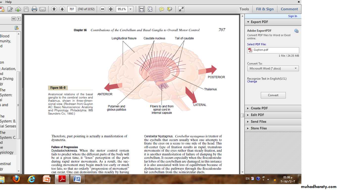
The basal ganglia receive most of their input signals from the cerebral cortex itself and also return almost all their output signals back to the cortex.On each side of the brain, these ganglia consist of the caudate nucleus, putamen, globus pallidus, substantia nigra, and subthalamic nucleus.
Function of the basal ganglia in executing patterns of motor activity.
The basal ganglia function is; in association with the corticospinal system to control complex patterns of motor activity.An example is the writing of letters of the alphabeta; cutting paper with scissors, hammering nails, shooting a basketball through a hoop, passing a football, throwing a baseball, the movements of shoveling dirt, most aspects of vocalization, controlled movements of the eyes, and virtually any other of our skilled movements.
Abnormal Function in the basal ganglia.
• Lesions in the globus pallidus frequently lead to spontaneous and often continuous writhing movements of a hand, an arm, the neck, or the face movements called Athetosis.
• A lesion in the subthalamus often leads to sudden flailing movements of an entire limb, a condition called Hemiballismus.
• Multiple small lesions in the putamen lead to flicking movements in the hands, face, and other parts of the body, called a Chorea.
• Lesions of the substantia nigra lead to the common and extremely severe disease of rigidity, akinesia, and tremors known Parkinson’s disease.
lesions in the putamen Chorea.
Lesions in the globus pallidus Athetosis.Lesions of the substantia nigra Parkinson’s disease.( rigidity. Tremer, akinesia)
A lesion in the subthalamus Hemiballismus.