The Sense of Hearing
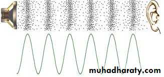
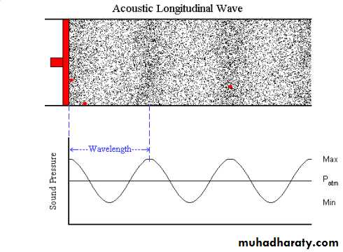
SoundIt is mechanical waves transmitted through a medium( air, water, and other matters). The waves are formed of positive and negative. There are 2 parameters which identify sound, the frequency and intensity.
The frequency is the number of cycles per second, and is measured by Hertz(Hz) , Human ear can generally hear sounds with frequencies between 20 Hz and 20,000Hz .
The intensity which is measured by the decibel(dB).
Anatomy of the ear
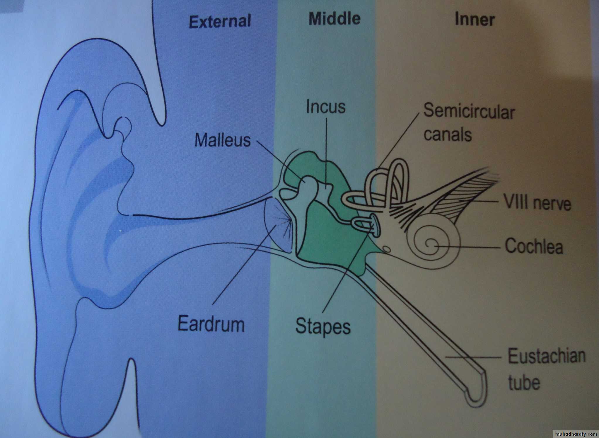
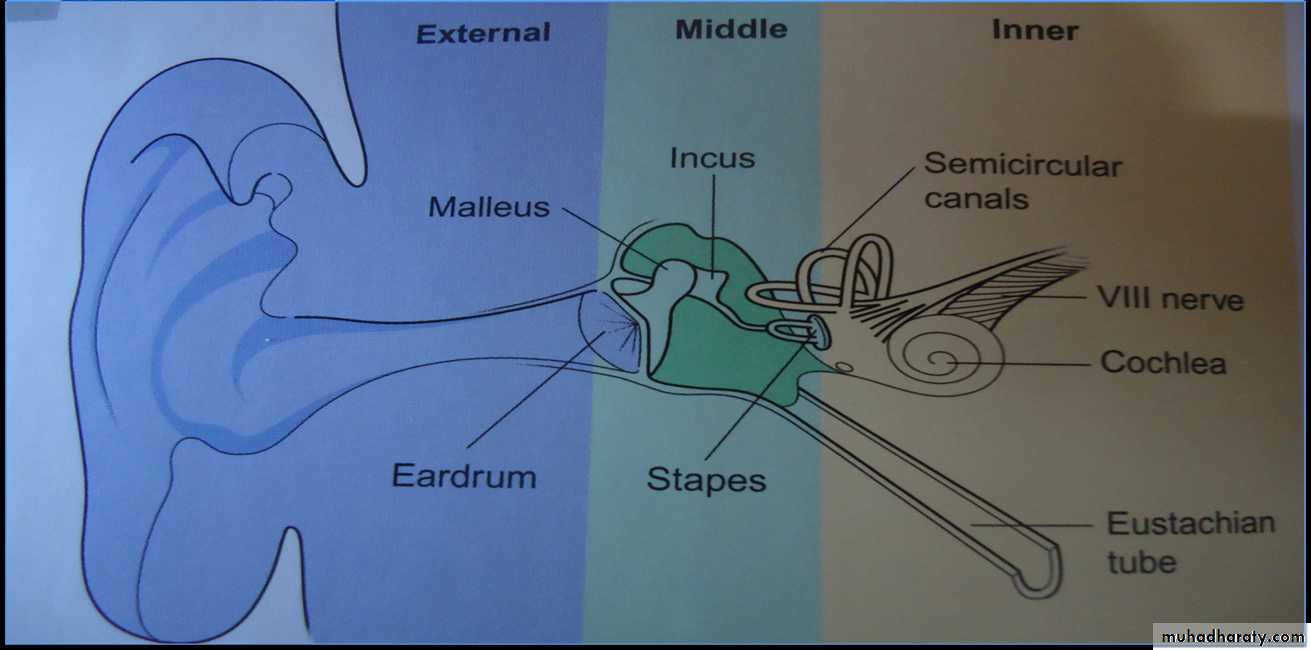
The ear divided into three partsThe external ear ,Middle ear ,Inner ear
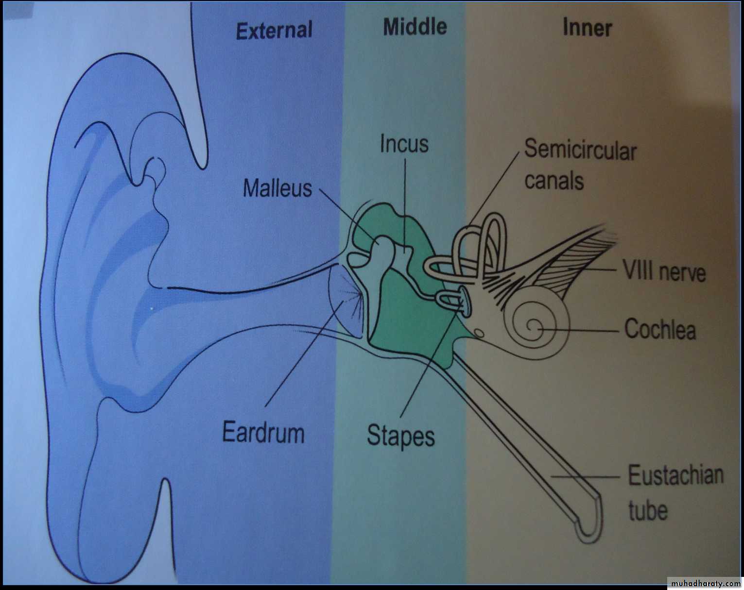
I-The external ear or Outer ear
1-The pinna (Auricle), It serves to collect the sound2-The external auditory meatus (EAM): It is approximately 7mm in diameter and 2.5 cm long, unless the canal is open, hearing will be dampened.
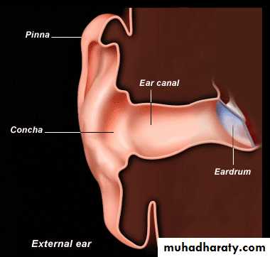
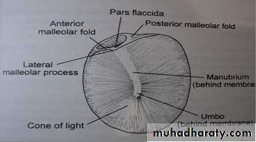
The tympanic membrane (TM) or (ear drum)
It is a window of the middle ear.It is thin, semitransparent membrane, makes the boundary between outer and middle ears.
It is 55 mm2 in surface area and is slightly concave .
The tip end of the handle of the malleus is attached to the center of the tympanic membrane, and this point of attachment is constantly pulled by the tensor tympani muscle, which keeps the tympanic membrane tensed
II- The middle ear
It is an air-filled cavity behind the tympanic membrane.occupied by three of the smallest bones of the body, known as the ossicles, the malleus, incus, and stapes. These ossicles convert the sound waves striking the eardrum into mechanical vibrations .
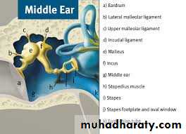
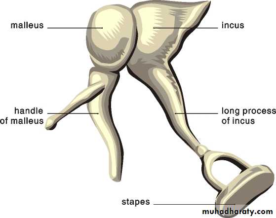
The malleus: the long process (the handle), is attached to the eardrum
The incus: is the bridge between the malleus and stapes.
The stapes: is the smallest bone in the human body. The head (caput) articulates with the incus, and the neck bifurcates to become the crura of stapes which converges on the footplate ,it rests on the oval window.
External Ear
Middle Earpinna
The external auditory meatus
Ear drumStapes
Semicircular canal
VIII Nerve
Eustachian tube
Eustachian tubeThe opening of the Eustachian tube is within the middle ear. The Eustachian tube connects from the chamber of the middle ear to the back of the nasopharynx. It is usually closed and opened during swallowing, yawning to equalize the pressure in middle ear.
The round window is a membrane-covered round opening between the cochlea and the middle ear. The round window is actually situated below and a little to the back of the oval window, from which it is separated by a rounded elevation, the promontory .
Round window
The articulation of the incus with the stapes causes the stapes to push forward on the oval window every time the tympanic membrane moves inward, and to pull backward every time the malleus and tympanic membrane moves outward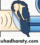
Impedance Matching
• The middle ear acts as an impedance matching device to transfer sound energy efficiently from air to a fluid medium in the cochlea. Because fluid has far greater inertia than air does, so increased amounts of force are needed to cause vibration in the fluid and overcome mismatched impedance• the system actually increases the force of movement this is done by several simple mechanisms:
lever principle
"There is a difference in the dimensions of the articulating ossicles. The length of long process of malleus is approximately 9 mm, while that of the long process of incus is about 7 mm. This leads to an increase in the force applied to the stapes footplate compared with that applied to the malleus by about 1.3 times and gains about 2 decibel (dB).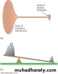
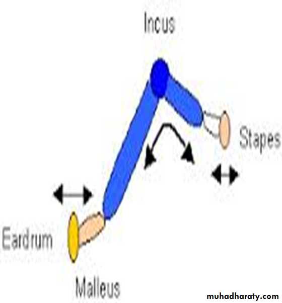
A lever amplifies an input force to provide a greater output force

the surface area
The surface area of the tympanic membrane is about 55 mm2, whereas the surface area of the stapes footplate averages 3.2 mm2.Pressure =force/surface area
This 17-fold difference in area times the 1.3-fold ratio of the lever system causes about 22 times as much total force to be exerted on the fluid of the cochlea as is exerted by the sound waves against the tympanic membrane.
Combination of all these results in a gain about 25- 27 dB.
Attenuation of Sound by Contraction of the Tensor Tympani and Stapedius Muscles
When loud sounds are transmitted through the ear to the central nervous system, a reflex occurs after a latent period of few milliseconds to cause contraction of the stapedius muscle and the tensor tympani muscle.The tensor tympani muscle pulls the handle of the malleus inward while the stapedius muscle pulls the stapes outward. These two forces oppose each other and thereby cause the ossicular system to increased rigidity, and reduce the conduction of low frequency sound,
The function of this mechanism is to
1-To protect the cochlea from damaging effect of loud sound.
2-To mask low-frequency sounds in loud environments.
3-decrease a person’s hearing sensitivity to his or her own speech.
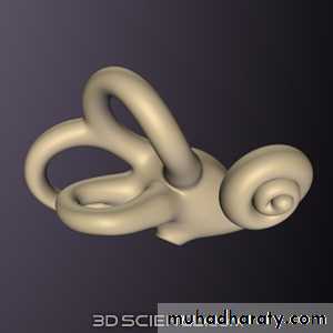
The inner ear(labyrinth)
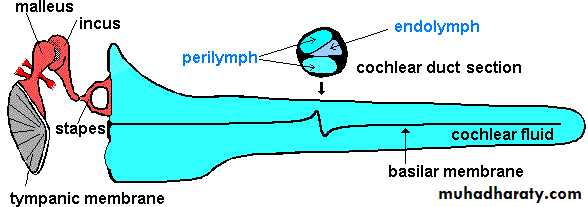
It is made of two parts one within the other. The bony labyrinth. Within it, there is membranous labyrinth which contain a fluid called endolymph. The membranous labyrinth is surrounded by perilymph there is no connection between endolymph and perilymph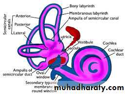
There are three major sections of the inner ear
1-The front portion is the snail-shaped cochlea, which functions in hearing.2-The semicircular canals, which helps maintain balance.
3-Interconnecting the cochlea and the semicircular canals is called the vestibule. It contains the sense organs responsible for balance
vestibule
semicircular canalscochlea
The cochlea
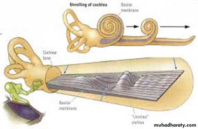
it is approximately 35 mm in length and coild 2.5 times into a snail shape about the size of large pea.
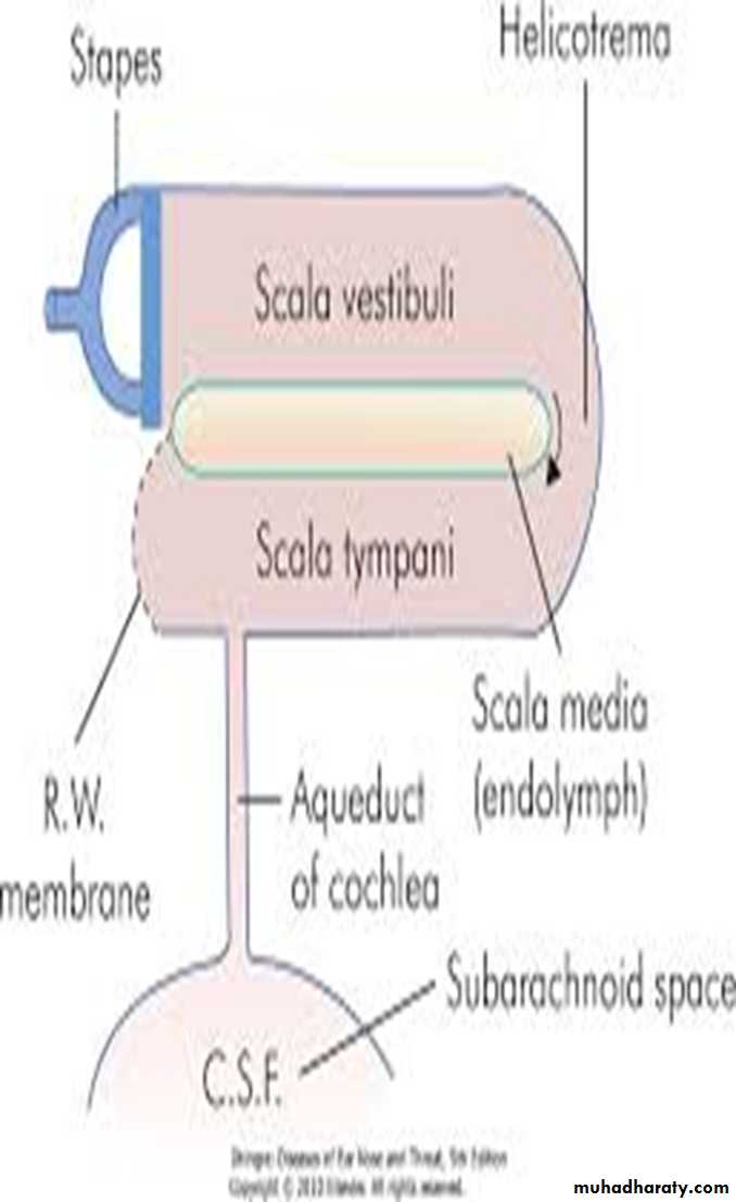
Within the cochlea are three fluid filled space the scala vestibuli, the scala media, and the scala tympani.
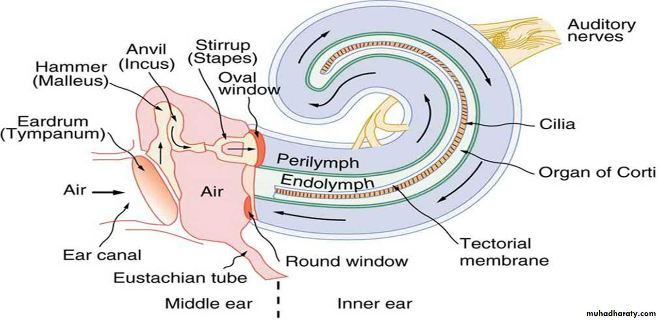
• The scala tympani and the scala vestibuli are filled with perilymph and communicate with each other at the helicotrema.
• At the base of the cochlea, the scala vestibule ends at the oval window.
• The scala tympani ends at the round window.
• The scala media is continuous with the membranous labyrinth and does not communicates with the other two scala. It is filled with endolymph
The cochlea
.
scala tympani
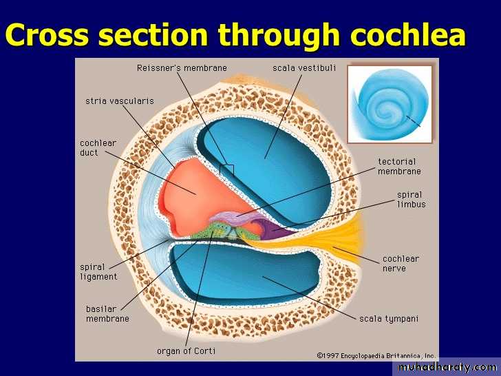
scala mediaOrgan of corti
Basilar membrane
Reissners membrane
Hair cells
Tectorial membrane
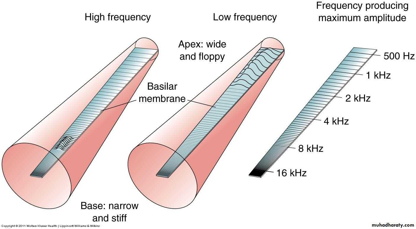
The Basilar Membrane:
is a fibrous membrane that separates the scala media from the scala tympani. These fibers are stiff, elastic reed like structures that are fixed at their basal ends but are free at their distal ends end, so they can vibrate like the reeds of a harmonica The lengths of the basilar fibers increase progressively beginning at the oval window and going from the base of the cochlea to the tip (resemble a harp).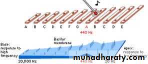
• At the basal end, it has short and taut fibers that vibrate best at a very high frequency, while the long, looser fibers near the tip of the cochlea vibrate best at a low frequency
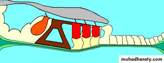
Organ of corti
lies on the surface of the basilar membrane. The actual sensory receptors in the organ of Corti are two specialized types of cells called hair cells ,The outer ends of the hair cells are fixed the reticular lamina, supported by triangular rods of Corti. The basilar fibers, the rods of Corti, and the reticular lamina move as a rigid unit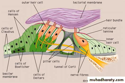
Rods of corti
Basilar membrane
The hair cells are mechanoreceptors that release a chemical neurotransmitter when stimulate by mechanical pressure two type of hair cell
A single row of internal (or “inner”) hair cells, and three or four rows of external (or “outer”) hair cells.
Their cilia(steriocilia) project into the tectorial membrane. The bases and sides of the hair cells synapse with a cochlear nerve . Between 90 and 95 per cent of these endings terminate on the inner hair cells
The outer hair cells, control the sensitivity of the inner hair cells at different sound pitches
Traveling Wave
The initial effect of a sound wave entering at the oval window is to cause the basilar membrane at the base of the cochlea to bend in the direction of the round window. However, the elastic tension that is built up in the basilar fibers as they bend toward the round window initiates a fluid wave that “travels” along the basilar membrane toward the helicotrema, and is called Traveling Wave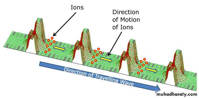
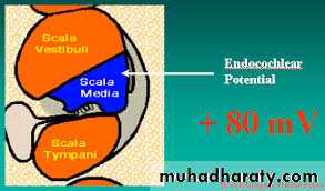
Endocochlear potential
The scala vestibuli and scala tympani contain perilymph which has a relatively low potassium
The scala media is filled with endolymph. contains a high concentration of potassium and a low concentration of sodium.
An electrical potential of about +80 millivolts exists all the time between endolymph and perilymph, with the positivity inside the scala media and the negativity outside.
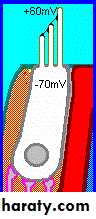
the hair cells project through the reticular lamina are bathed by the endolymph of the scala media, whereas perilymph bathes the lower bodies of the hair cells. Furthermore, the hair cells have a negative intracellular potential of –70 millivolts with respect to the perilymph but –150 millivolts with respect to the endolymph at their upper surfaces where the hairs project. this high electrical potential at the tips of the stereocilia sensitizes the cell by increasing its ability to respond to the slightest sound.
The auditory pathway
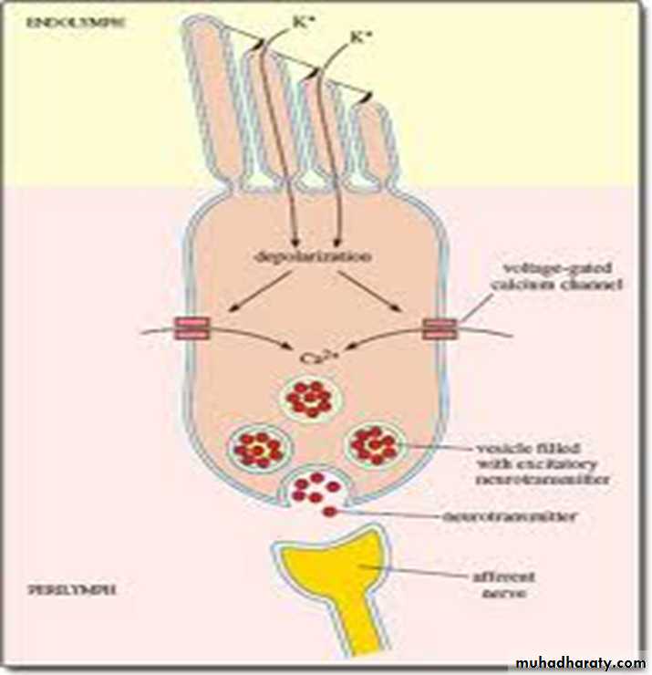
Sound waves moving through fluid cause vibration of the basilar membrane which push the hair cell. This causes a mechanical transduction that opens 200 to 300 cation-conducting channels, allowing rapid movement of potassium ions from the scala media fluid move into the stereocilia, and causes depolarization of the hair cell and, neurotransmitter is released by the hair cells at these synapses which in turn, stimulates the cochlear nerve endings.In this way sound waves are transformed into nerve impulses.

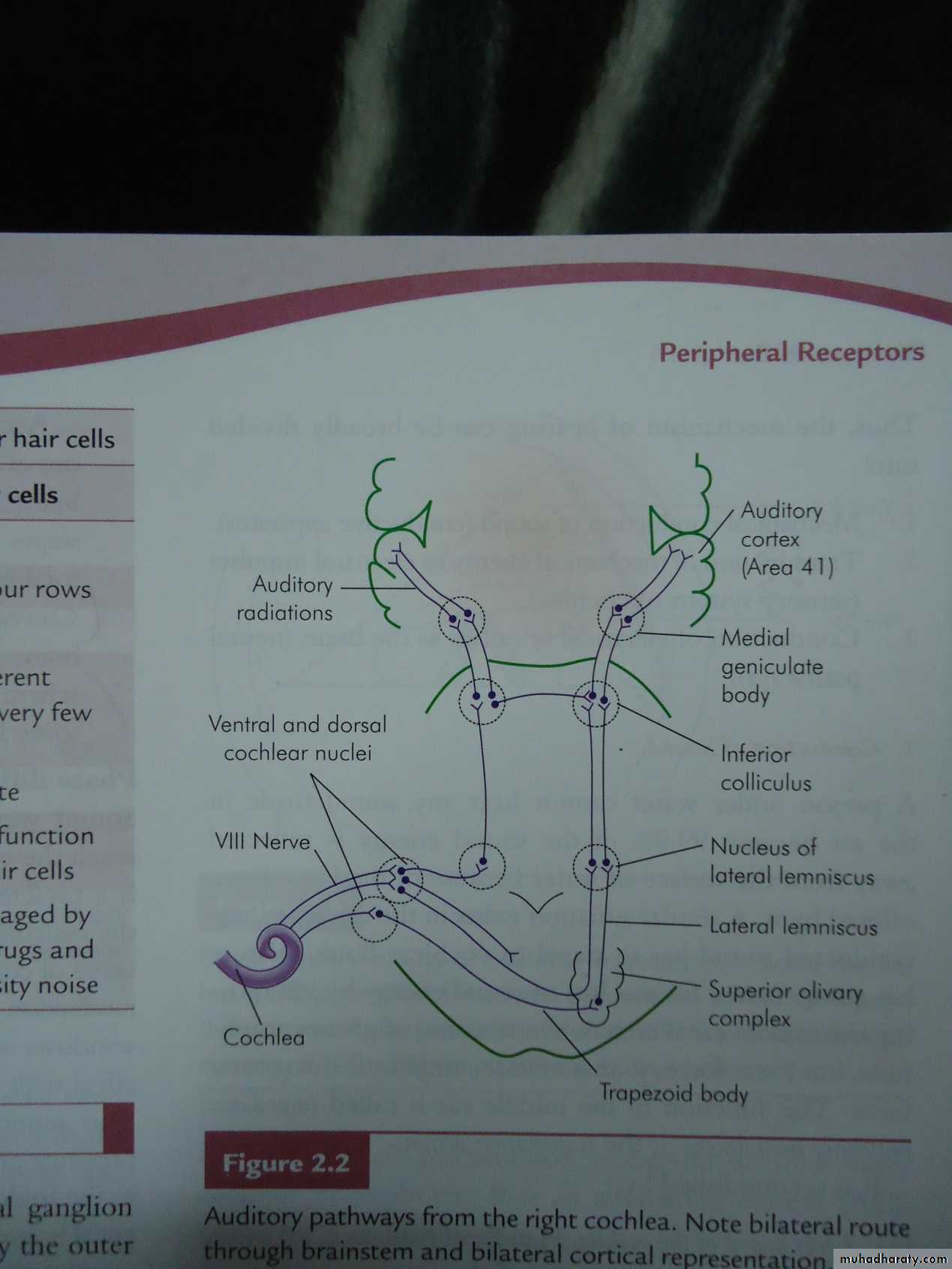
VIII nerve
Superior olivary complexLateral lemniscus
Inferior colliculus
Medial geniculate body
Auditory cortex
Cochlear nuclei
Cochlea
We have the primary auditory cortex and the auditory association cortex (secondary auditory cortex).The primary auditory cortex is directly excited by projections from the medial geniculate body whereas the auditory association areas are excited secondarily by impulses from the primary auditory cortex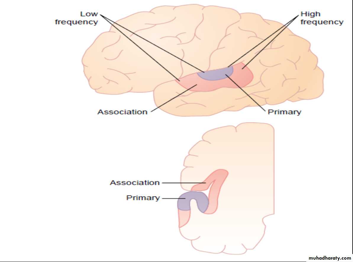
Determination of Sound Frequency
1- Place Principle: low-frequency sounds cause maximal activation of the basilar membrane near the apex of the cochlea, and high-frequency sounds activate the basilar membrane near the base of the cochlea. Intermediate frequency sounds activate the membrane at intermediate distances between the two extremes.2- Frequency principle or volley principle, by which, low frequency sounds from 20 to 2000 Hz can cause volleys of nerve impulses synchronized at the same frequencies. These volleys are transmitted by the cochlear nerve into the cochlear nuclei of the brain. It is further suggested that the cochlear nuclei can distinguish the different frequencies of the volleys .
Determination of Loudness
Loudness is determined by three ways.1- The amplitude of vibration of both the basilar membrane and hair cells increases, so that the hair cells excite the nerve endings at more rapid rates.
2- As the amplitude of vibration increases, it causes more and more of the hair cells to become stimulated; thus causing spatial summation of impulses.
3-The outer hair cells do not become stimulated significantly until vibration of the basilar membrane reaches high intensity
Localization of the sound
Human beings localize sound by different mechanisms:1-Comparing arrival-time differences, time lag. 2- The difference between the loudness of the sounds in the two ears
3-The pinna of ear plays a role in sound localization
Hearing Loss
It is a general term for the complete or partial loss of the ability to hear by one or both ears. It is a major public health problemTypes of Hearing Loss
Conductive hearing loss is a problem in the outer or middle ear.There is a block of sound conduction to the inner ear .
Sensorineural hearing loss (SNHL) is a problem with the inner ear or the auditory nerve and higher centers.
(a)deafness for low-frequency
(b) deafness for all frequencies
(c)-high frequency hearing loss(Presbycusis)
Mixed type of hearing loss
Audiometer
It is a device for determining the threshold of hearing and the nature of hearing disabilities Pure tone audiometry (PTA) is described as the gold standard for assessment of hearing level
Pure tones at various frequencies are generated, and their levels are increased and decreased until thresholds are found.