Group c
Tutorial
NO.2
د
.
ظاهر
19/10/2017
Oxygen Therapy and Airway Management, Ventilator therapy
Oxygen content of blood:
The theoretical maximum oxygen carrying capacity is 1.39 ml O2/g Hb,
but direct measurement gives a capacity of 1.34 ml O2/g Hb.1.34 is also
known as Hüfner’s constant.
The oxygen content of blood is the volume of oxygen carried in each 100
ml blood.
It is calculated by: (O2 carried by Hb) + (O2 in solution) = (1.34 x Hb x
SpO2 x 0.01) + (0.023 x PaO2)
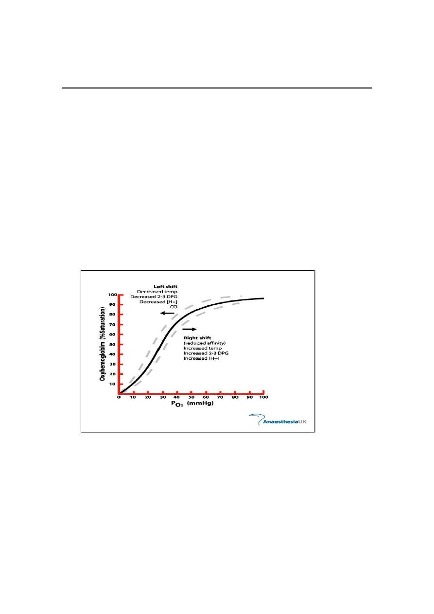
Oxygen dissociation curve (ODC)
3

Clinical Conditions With Increased Risk of Hypoxia:
Myocardial infarction
Acute pulmonary disorders
Sepsis
Drug overdose
Liver failure
Head trauma
CHF
Hypovolemic shock
Blunt chest trauma
Acute neuromuscular disease
Acute abdomen (splinting)
Acute pancreatitis
Spinal cord injury
Indications for Oxygen Therapy:
Tachypnea
Cyanosis
Restlessness
Disorientation
Cardiac arrhythmias
Slow bounding pulse
Tachycardia
Hypertension
Dyspnea
Coma
Labored breathing (use of accessory muscles, nasal flaring)
Lethargy
Tremors/seizure activity
Oxygen Therapy:
“Generally speaking”, a patient who is breathing less than 12 and more
than 24 /minute needs oxygen of some kind

Oxygen therapy To ensure safe and effective treatment
Oxygen is required for the functioning and survival of all body tissues
and deprivation for more than a few minutes is fatal.
In immediately life threatening situations oxygen should be
administered.
Hypoxaemia.
Acute hypotension.
Breathing inadequacy.
Trauma.
Acute illness.
CO poisoning.
Severe anaemia.
During the peri-operative period.
Oxygen therapy To ensure safe and effective treatment:
Oxygen is a prescription drug.
Prescriptions should include :
Flow rate.
Delivery system.
Duration.
Instructions for monitoring. :Monitoring resps oxygen sats not
definitive tool need to be looking at other things acccessory
muscles etc
Oxygen therapy:
Oxygen therapy Humidification Is recommended if more than 4
litres/min is delivered.
Helps prevent drying of mucous membranes.
Helps prevent the formation of tenacious sputum.
Oxygen concentrations will be affected with all delivery systems if
not fitted correctly or tubing becomes kinked and ports
obstructed.
Methods of Oxygen Delivery:
Most common methods of oxygen delivery include
Nasal Cannula
Venturi Mask
100% Non-Rebreather Mask
Mechanical Ventilation
Hyperbaric Oxygen Therapy(HBOT)
Oxygen Delivery Methods:
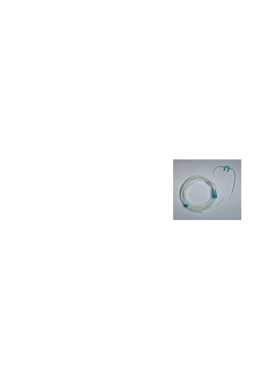
Nasal Cannula
Comfortable, convenient, mouth breathing will not effect % of O2
delivered
Liters/min = %
2 l/m = 24-28%
3 l/m = 28-30%
4 l/m = 32-36%
5 l/m = 36-40%
6 l/m = 40-44%
Cannot administer > 6 liters/minute (44%)
Nasal Cannula
Provides limited oxygen concentration,Used when patients cannot
tolerate mask,Prongs and other uses,Concentration of 24 to 44%
Flow rate set between 1 to 6 liters,For every liter per minute of flow
delivered, the oxygen concentration the patient inhales increases by 4%

Oxygen therapy:
Simple facemask Easy to use.
Allows administration of variable concentration dependant on flow of
fresh gas up to 40%.
Nasal cannulae Easy to use. Well tolerated. Comfortable for long
periods. Patient can eat and talk easily.
Possible to deliver oxygen concentrations of 24-40% at flow rates of 1-6
litres/min.
Flow rates in excess of 4 litres/min might cause discomfort and
drying of mucous membranes and are best avoided.
Flow Rate: 10 L/Min
O2 Conc.: 40 – 60 %
Use: moderate FiO2, mouth breathers
Venturi Mask:
FiO2 Delivery
Blue 24% Yellow 28%
White 31% Green 35%
Pink 40%
Provides precise concentrations of oxygen
Entrainment valve to adjust oxygen delivery
Mostly used in the hospital setting for COPD patients
Concerns
Tight seal is a must Interferes with eating/drinking Condensation
collection
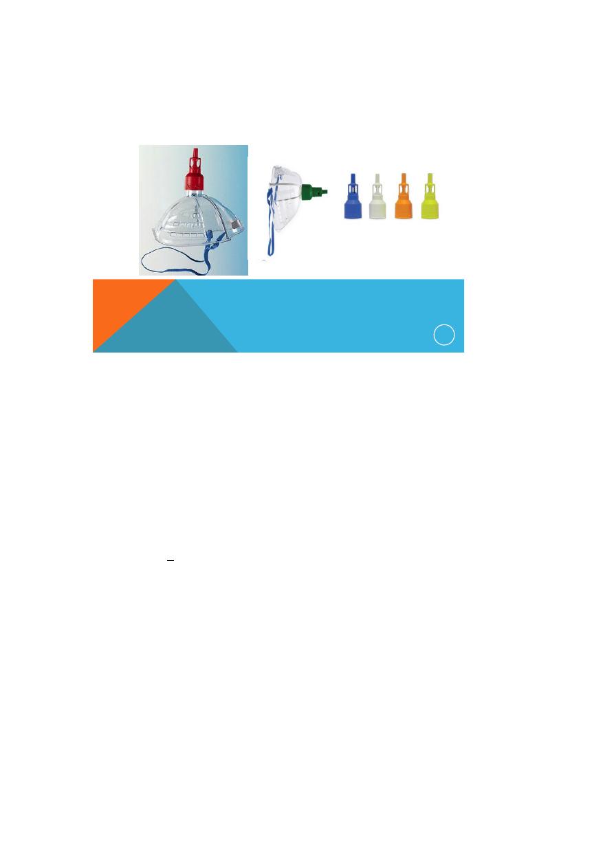
VENTURI MASK
15
Red 40% 10/L/M
Blue 24% 2/L/M Yellow 35% 8/L/M
White28% 4/L/M Green 60% 15/L/M
Orange 31% 6/L/M
Oxygen Delivery Methods
100% Non-Rebreather
Delivery percentages
6 l/min = 55 – 60 %
8 l/min = 60 – 80 %
10 l/min = 80 – 90 %
>12 l/min = 90 + %
Benefit:
Has a one way expiratory valve that prevents re-breathing expired
gases
Concern
May lead to O2 toxicity
100% Non-Rebreather Mask
17
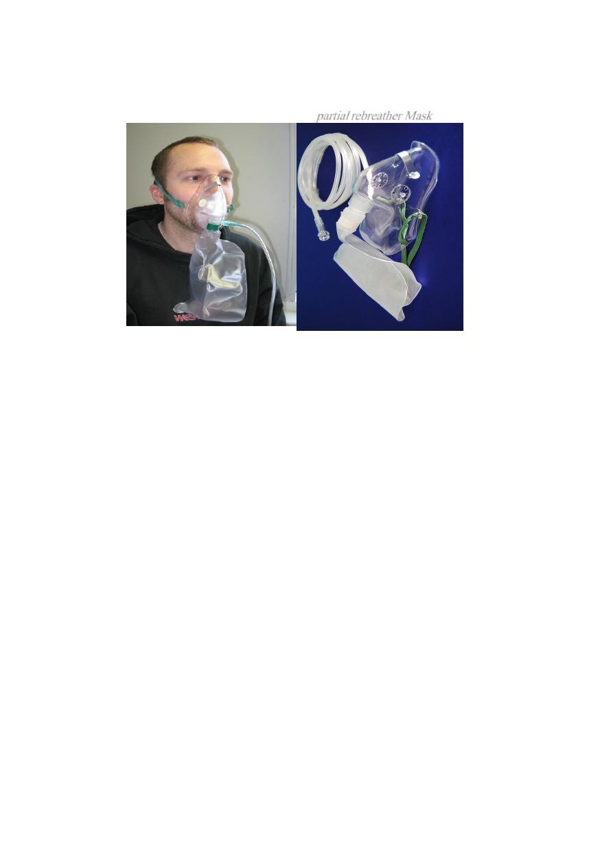
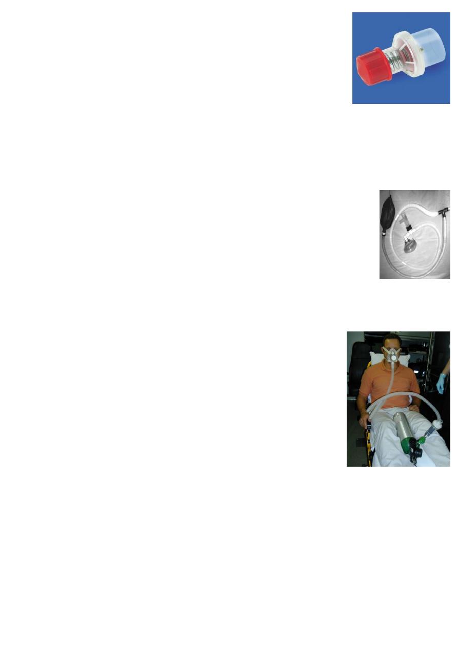
partial rebreather Mask
Oxygen therapy:
Non-rebreathing mask Allows the delivery of high concentrations
of oxygen (85% at 15 litres/min).
Has a reservoir bag to entrain oxygen. One way valves prevent
room and expired air from diluting the oxygen concentration. A
tight seal is essential.
Reservoir bag must be seen to expand freely.
Oxygen Delivery Methods Mechanical Ventilation
Allows administration of 100% oxygen
Controls breathing pattern for patients who are unable to
maintain adequate ventilation
Is a temporary support that “buys time” for correcting the
primary pathologic process

Indications for Mechanical Ventilation:
Mechanical Failure
Ventilatory Failure
Oxygenation Failure
General Anesthesia
Post-Cardiac Arrest
Mechanical Ventilation:
Two categories of ventilators
Negative pressure ventilators
Iron lung
Cuirass ventilator
Positive pressure ventilators
Two categories
Volume-cycled (volume-preset)
Pressure-cycled (pressure-preset)
Mechanical Ventilation PEEP;
Description
Maintains a preset positive airway pressure at the end of
expiration
Increases PaO2 so that FiO2 can be decreased
Increases DO2 (amt of delivered O2 to tissue)
Maximizes pulmonary compliance
Minimized pulmonary shunting
Indications
PaO2 < 60 on FiO2 > 60% by recruiting dysfunctional alveoli
Increases intrapulmonary pressure after cardiac surgery to
decrease intrathoracic bleeding (research does not support
this idea)
Advantages
Improves PaO2 and SaO2 while allowing FiO2 to
be decreased
Decreases the work of breathing
Keeps airways from closing at end expiration (esp. in pts
with surfactant deficiency)
Disadvantages
Increased functional residual capacity (increases risk for
barotrauma)
Can cause increased dead space and increased ICP
In pts with increased ICP, must assure CO2 elimination
Contraindicated: hypovolemia, drug induced low cardiac
output, unilateral lung disease, COPD
Mechanical Ventilation CPAP
Description
Constant positive pressure is applied throughout
the respiratory cycle to keep alveoli open
Indications
To wean without having to remove the
ventilator and having to connect to additional
equipment
Advantages
– Takes advantage of the ventilator alarm systems providing
psychological security of the ventilator being there
Disadvantages
– Patient may sense resistance as he breathes through the
ventilator tubing

Mechanical Ventilation Complications:
Respiratory arrest from disconnection
Respiratory infection (VAP)
Acid-base imbalances
Oxygen toxicity
Pneumothorax
GI bleeding
Barotrauma
Decreased cardiac output
Ventilator Weaning;
Vital Capacity at least 10 – 15 ml/kg
Tidal Volume > 5 ml/kg
Resting minute volume > 10 L per minute
ABG’s adequate on < 40% FiO2
Stable vital signs
Intact airway protective reflexes (strong cough)
Absence of dyspnea, neuromuscular fatigue, pain, diaphoresis,
restlessness, use of accessory muscles
Hyperbaric Oxygen Therapy (HBOT):
Hyperbaric Oxygen Therapy
Uses a special chamber, sometimes called a pressure chamber, to
allow a person to get high levels of oxygen in the blood.
This means that the air inside the pressurized chamber is typically
2 1/2 times greater than normal atmospheric pressure.
This leads to make the blood carrying larger amounts of oxygen,
and bringing this oxygen to organs and tissues in the body.
By doing so, wounds, particularly infected wounds, can heal more
readily.
HBOT— What is it USED for?
Decompression sickness
Arterial gas embolism
Carbon monoxide poisoning

Osteomyelitis
Skin grafts
Burns
Necrotizing fascitis
Anemia
Gas gangrene
Chronic non-healing wounds
Sports injuries
and more…
Analysis Arterial Blood Gas results:
If you can remember the following pyramid points and steps, you will
be able to analyze any blood gas report.
Pyramid points:
In acidosis, the PH is down.
In alkalosis, the PH is high.
The respiratory function indicator is the PCO2.
The metabolic function indicator is the HCO3.
Normal blood gas value:
PH:
7.35-7.45
PCO2:
35-45 mmHg
HCO3:
22-27meq/liter
PO2 :
80-100

Pyramid steps:
Pyramid step 1:-
look at the blood gas report. Look at the PH, is it up or down; if it
is up; it reflects alkalosis. If it is down; it reflects acidosis.
Pyramid step 2:-
look at the PCO2, is it up or down; if it reflects an opposite
response to the PH, then you know that the condition is a respiratory
imbalance.
If it does not reflect an opposite response to the PH; then move on to
pyramid step 3.
Pyramid step 3:-
look at the HCO3. Does the HCO3 reflect a corresponding response
with the PH; if it does, then the condition is a metabolic imbalance.
Pyramid step 4:-
Remember, compensation has occurred if the PH is in a normal
range of 7.35-7.45. If the PH is not within normal range, look at the
respiratory or metabolic function indicators.
Respiratory Imbalances :
if the condition is a respiratory imbalance look at the HCO3 to
determine the state of compensation.
if the HCO3 is normal, then the condition is uncompensated.
if the HCO3 is abnormal, then the condition is partial compensation.
Metabolic Imbalance :
If the condition is metabolic imbalance, look at the PCO2 to
determine the state of compensation.
If the PCO2 is normal, then the condition is uncompensated.
If the PCO2 is abnormal, then the is partial compensation.
