
Emergency of Diabetes
Mosul Medical College
Department of Medicine
supervised by:
DR.MOHAMMED KHALID

Case Scenario
A 20-year-old woman is evaluated in the emergency
department for polyuria, polydipsia, polyphagia, and an
unintentional 5.4-kg (11.9-lb) weight loss over the past
month. She has had increasing lethargy over the last 24 hours.
Her medical history and family history are unremarkable. She
takes no medications.

On physical examination,
temperature is 37.5 °C , blood pressure is 98/52 mm Hg, pulse
rate is 120/min, and respiration rate is 30/min. BMI is 17.
She is lethargic with dry mucous membranes, tachypnea, and
tachycardia. Chest auscultation is clear. Abdominal
examination shows diffuse mild tenderness and normal bowel
sounds. There is no rebound tenderness or guarding with
palpation.

Laboratory studies:
• Hemoglobin= 17 g/dL (170 g/L)
• Leukocyte count= 14,200/µL (14.2 × 10
9
/L)
• Blood gases, arterial::
pH= 7.25
PCO
2
= 21 mm Hg
Creatinine= 1.3 mg/dL
Electrolytes
Sodium= 130 mEq/L
Potassium= 3.0 mEq/L
Chloride= 99 mEq/L
Bicarbonate= 9 mEq/L
• Glucose= 620 mg/dL (34.4 mmol/L)

An electrocardiogram shows sinus tachycardia 120/min.
Chest radiograph is normal.
Your differential diagnosis ?

Diabetic ketoacidosis

INTRODUCTION
Diabetic ketoacidosis (DKA) is an acute, major, life-threatening
complication of diabetes that mainly occurs
In patients with type 1 diabetes, but it is not uncommon in some
patients with type 2 diabetes-20%.
This condition is a complex disordered metabolic
state characterized by:
hyperglycemia: blood glucose level > 200 mg/ dl
Ketoacidosis: ketonuria > ++ on standard urine sample.
Metabolic acidosis: PH < 7.3, s. bicarbonate < 15

Pathophysiology
DKA typically occurs in the setting of hyperglycemia with
relative or absolute insulin deficiency and an increase in
counterregulatory hormones.
Sufficient amounts of insulin are not present to suppress
lipolysis and oxidation of free fatty acids, which results in
ketone body production and subsequent metabolic acidosis.
DKA occurs more frequently with type 1 diabetes, although
10% to 30% of cases occur in patients with type 2 diabetes.

Predisposing Factors
Several risk factors can precipitate the development of extreme
hyperglycemia:
• infection(UTI).
• intentional or inadvertent insulin therapy omission.
• myocardial infarction.
• Stress.
• trauma.
• confounding medications, such as glucocorticoids or atypical
antipsychotic agents.

Clinical Presentation
The most common early symptoms of DKA are the insidious increase in
polydipsia and polyuria. The following are other signs and symptoms of
DKA:
-may be the 1
st
presentation
• Malaise, generalized weakness, and fatigability
• Nausea and vomiting; may be associated with diffuse abdominal pain,
decreased appetite, and anorexia
• Rapid weight loss in patients newly diagnosed with type 1 diabetes
• History of failure to comply with insulin therapy or missed insulin
injections due to vomiting or psychological reasons or history of
mechanical failure of insulin infusion pump
• Decreased perspiration
• Altered consciousness (eg, mild disorientation, confusion)

Signs and symptoms of DKA associated with
possible intercurrent infection are as follows
:
• Fever
• Coughing
• Chills
• Chest pain
• Dyspnea
• Arthralgia
• Urinary symptoms
Hormones
affect insulin
level
Cortisone
Nor
adrenaline
Gulcagon

On examination
• ill appearance
• Dry skin
• Labored respiration
• Dry mucous membranes
• Decreased skin turgor
• Decreased reflexes
• Characteristic acetone (ketotic) breath odor
• Tachycardia
• Hypotension
• Tachypnea
Investigations:
•Serum glucose levels
•Serum electrolyte levels
•Amylase and lipase levels
•Urine dipstick
•Ketone levels
•ABG measurements
•CBC count
•BUN and creatinine levels
•C-RP
•Urine and blood cultures if intercurrent infection is suspected
•ECG(hyper-k+=peaked T wave+no ST segment+widening of QRS)
•Chest radiography: to rule out pulmonary infection
•Head CT scanning: to detect early cerebral edema.
normal urea ( < 40 mg/dl)
Normal creatinine( <1.2 mg/dl)
Management:
Managing DKA IN an intensive care unit during the first
24-48 hours always is advisable.
Plan for therapy:
When treating patients with DKA, the following points must be considered
and closely monitored:
• Correction of fluid loss with intravenous fluids
• Correction of hyperglycemia with insulin
• Correction of electrolyte disturbances, particularly potassium loss
• Correction of acid-base balance
• Treatment of concurrent infection, if present

Laboratory studies for diabetic ketoacidosis
(DKA) should be scheduled as follows:
• Blood tests for glucose every 1-2 h until patient is stable, then every 4-6 h
• Serum electrolyte every 1-2 h until patient is stable, then every 4-6 h
• Initial blood urea
• Initial arterial blood gas (ABG) measurements, followed with bicarbonate
as necessary
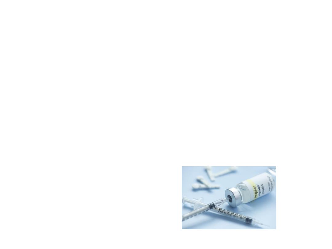
Insulin Therapy:
Using soluble ( Short acting ) insulin given either:
1. I.V infusion ( prefered method ):
o Bolus: 0.1 unit/kg. I.V direct
o then maintain continuous I.V infusion of 0.1 unit/kg./hr using syringe
pump.
2. I.M:
o Bolus: 10-20 units
o Followed by 5 units hourly.

Target blood sugar:
Falling 55-110 mg/dl per hr.
(3-6 mmol/l)
• Rapid decline → cerebral edema
• Failure to reach the target → require reassessment of insulin therapy.
Shift to subcutaneous insulin when the patient vomiting stopped and
become biochemically stable.
Fluid Replacement:
Average of 6 litres fluid deficit exist
3 L are extracellular replaced by 0.9% isotonic saline.
3 L are intracellular replaced by dextrose
Timing and amount as following:
• 1
st
hr: using normal (isotonic) saline
systolic BP > 90 mmHg → 1 L
systolic BP < 90 mmHg → 2 L
Set 2 wide bore IV line initially

• Then as :
1 L OVER 2 hrs
1 L OVER 2 hrs
1 L EVERY 6 hrs
Shift to 10% dextrose fluid whenever blood sugar level become < 250
mg/dl (14mmol/l).
Note: be cautious with elderly, pregnant, those with heart or renal
failure.

Potassium Replacememt
According to serum potassium level as:
> 5.5 mmol/l → non to be given
3.5 – 5.5 (mmol/l) → 40 meq/l
be cautious in replacing K usually hyperkalemia occurs initially due to
prerenal failure secondary to dehydration for that reason K is not
recommended to be given in the first hour of therapy.

Other
Acidosis: is usually corrected with the time by adequate fluid and insulin
replacement. Bicarbonate therapy is not recommended as it can induce
cerebral edema
Infection: should be treated by antibiotcs accordingly
Brain edema: is the leading cause of death in DKA, it can exist in spite of
metabolic stablisation. It should be treated by mannitol solution 20%
(7 ml/ kg.)

Hypoglycemia
Means blood glucose level < 63 mg/ dl (3.5 mmol/l) which is a common
complication in diabetes [ those on insulin therapy or on oral insulin
secretagoues especially sulphonylurea as Glibenclimide].
Risk factors of
sever
hypoglycemia :
1- strict glycemic control
2- extreme of age( elderly & young)
3- renal impairment
4- impaired awareness of hypoglycemia
5- long duration of DM
6- pevious history of hypoglycemia
7-ESRF (bz of insulin retention)

Causes of hypoglycemia :
1- missed or inadequate meal
2- error in therapy or poorly designed regimen
3- exercise
4- alcohol
5- lipohypertrophy at site of insulin injection.
6- factitious
7- breastfeeding

Clinical presentation:
I.
Autonomic symptoms: sweating, hunger, anxiety, trembling.
II.
Neuroglycopenic symptoms: confusion, inability to concentrate,
drowsiness, incoordination, slurring of speech, coma.
III.
Non specific: nausea, headache
Nocturnal hypoglycemia is common and usually not awake the person,
described as poor sleep, morning headache, vivid dreams. The partner
may notice sweating, twitching and seizure. It can be fatal in rare cases
( dead-in-bed syndrome)

Spontaneous hypoglycemia
means hypoglycemia that exist in non diabetic person (uncommon
condition).
---------------------------------------
Any Hypoglycemia can be Confirmed by whipple s criteria:
• symptoms of hypoglycemia
• low blood glucose
• symptoms resolved by correction of blood sugar.
Assessed by doing serum insulin & C-peptide level.

Management of Hypoglycemia:
Mild cases: oral 10-15 gm. Glucose followed by snack of complex
carbohydrate content.
Severe cases:
1- I.V hypertonic glucose 50% (30 ml) or 70 ml 20 %
2- I.M glucagon 1 mg
3- if the patient is conscious; 25 gm of oral refined sugar.

if the patient fail to respond
so we should exclude:
1- cerebral edema
2- alcohol intoxicaion
3- post ictal state
4- cerebral hemorrhage

Non-Ketonic hyper Osmolar Diabetic Coma:
• This condition is describe by sever hyper-glucaema (50
mmol/L(900mg/dL)) , without significant hyper-ketonaemia
or acidosis.
•
Sever dehydration and pre renal uremia are common.
Its usually affect eldelary patient , Many with previously
undiagnosed diabetes. Its carry high mortality rate (40%).
The treatment differ from Keto-aci-dosis in two main
respects:
First: the patients are more sensitive to insulin, they
required half dose of insulin(1-3 units)/hr.
secondly: the plasma osmolarity should be measured.

It can be calculated by using this formula:
Plasma osmolarity= 2(Na+)
+
2(k+)
+
Blood glucose
+
Blood urea {All in
mmol/L/Kg}
The conscious is depressed when it is high 340 mmol/L/Kg
Normal Value ((280-300)mmol/L/Kg).
The patien should be given 0.45% of saline until the osmolarity approach
normal when isotonic saline(0.9%) should be substituted.
Thrombo-embolic complications are common and heparin should be givin
1cc (5000 u.)*2 S.C .

Lactic Acidosis:
• The coma in Lactic acidosis is likely to be in diabetic
patient on Metformin therapy, for type II D.M.
The patient is very ill and over breathing(sever acidosis)
but not profoundly dehydrated, like in diabolic-keto-
acidosis, and the patient smell dose not smell of acetone
and ketone, urea is mild or even abscent.
•
The plasma bicarbonate and PH are markedly reduced
(sever acidosis) (PH<7.2) and the anoin gap is increased.
The diagnosis is confirm by high does of lactic
acid(>5.0mmol/L) in the blood.
The treatment is I.V sodium bicarbonate sufficient to
raise the artrial PH above 7.2 along with insulin and
glucose.
Mortality is high>50% in spite of treatment.