
1
Course: Microbial Physiology
Lecturer: Dr. Weam Saad
Lecture: Prokaryotes Growth
Prokaryotes
Growth
Growth is an increase in cell mass by forming new protoplasm from nutrients
in the environment. In bacteria, growth involves increase in cell components,
number of ribosomes, duplication of the bacterial chromosome, synthesis of
new cell wall and plasma membrane, formation of the two chromosomes,
septum formation, and cell division. This asexual process of reproduction is
called binary fission.
Growth can be measured by: changes in cell mass and changes in cell
numbers.
Methods for Measurement of Cell Mass
Methods for measurement of the cell mass involve both direct and indirect
techniques.
1. Direct physical measurement of dry weight, wet weight, or volume of cells
after centrifugation.
2. Direct chemical measurement of some chemical component of the cells such
as total N, total protein, or total DNA content.
3. Indirect measurement of chemical activity such as rate of O
2
or
CO
2
production or consumption, production or consumption, etc.
4. Turbidity measurements or optical density using instruments of a suspension
of bacterial cells. The method is simple, but the sensitivity is limited to about
107 cells per ml for most bacteria.

2
Methods for Measurement of Cell Numbers
1. Direct microscopic counts are possible using special slides known as
counting chambers. Dead cells cannot be distinguished from living ones.
2. Electronic counting chambers count numbers and size of cells.
3. Indirect viable cell counts, also called plate counts, involve plating
(spreading) a sample of a culture on a nutrient agar surface. The sample or cell
suspension can be diluted in a nontoxic diluent (e.g. saline) before plating. Each
viable (still alive) unit grows and forms a colony. Each colony that can be
counted is called a colony forming unit (cfu) and the number of cfu's is the viable
number of bacteria in the sample.
Advantages of the technique is a single cell can be detected. The Disadvantages
are:
(1) Only living cells develop colonies that are counted
(2) Multi-cells form a single colony.
(3) Colonies will form only when cultural conditions are suitable or good for
growth.
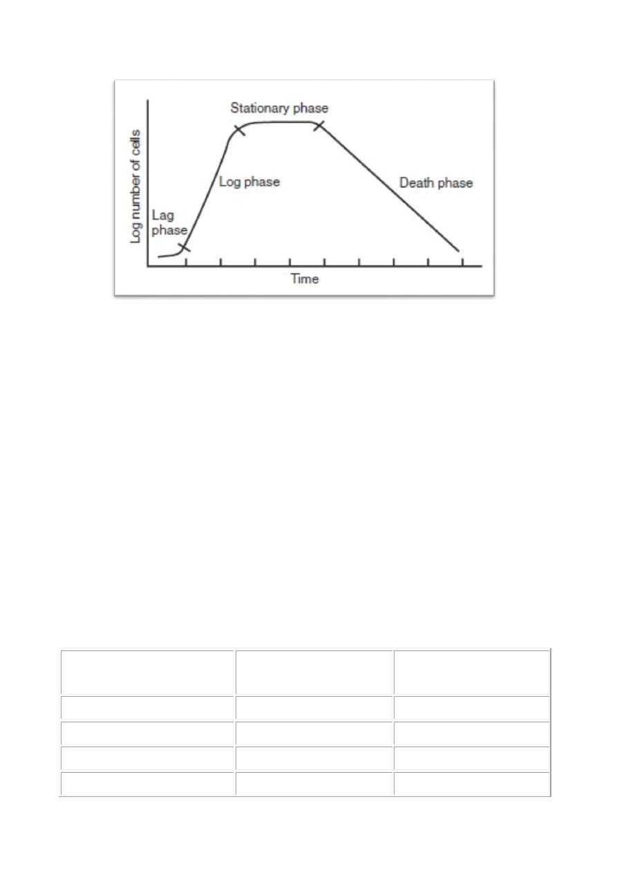
The Bacterial Growth Curve
In the laboratory, under good conditions, a growing bacterial population doubles
by geometrically: 1, 2, 4, 8, etc. or 20, 21, 22, 23.........2n (n = the number of
generations). This is called exponential growth.
The typical bacterial growth curve of batch cultures (growth cycle of
bacteria)
1. Lag Phase. Immediately after inoculation of the bacterial cells into fresh
medium, there is no cell division occurring, the cells grow in volume and
mass, synthesizing enzymes, proteins, RNA, etc., and increasing in

3
metabolic activity. The length of the lag phase depends on many factors
including:
the size of the inoculum
time to recover from physiacal damage or shock in the transfer
time required for synthesis of essential coenzymes or division
factors
time required for synthesis of new (inducible) enzymes that are
necessary to metabolize the new substrates in the medium.
2. Exponential (log) Phase. The exponential phase of growth is a balanced
growth and all the cells are dividing by binary fission, and are growing
geometrically. The cells divide at a constant rate and consume of the growth
medium by help of good standard conditions of incubation.
The rate of exponential growth of a bacterial culture is the generation time, also
the doubling time of the bacterial population. Generation time (G) is defined as
the time (t) per generation (n = number of generations). Hence, G=t/n is the
equation from which calculations of generation time (below) derive.
3. Stationary Phase. In closed system such as a test tube or flask the bacterial
growth is limited by the factors:
The available nutrients finished
Accumulation of metabolites wastes or end products
No enough biological space
During the stationary phase in batch cultures, number of cells are dying =
number of cells are dividing. Bacteria usually produce secondary metabolites,
such as antibiotics during the stationary phase of the growth cycle, also spore-
forming bacteria will start sporulation process.
4. Death Phase. Decrease the viable cells geometrically (exponentially).
4
Growth Rate and Generation Time
The generation time bacterial growth is the rate during the phase of exponential
growth, under standard nutritional conditions (culture medium, temperature,
pH, etc.). Generation times for bacteria is different from 12 minutes to 24 hours
or more.
The generation time for E. coli in the laboratory is 15-20 minutes, but in the
intestinal tract, the coliform's generation time is 12-24 hours. Rhizobium have
longer generation times. Many lithotrophs, such as the nitrifying bacteria, also
have long generation times. Some bacteria that are pathogens, such
as Mycobacterium tuberculosis and Treponema pallidum, have long generation
times and it is factor of virulence.
Generation times for some bacteria under standared conditions of
growth.
Bacterium
Medium
Generation
Time
(minutes)
Escherichia coli
Glucose-salts
17
Bacillus megaterium
Sucrose-salts
25
Streptococcus lactis
Milk
26
Streptococcus lactis
Lactose broth
48

5
Staphylococcus aureus
Heart infusion broth
27-30
Lactobacillus acidophilus Milk
66-87
Rhizobium japonicum
Mannitol-salts-yeast
extract
344-461
Mycobacterium
tuberculosis
Synthetic
792-932
Treponema pallidum
Rabbit testes
1980
Calculation of Generation Time
The increase in a bacterial population is by binary fission. If we start with one
cell, when it divides, there are 2 cells in the first generation, 4 cells in the second
generation, 8 cells in the third generation, and so on. The generation time is the
time required for the cells (or population) to divide.
G (generation time) = t (time, in minutes or hours)/n (number of generations)
G = t/n
G = generation time (time for the cells to divide)
t = time interval in hours or minutes
B = number of bacteria at the beginning of a time interval
b = number of bacteria at the end of the time interval
n = number of generations (number of times the cell population doubles during
the time interval)
b = B x 2n (This equation is an expression of growth by binary fission)
n is calculated as the following:
logb = logB + nlog2

6
n = logb - logB
log2
n = logb - logB
.301
n = 3.3 logb/B
G = t/n
G = t
3.3 log b/B
Example: What is the generation time of a bacterial population that increased
from 10,000 cells to 10,000,000 cells in four hours of growth?
G = t
3.3 log b/B
G = 240 minutes
3.3 log 10
7
/10
4
G = 240 minutes
3.3 x 3
G = 24 minutes
Continuous Culture of Bacteria
In batch cultures nutrients are not renewed, exponential growth is limited to a
few generations. Bacterial cultures can continue in the exponential growth for
long periods of time using a system of continuous culture. It is occur in a device
called a chemostat to keep a bacterial population in similar conditions to
bacterial growth in natural environments.
In a chemostat, the growth chamber is connected to a reservoir of medium
(sterile tank for storage of media) and fresh medium is continuously supplied
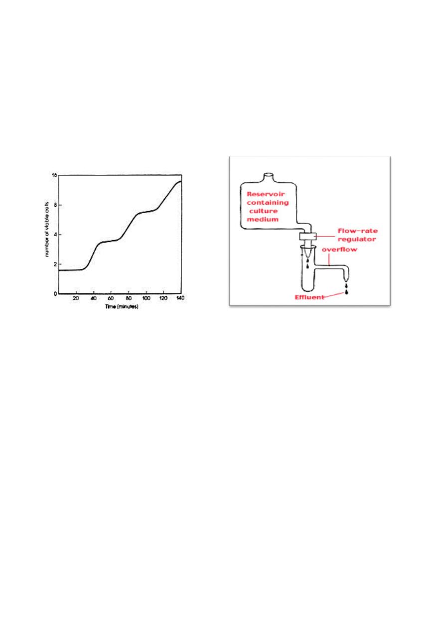
7
from the reservoir and enter into the growth chamber at a rate that limits the
growth of the bacteria. The rate of addition of the fresh medium determines the
rate of growth because the fresh medium always contains a limiting amount of
an essential nutrient. Thus, the chemostat avoid factors that start staitionary
phase like the accumulation of toxic substances, and the accumulation of
bacterial cells.
Control Microbial Growth
The control of microbial growth is necessary in agriculture, medicine, and food
science have been made through study of this area of microbiology. It means to
prevent growth of microorganisms by:
(1) killing microorganisms.
(2) inhibiting the growth of microorganisms.
Control of growth usually involves the use of physical or chemical agents which
either kill or prevent the growth of microorganisms. Agents which kill cells are
called cidal agents; agents which inhibit the growth of cells (without killing
them) are static agents. Thus the term bactericidal is killing bacteria
and bacteriostatic is inhibiting the growth of bacterial cells. Example:
the bactericide kills bacteria, a fungicide kills fungi, and so on.
Sterilization is the complete destruction or killing all viable microorganisms
using heat, radiation or chemicals, or physical removal of cells.

8
Methods of Sterilization
2. Heat: most important and widely used. Endospores of bacteria are can
resist.
3. Incineration: burns and destroy microorganisms (more than 500
o
). Used
for needles, inoculating wires and glassware.
4. Boiling: 100
o
for 30 minutes. Kills everything except some endospores
(Actually, for the purposes of purifying drinking water 100o for five
minutes is enough but some Giardia cysts can survive). To kill endospores
needs very long boiling or intermittent boiling.
5. Autoclaving (steam under pressure or pressure cooker): 121
o
for 15
minutes. Good for sterilizing for anything, heat-labile substances will be
denatured or destroyed.
6.
Dry heat (hot air oven): 160
o
/2hours or 170
o
/1hour. Used for glassware,
metal, and objects that do not melt.
7. Pasteurization (batch method) 63
o
/30 minutes heating (mild heat), kills
most vegetative bacterial cells including pathogens such as streptococci,
staphylococci and Mycobacterium tuberculosis and reduce the number
of microorganisms Usually used in food and milk products
8. Irradiation: destroys nucleic acids. Ultraviolet light is usually used
(commonly used to sterilize the surfaces of objects), x-rays and
microwaves are used in canned food.
9. Filtration: it is the physical removal (exclusion) of all cells in a liquid or
gas, especially important to sterilize solutions which are sensitive and
denature by heat (e.g. antibiotics, drugs, amino acids, vitamins, etc.)
10. Chemical and gas: (formaldehyde, glutaraldehyde, ethylene oxide) toxic
chemicals kill all forms of life in the gas chamber.
Control of Microbial Growth by Physical Agents
1. Heat. The time required to kill depends on the number of organisms,
species, pH, and temperature. Low temperature (refrigeration and
freezing) used in control because most microorganisms do not grow at
zero
o
. Store foods at low temperatures to slow rate of growth and and
avoid spoilage (e.g. milk). Drying (removal of H
2
O): Most

9
microorganisms cannot grow at reduced water activity (Aw < 0.90), this
method is used to preserve foods for storage (e.g. fruits, grains, etc.).
Methods involve removal of water from product by heat, evaporation,
freeze-drying, and addition of salt or sugar e.g. jams and pickle.
2. Irradiation (microwave, UV, x-ray): destroys microorganisms during
sterilization.
Control of microbial growth by chemical agents
Antimicrobial agents are chemicals that kill or inhibit the growth
microorganisms. Antimicrobial agents include chemical preservatives and
antiseptics and drugs used in the treatment of infectious diseases. Antimicrobial
agents may be of natural or synthetic, and they are static or cidal for
microorganisms.
Types of antimicrobial agents
1. Antiseptics: microbicidal agents can be applied to the skin and mucous
membrane, e.g. silver nitrate, iodine solution, alcohols, detergents.
2. Disinfectants: Agents that kill microorganisms, but not their spores, not
safe for living tissues; they are used for tables, floors. Examples: chlorine,
hypochlorites, chlorine compounds and ammonium compounds.
3. Preservatives: used to inhibit the growth of microorganisms in foods,
nontoxic for human. Examples; calcium propionate, sodium benzoate,
formaldehyde, nitrate, sulfur dioxide.
4. Chemotherapeutic agents: synthetic antimicrobial agents used in the
treatment of microbial or viral disease (infectious diseases). Examples:
sulfonilamides, isoniazid, ethambutol, chloramphenicol.
5. Antibiotics: antimicrobial agents produced by microorganisms that kill or
inhibit other microorganisms, used to kill or inhibit infectious Bacteria.
Antibiotics are low molecular-weight molecules produced as secondary
metabolic products, mainly by microorganisms that live in the soil during
the processes of sporulation. Penicillium and Cephalosporium , which the
beta-lactam antibiotics. In the Bacteria, the Actinomycetes and
Streptomyces species, produce many antibiotics including the

11
aminoglycosides (e.g. streptomycin), macrolides (e.g. erythromycin), and
the tetracyclines. Endospore-forming Bacillus species produce
polypeptide antibiotics such as polymyxin and bacitracin.
Mechanism of action for Antimicrobial Agents Used in the Treatment of
Infectious Disease
The most important thing is the selective toxicity of the antimicrobial agent (kill
microorganisms and not toxic on human or animal). The range of bacteria or
other microorganisms that are affected by a certain antibiotic are is expressed as
its spectrum of action. Antibiotics are effective against prokaryotes (kill or
inhibit) a wide range of Gram-positive and Gram-negative bacteria are called
broad spectrum. If effective mainly against Gram-positive or Gram-negative
bacteria, they are called narrow spectrum
1. Cell wall synthesis inhibitors Cell wall synthesis inhibitors inhibit the
synthesis of bacterial peptidoglycan e.g. Penicillin and Cephalosporin are
2. Cell membrane inhibitors they inhibit the function of bacterial membranes.
E.g.Polymyxin, produced by Bacillus polymyxis.
3. Protein synthesis inhibitors, their action is inhibition of some step in the
process of translation in tRNA and 70S ribosomes by binding to bacterial
ribosomes and preventing the initiation of protein synthesis. e.g.
tetracyclines, chloramphenicol, streptomycin , gentamicin, erythromycin and
streptomycin.
4. Effects on Nucleic Acids (the synthesis of DNA or RNA or bind to DNA or
RNA so the messages cannot be read. Only two nucleic acid are used, they have
selective activity against prokaryotes nalidixic acid and rifamycins. Because
most these Nucleic Acids drugs are not selective and affect human cells and
bacterial cells.
5. Competitive Inhibitors. Some are bacteriostatic and some are bactericidal.
They are inhibitors of the bacterial enzymes required for the synthesis of
important growth molecules. E.g. Sulfonamides inhibit folic acid formation
which is essential for 1-carbon transfer reactions.

11
Mechanisms of Bacterial resistance to antibiotics
Acquired resistance bacteria can develop resistance to antibiotics, e.g. bacterial
population was sensitive to antibiotics become resistant by changes in the
bacterial genome by two genetic processes in bacteria:
(1) Mutation and selection (called vertical evolution or natural selection), only
resistant bacteria can grow.
(2) Exchange of genes between strains and species (called horizontal
evolution). Bacteria are able to exchange genes in nature by three processes:
conjugation, transduction and transformation. Conjugation involves cell-to-
cell contact as DNA crosses a sex pilli from donor to recipient. The transduction,
a virus transfers the genes between bacteria. In transformation, DNA is acquired
directly from the environment after released from another cell. Genetic
recombination follow the transfer of DNA from one cell to another leading to
formation of recombinant DNA.
The medical problem of bacterial drug resistance is that antibiotic becomes
useless in the treatment of infectious disease caused by that pathogen, e.g.
natural penicillins have become useless against Staphylococcus aureus and
replaced by other antibiotics.