Respiratory
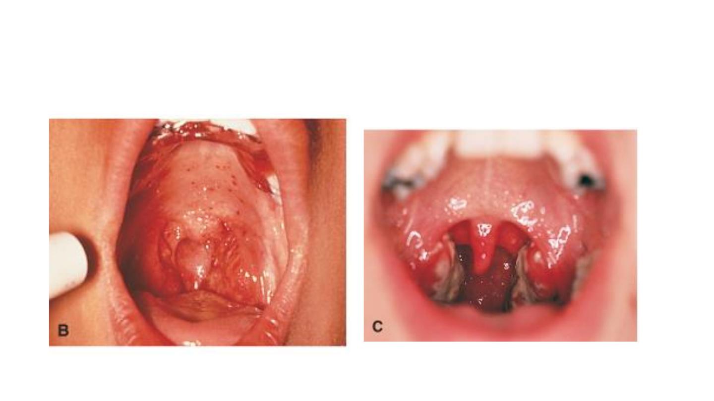
Streptococus pharyngitis
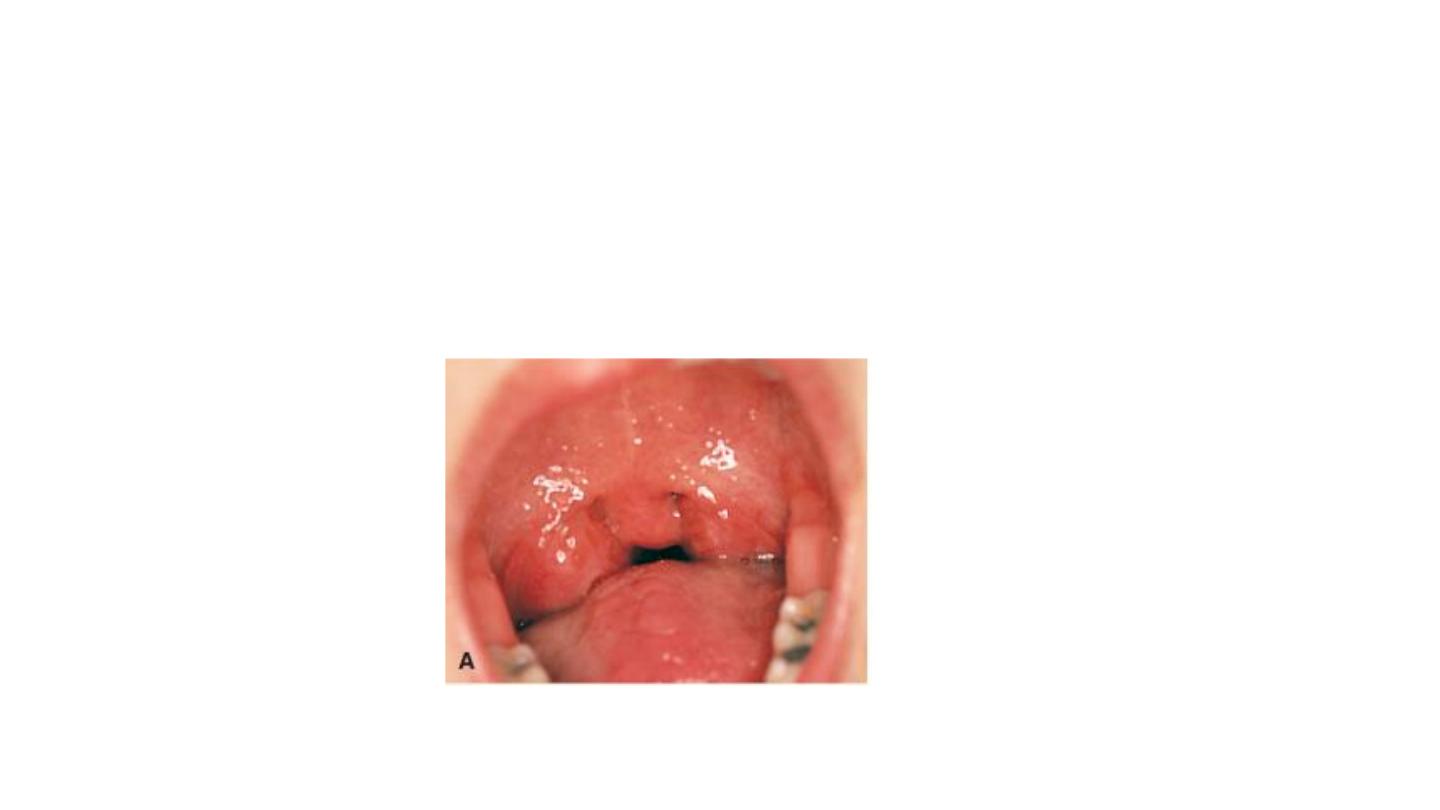
Viral pharyngitis
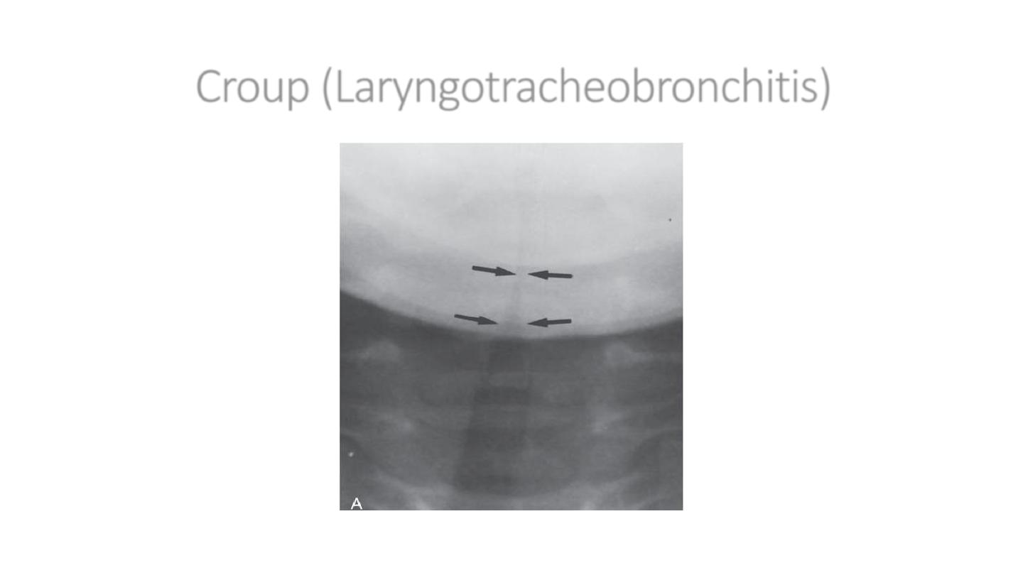
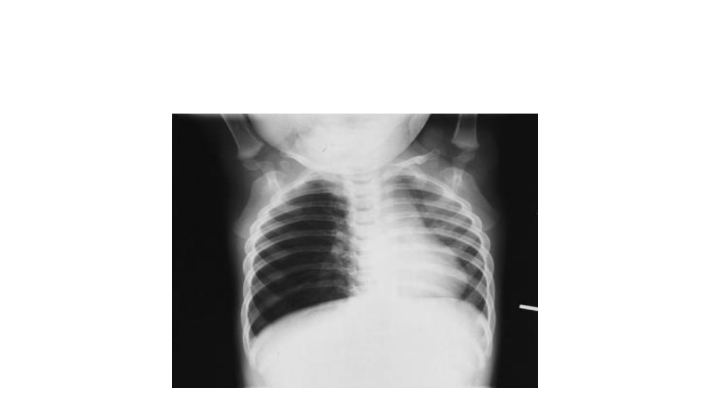
Croup (Laryngotracheobronchitis)
Steeple sign
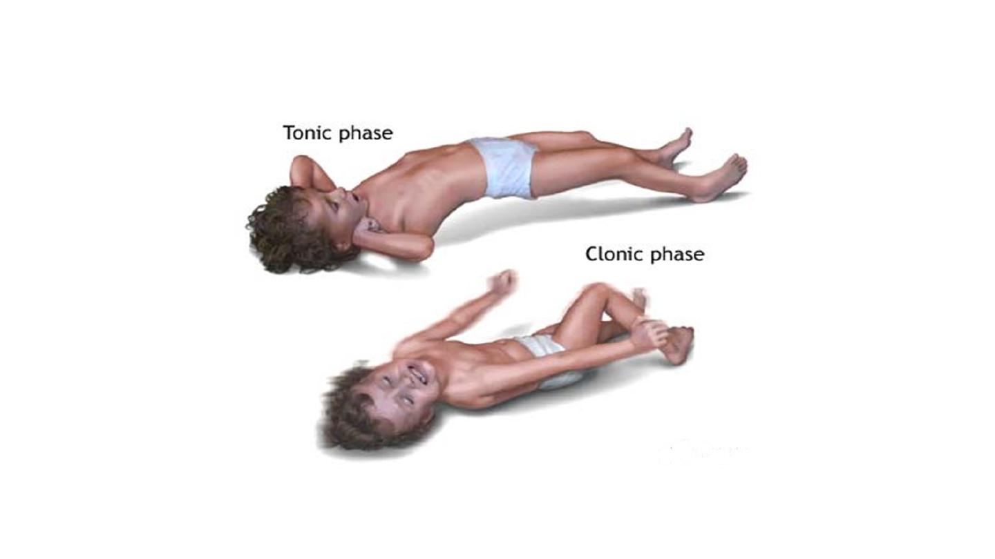
Asthmatic pt
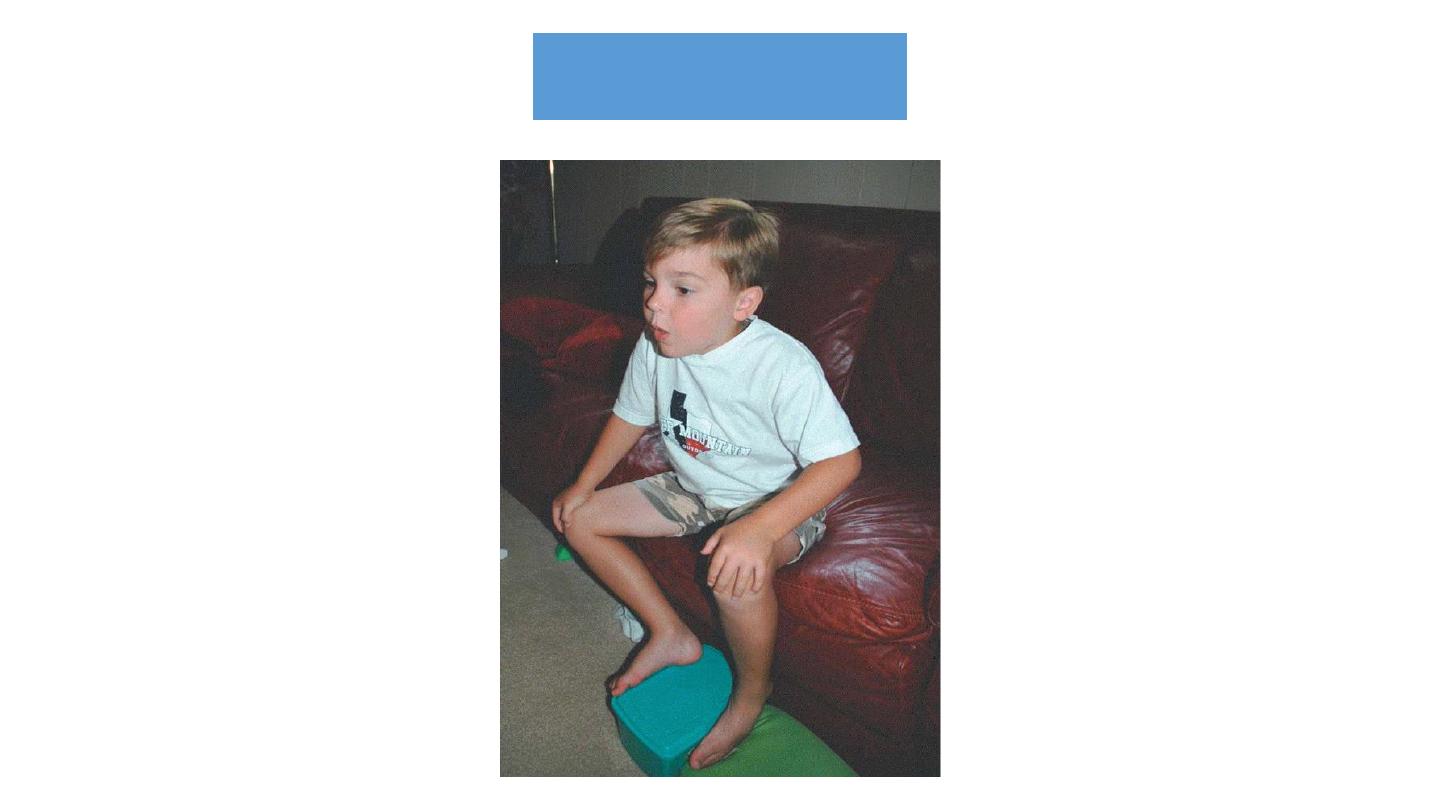
Tripod position
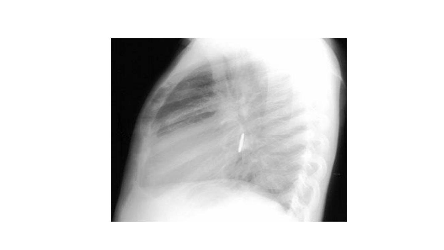
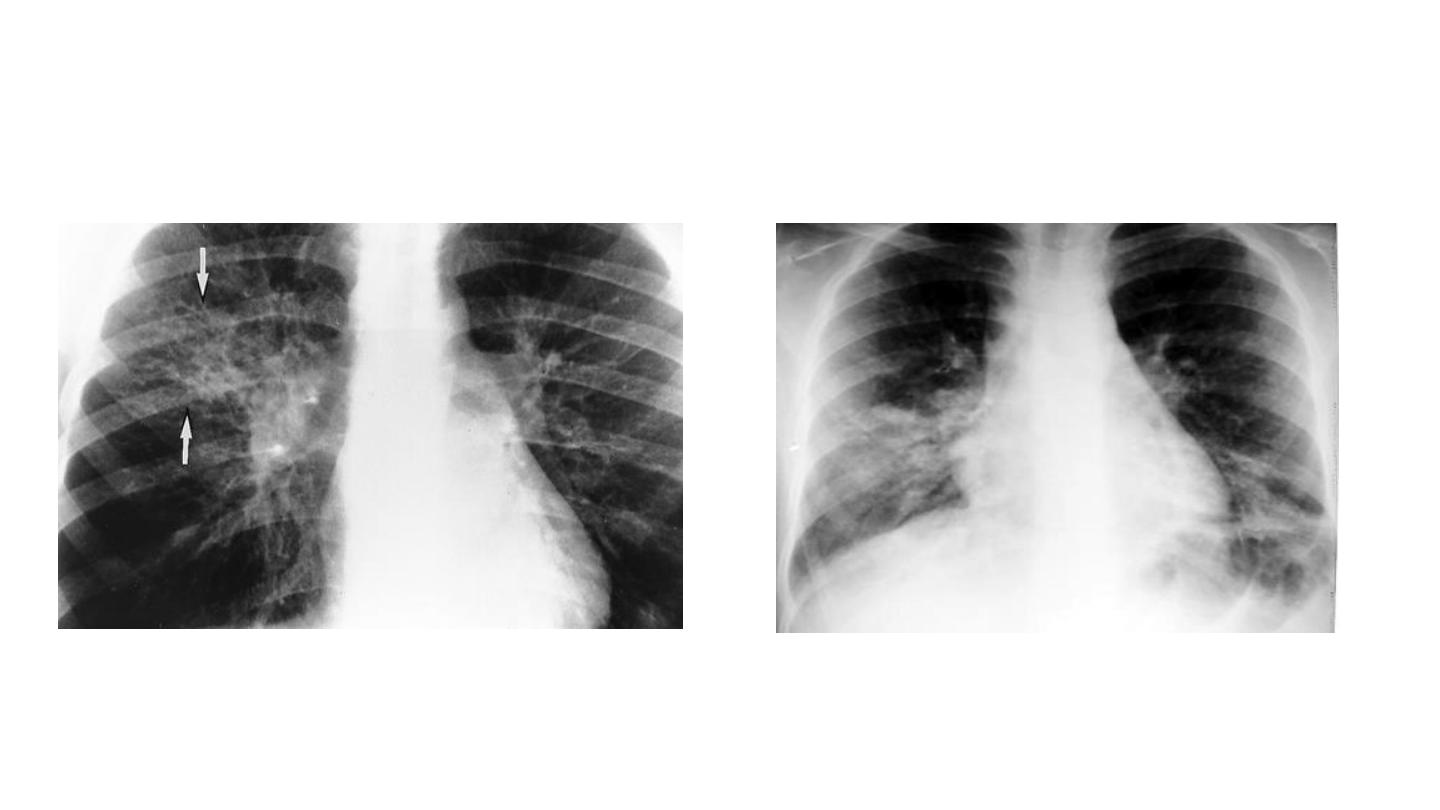
Expiratory chest radiograph in a 12-month-old boy with a 2-month history of wheezing
demonstrates continued hyperlucency and hyperexpansion of the right hemithorax.
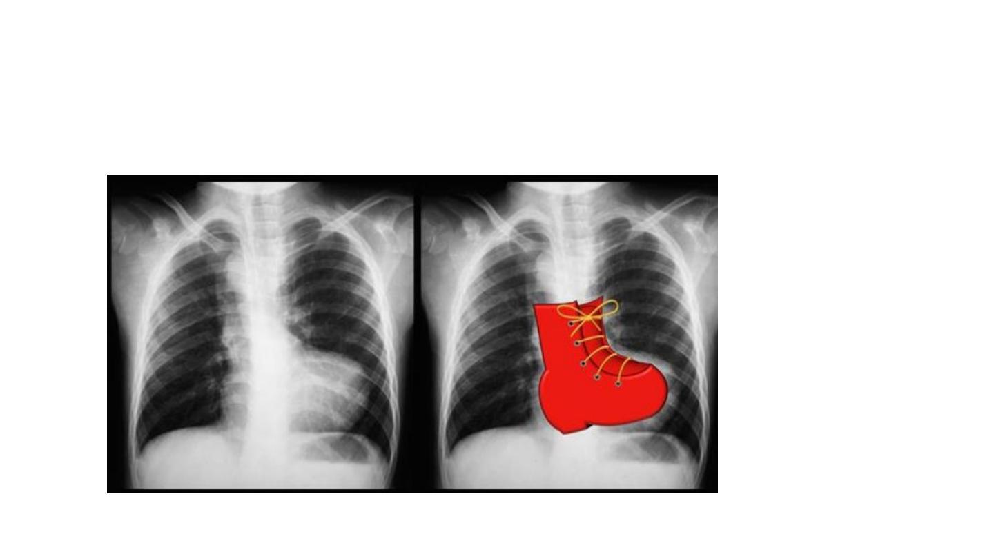
Foreign body
Foreign body
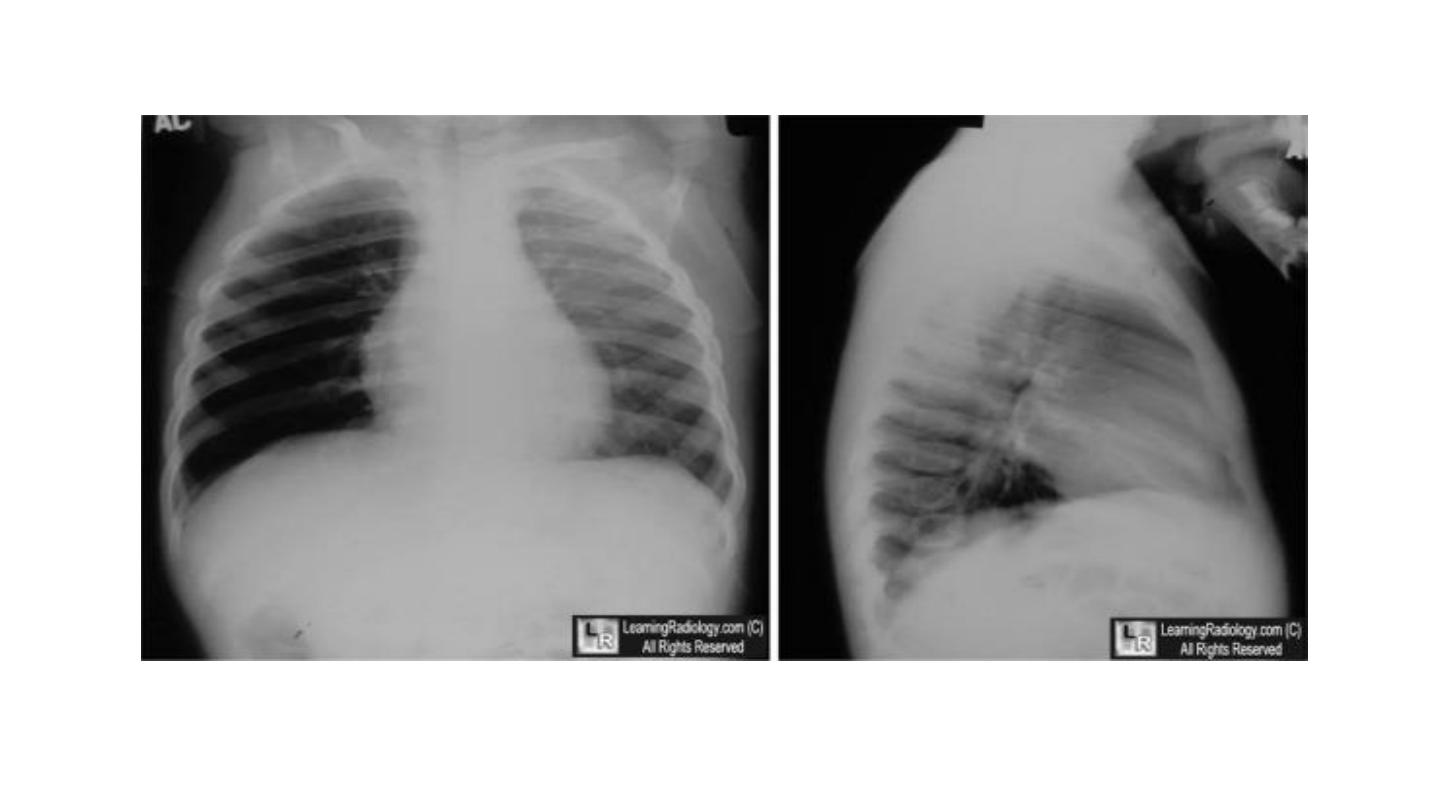
Foreign body
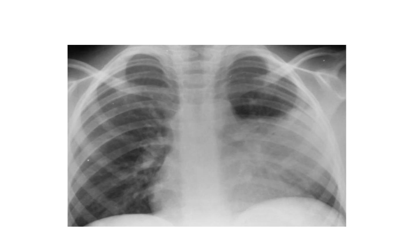
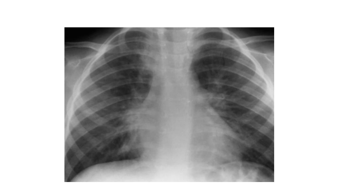
Lobar Pneumonia
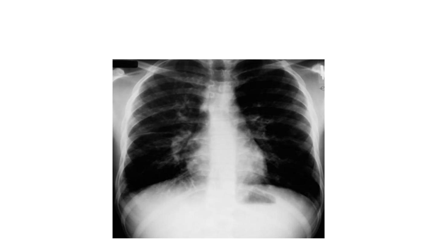
Bronchopneumonia
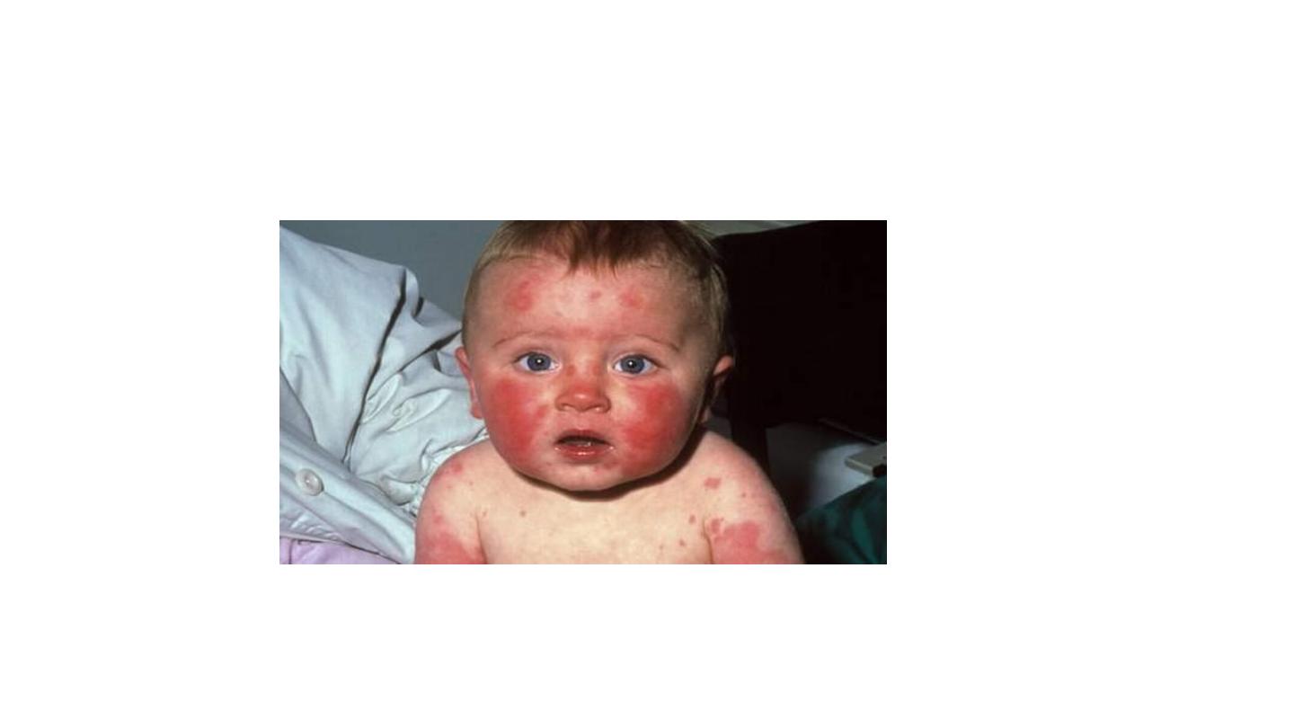
Mycoplasma pneumoniae infection (atypical
pneumonia)

• Whooping cough
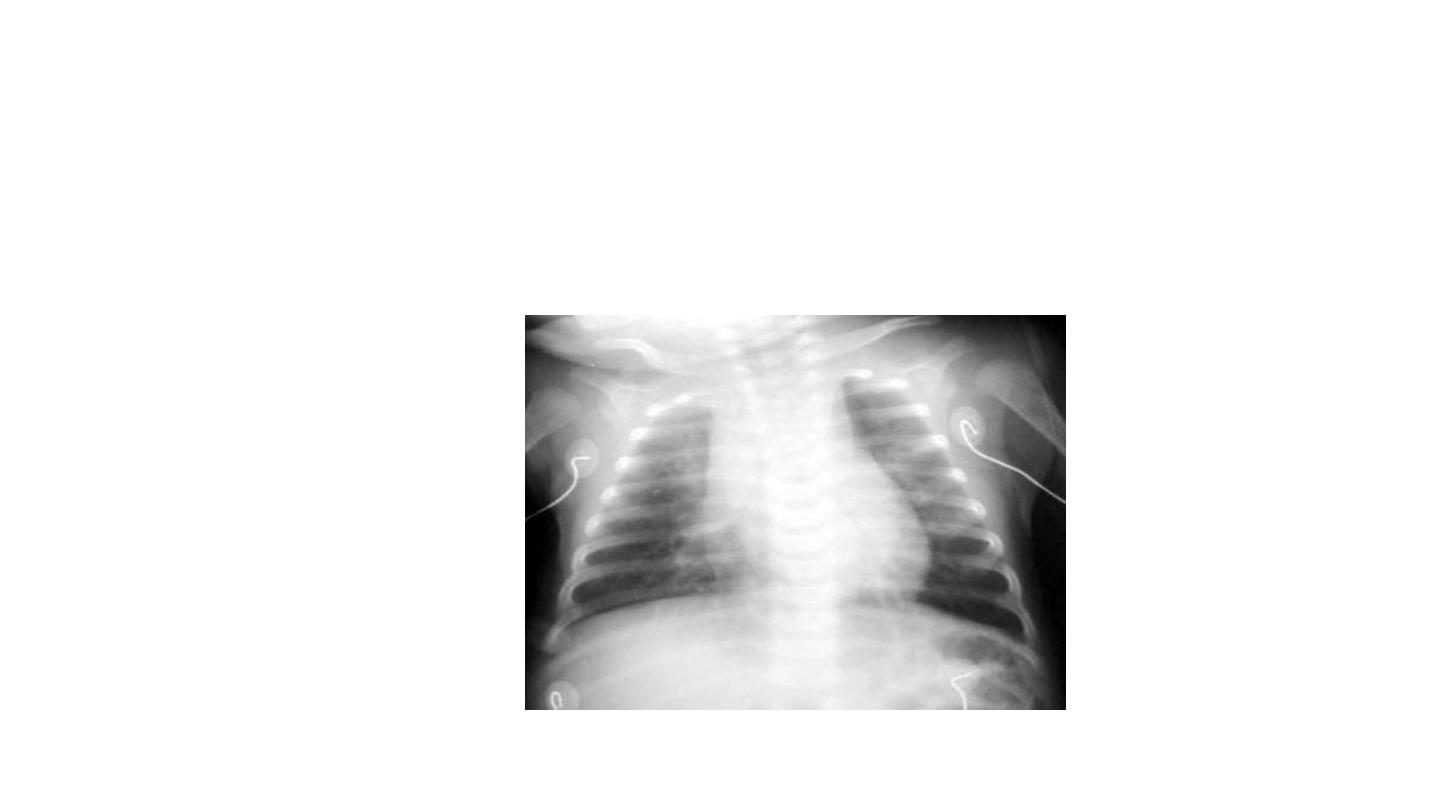
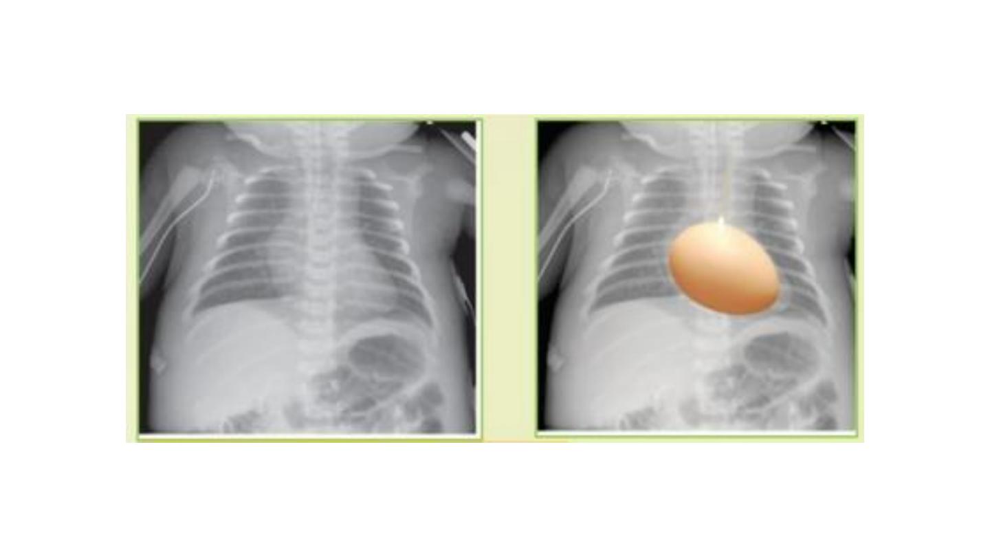
"shaggy heart" on CXR in a patient with
Bordetella pertussis pneumonia.
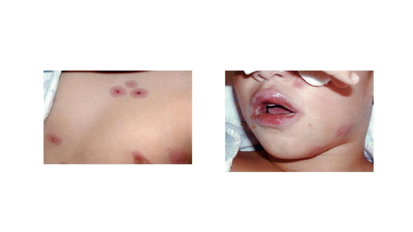
Stevens-Johnson-like syndrome
associated with Mycoplasma pneumoniae infection
Lower lobe pn
Hilar involvement

Neonatology
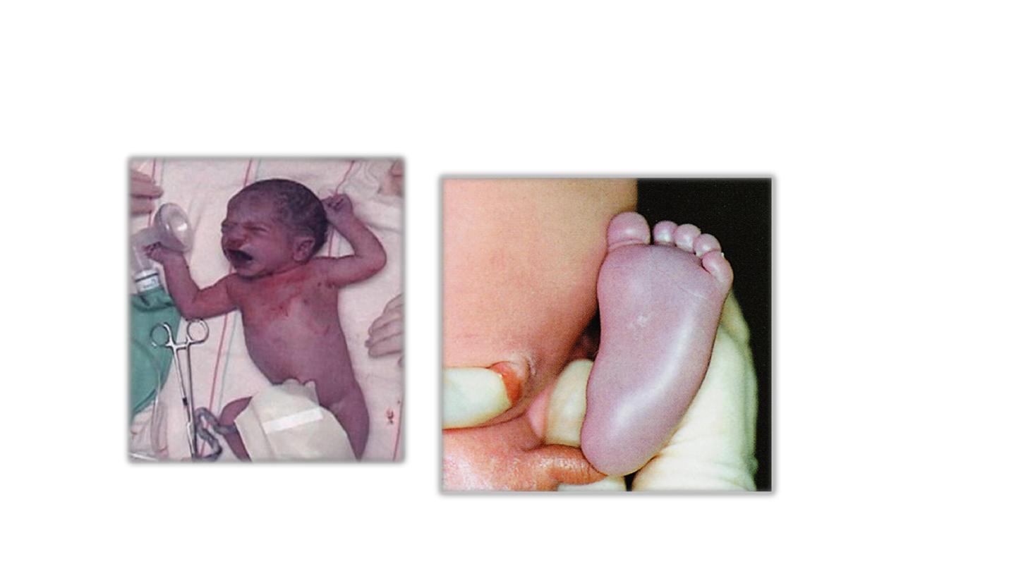
Acrocyanosis
Central cyanosis
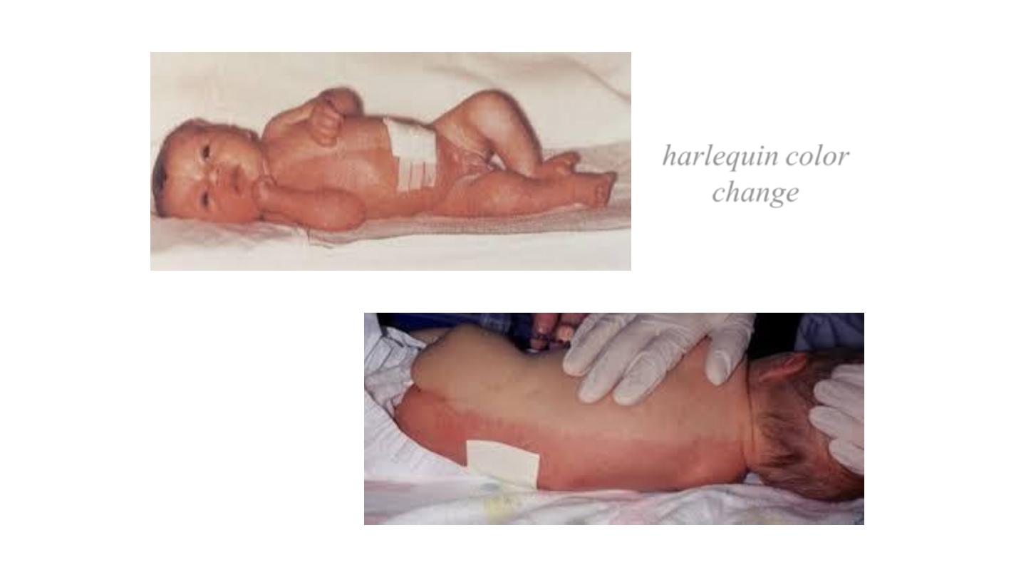
harlequin color
change
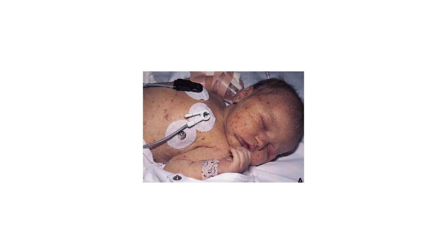
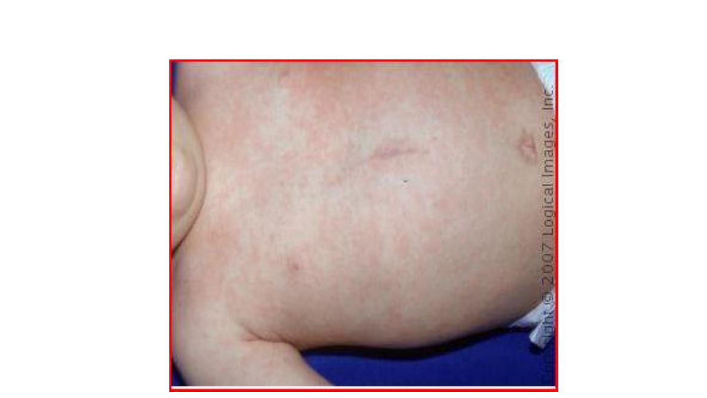
Petechiae
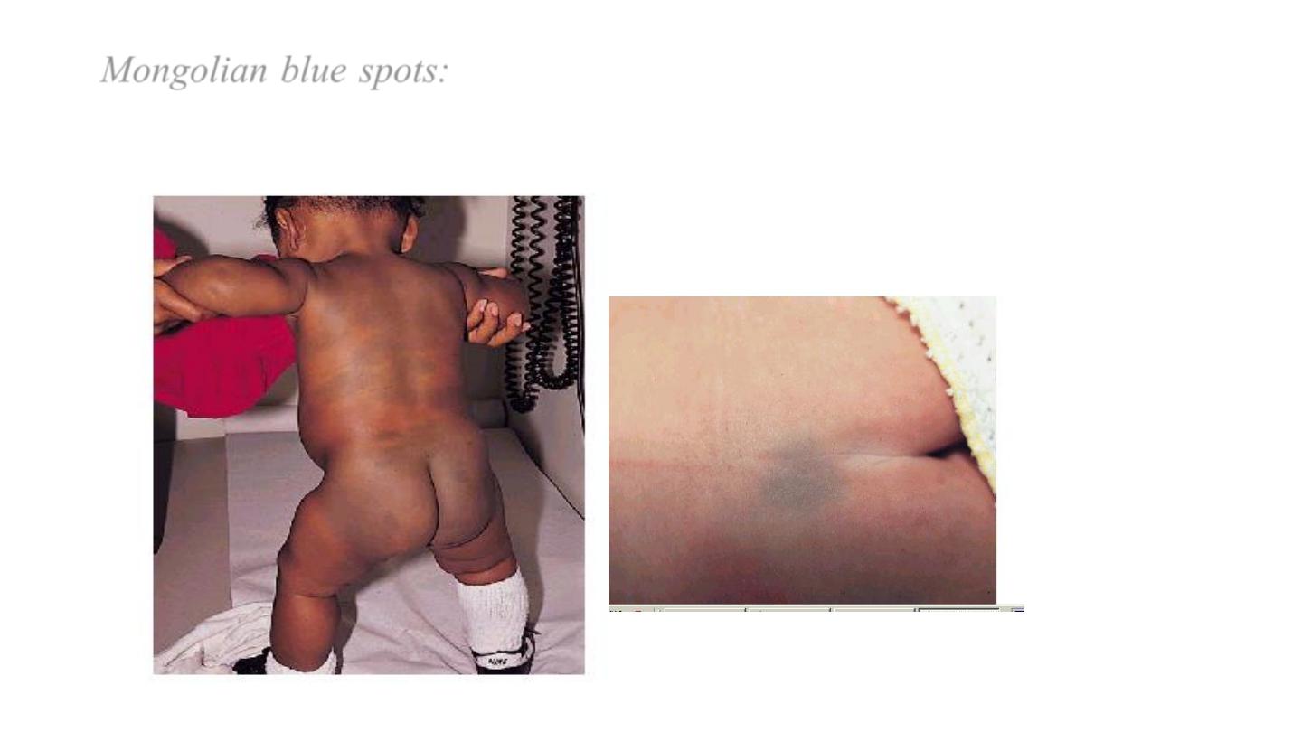
Mongolian blue spots:
are blue well demarcated areas of pigmentation
are seen over the buttocks, back and sometimes other parts of the body,
they tends to disappear within the first year of life.
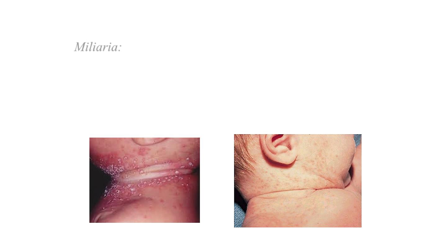
Miliaria:
erythematous minute papulovesic-ular lesions
may impact a prickly sensation the lesions are usually
located to sites of occlusion or to flexural areas such as
the neck, groin, and axilla. It is due to retention of
sweat in occluded sweat ducts.
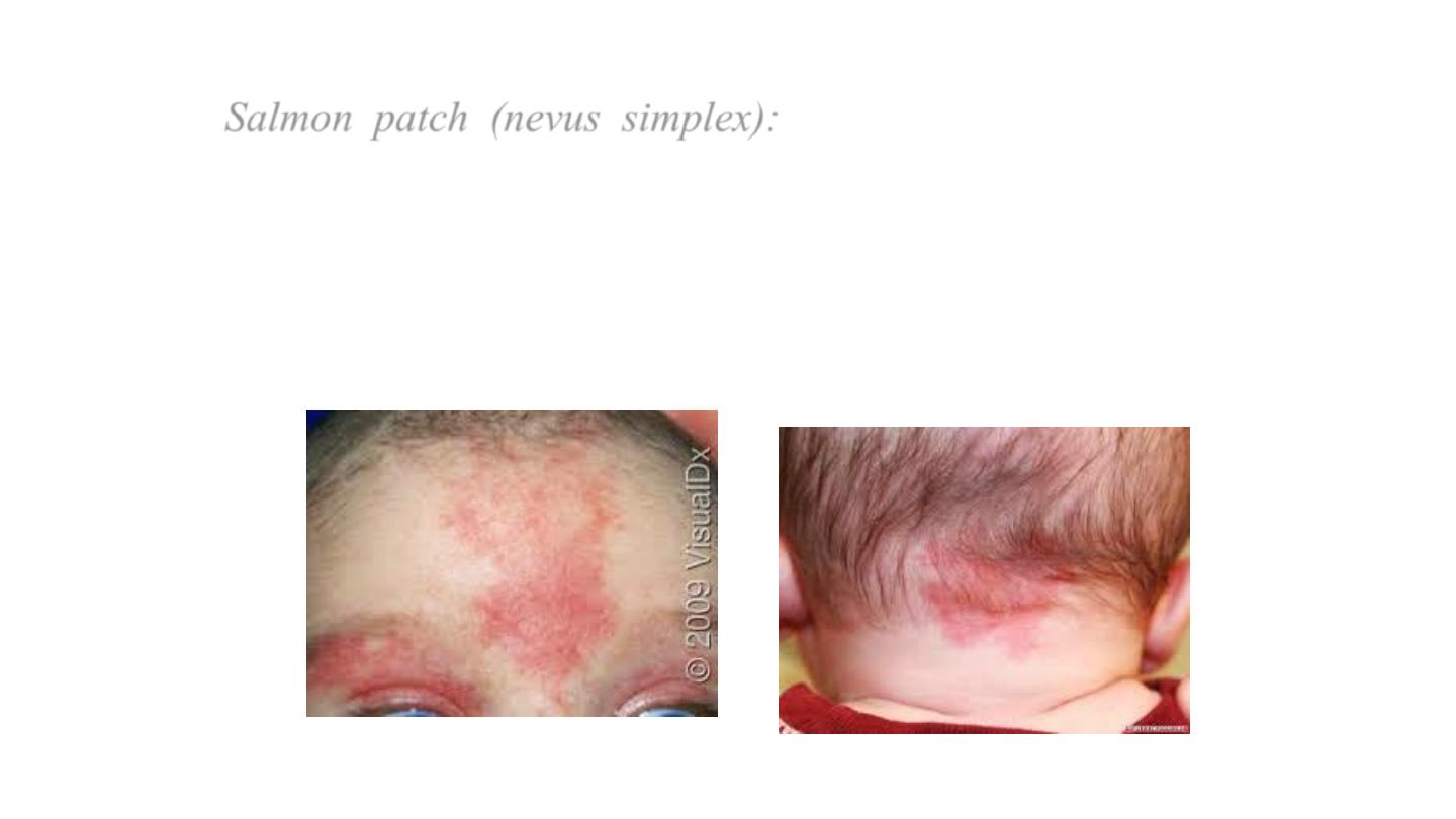
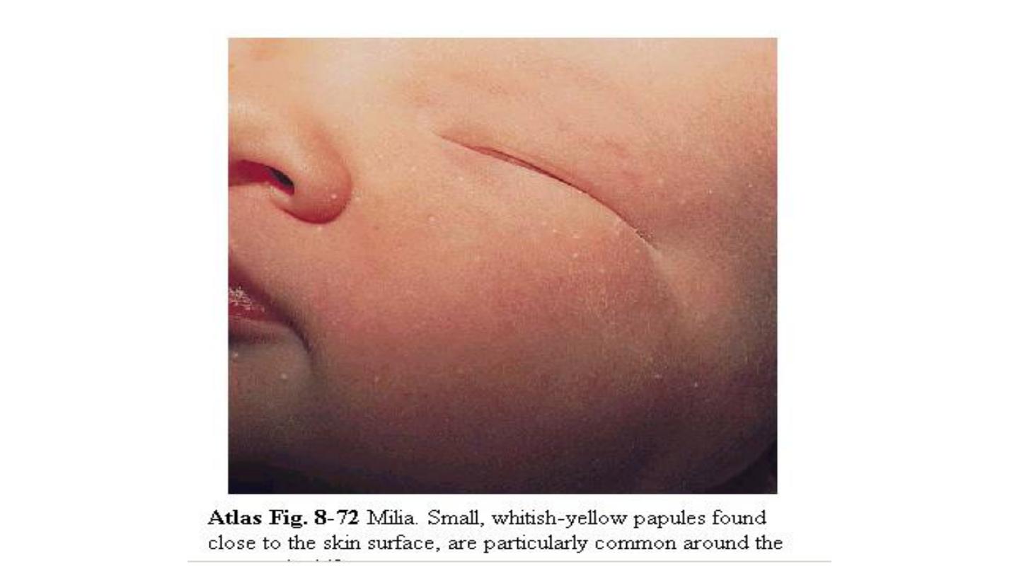
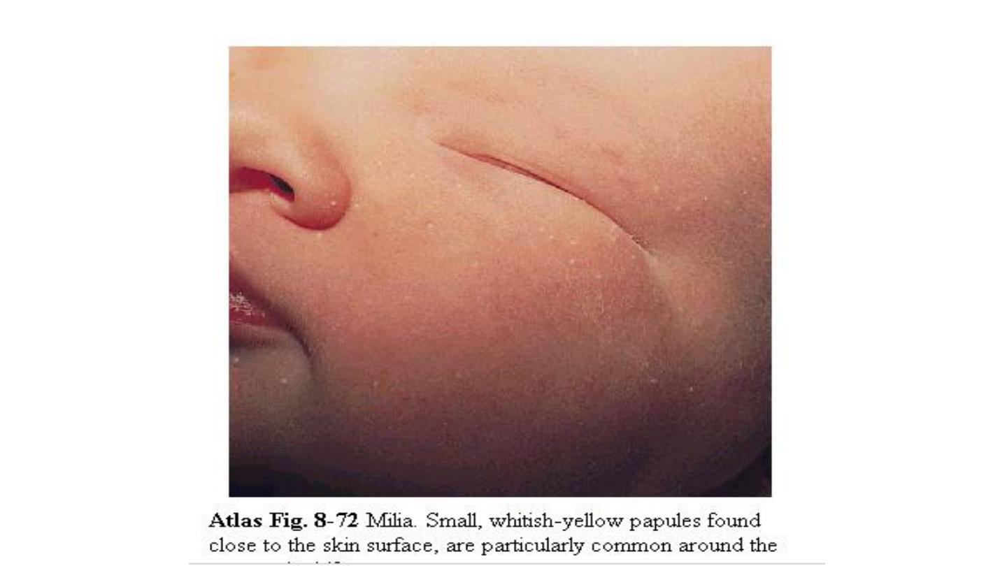
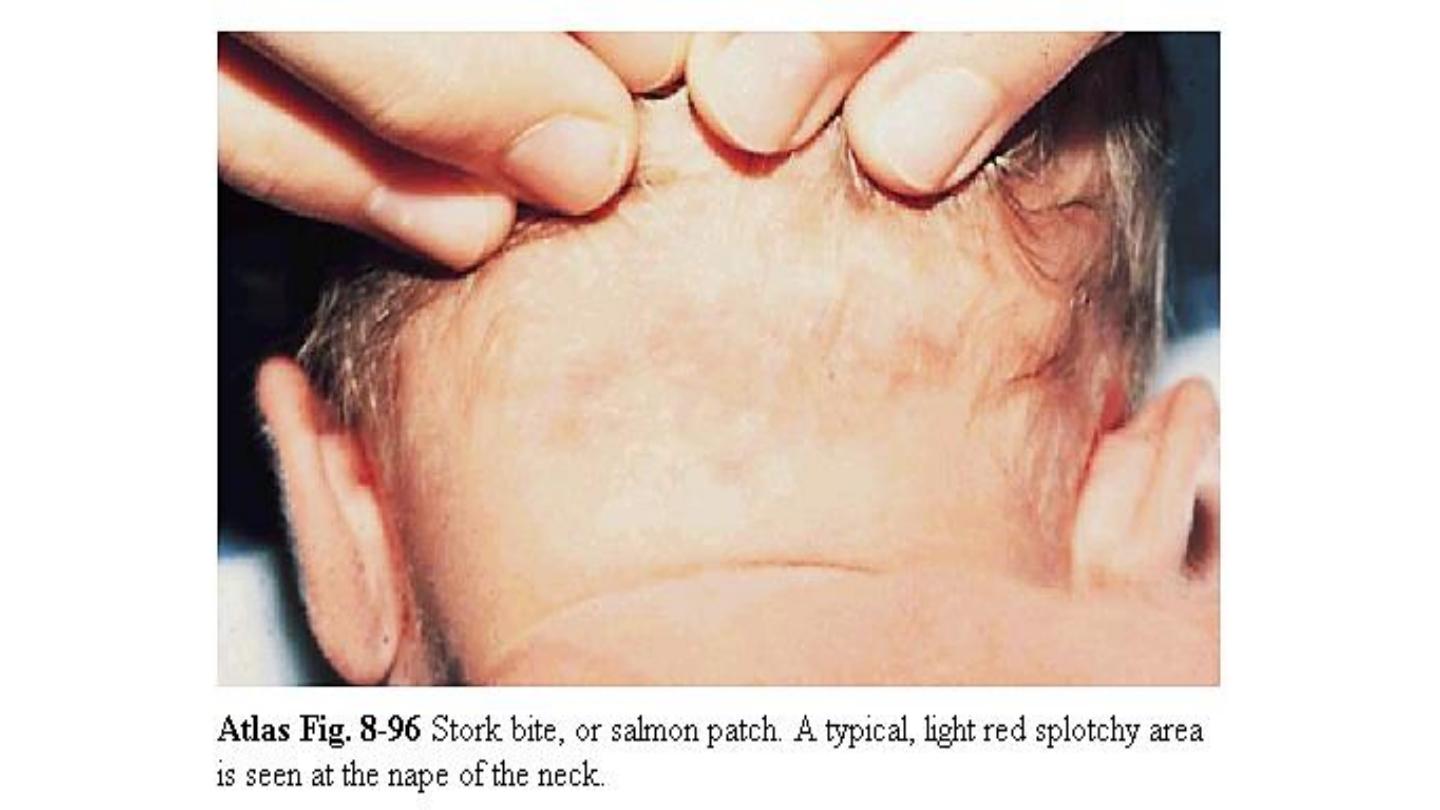
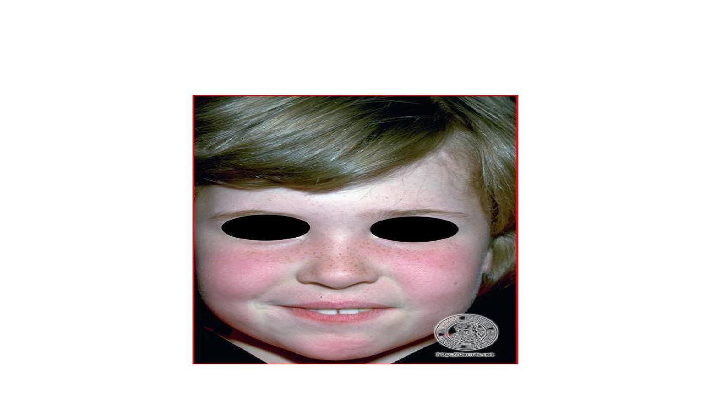
Salmon patch (nevus simplex):
are small
pale pink ill
defined flat vascular lesions that occur mostly on the
glabella, eye lid, upper lip & nuchal area of normal NBB,
they may persist for several months & become more
visible with crying

See neonatal examination lecture plz
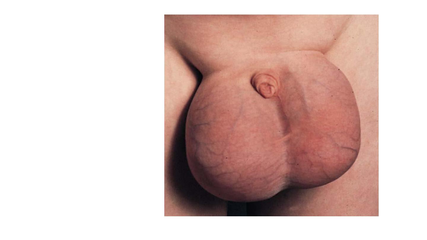
Caput succedaneum
cephalhematoma

Erb’s palsy

opisthotonos
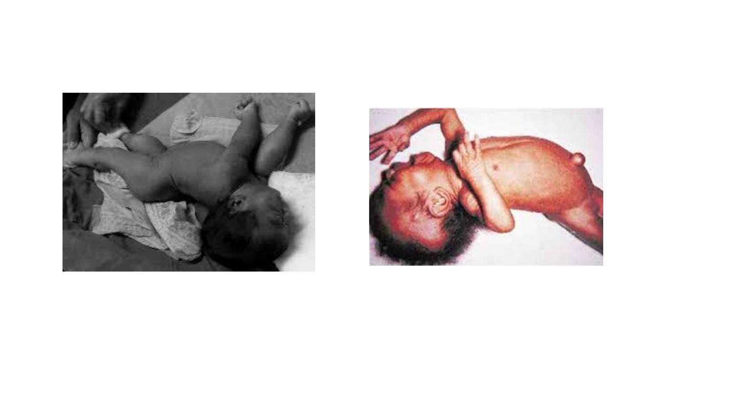
opisthotonos

Cardiology
RVH LVH
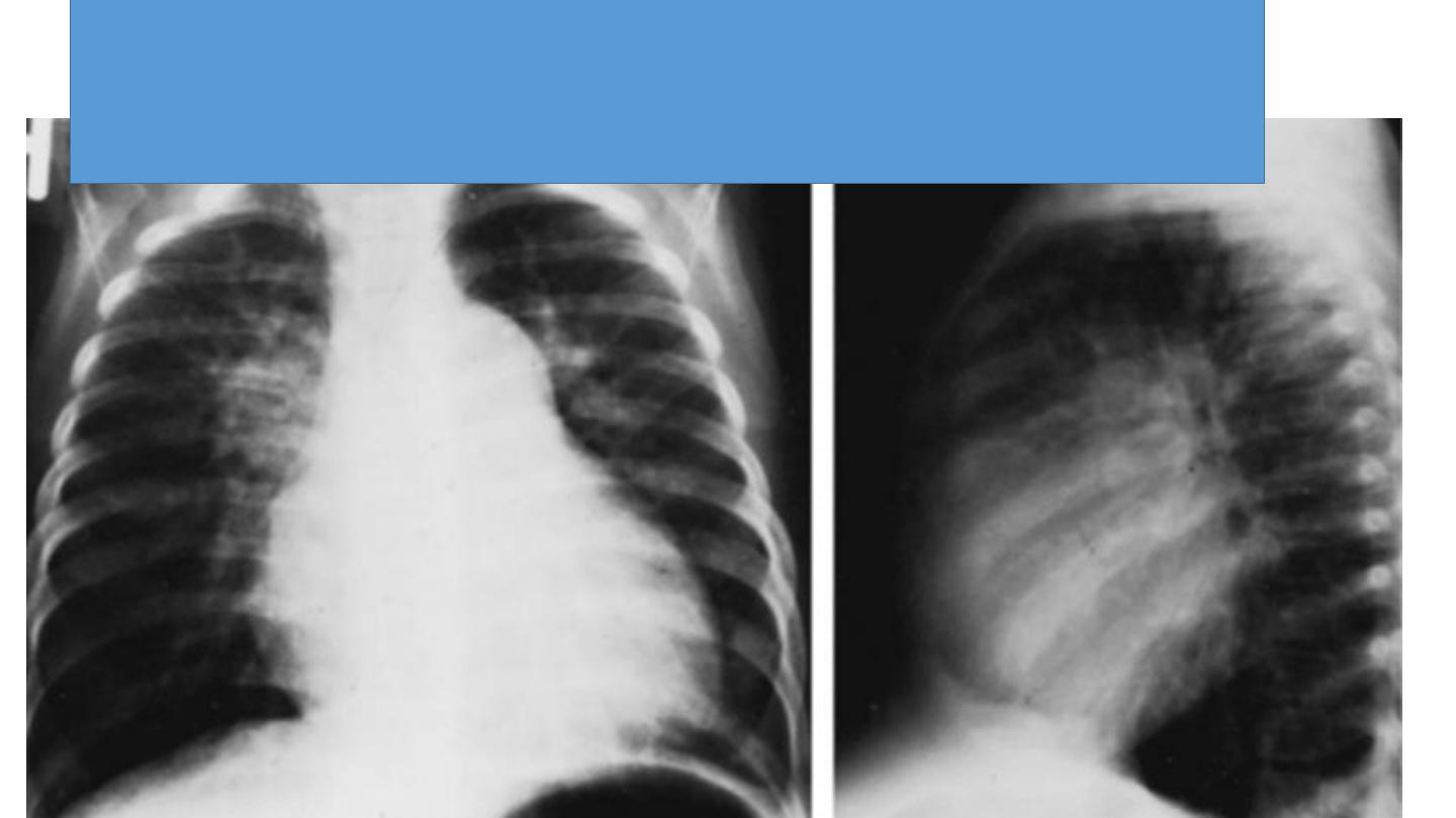
CXR of 6 years old child PA and lateral views showing
cardiac enlargement and increased pulmonary markings
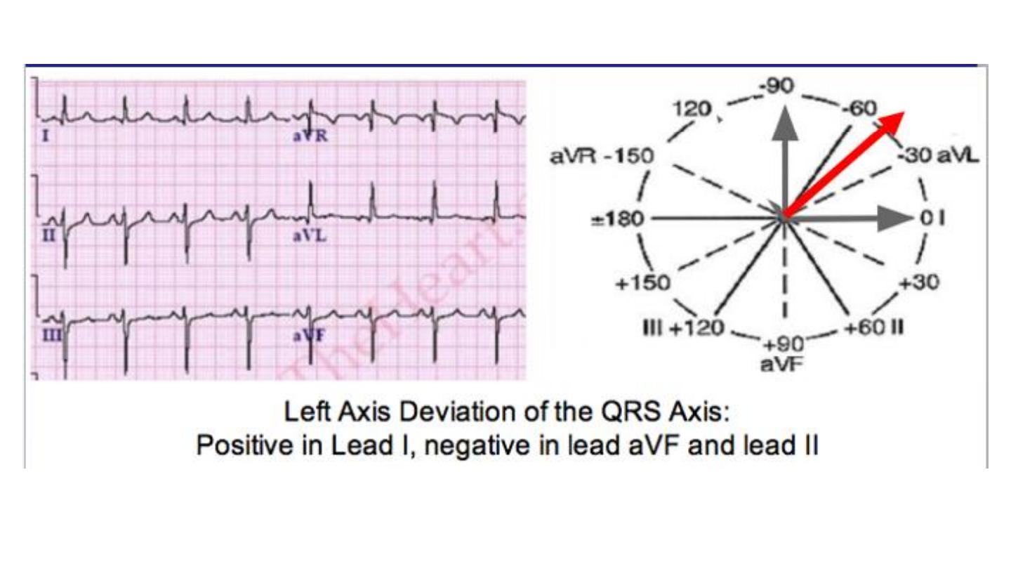
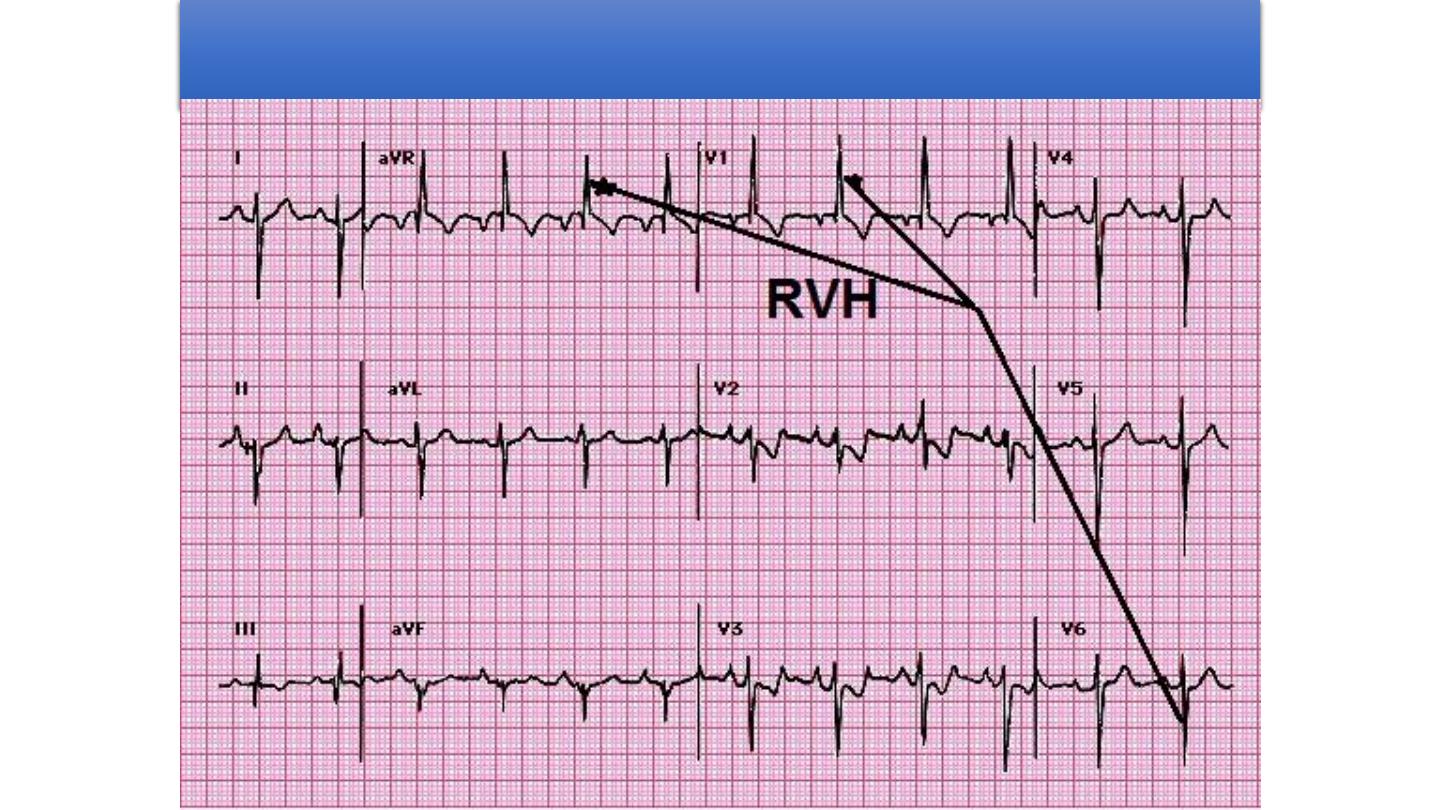
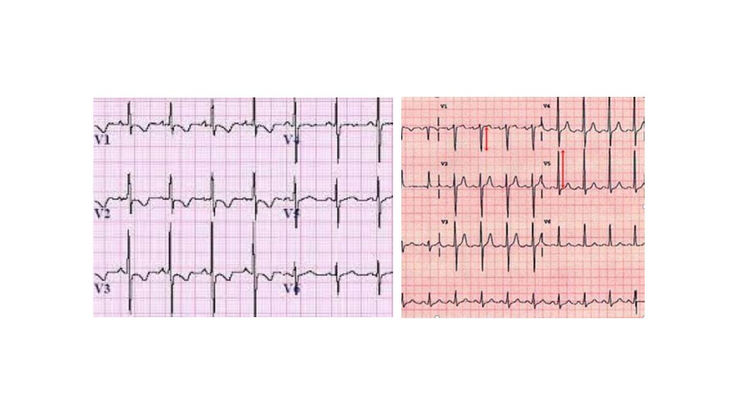
2-The ECG usually has right axis deviation and right
ventricular hypertrophy.
TGA

CNS
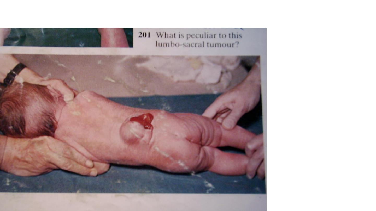
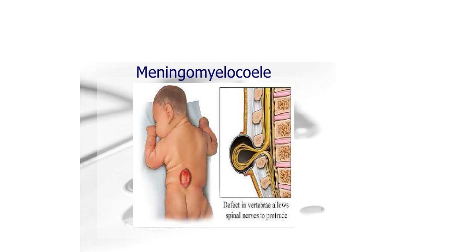
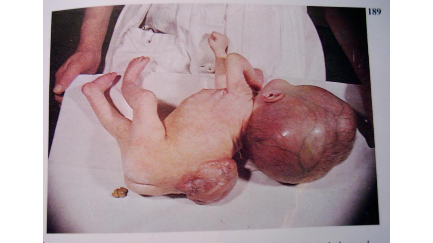
Myleomengiocele (spina bifida)
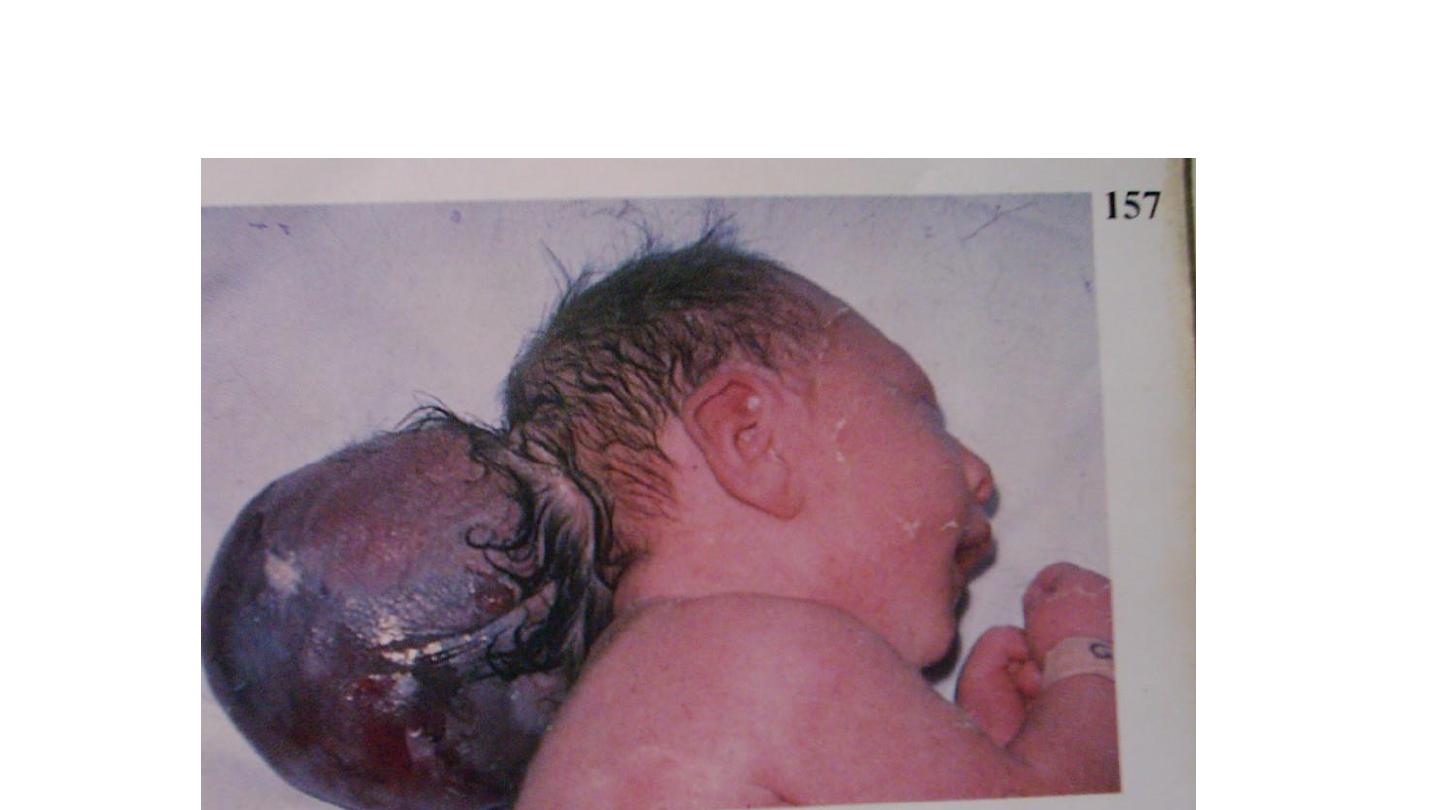
encephalocele
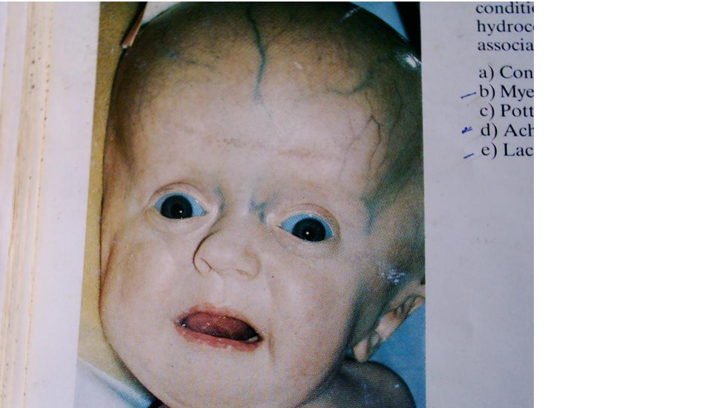
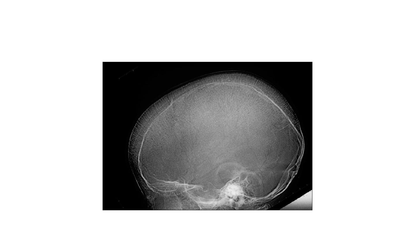
hydrocephaly
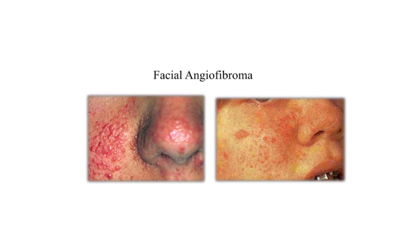
neurofibromatosis
Facial Angiofibroma


Sturge weber syndrome
• Weding hofman

Duchen muscle dystrophy

infections
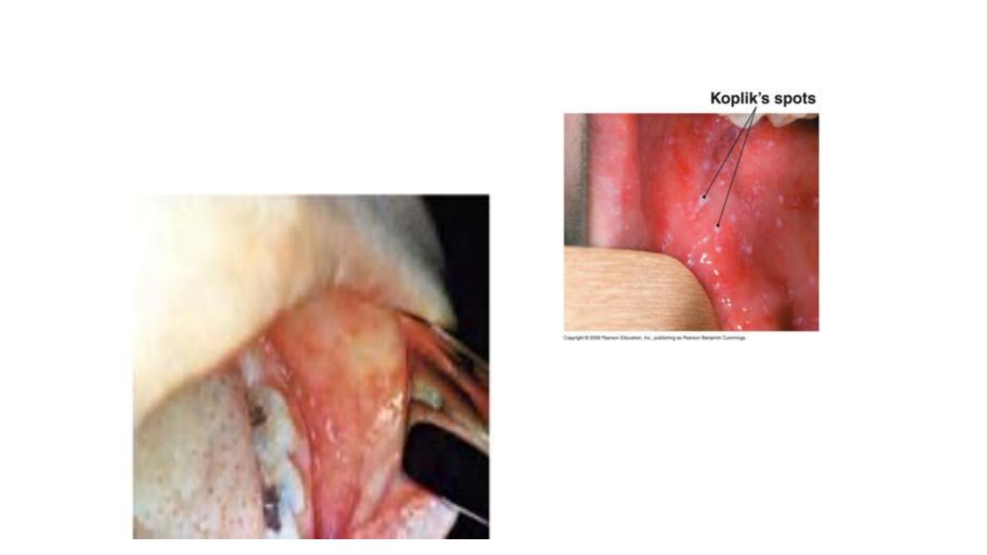
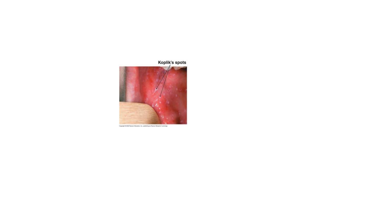
KOPLIK SPOTS

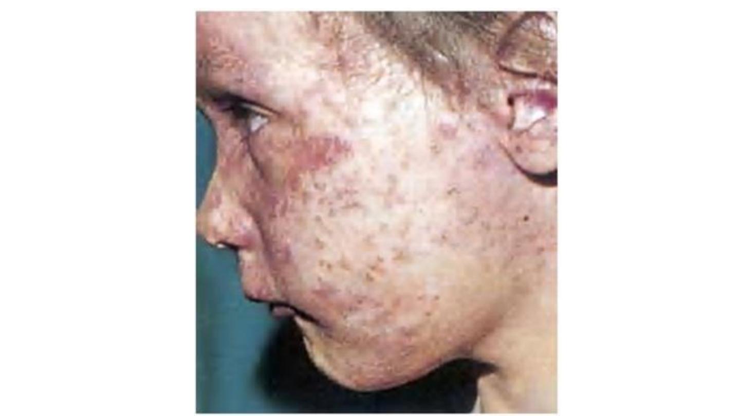
MEEASLES
Rubella’

mumps

mumps
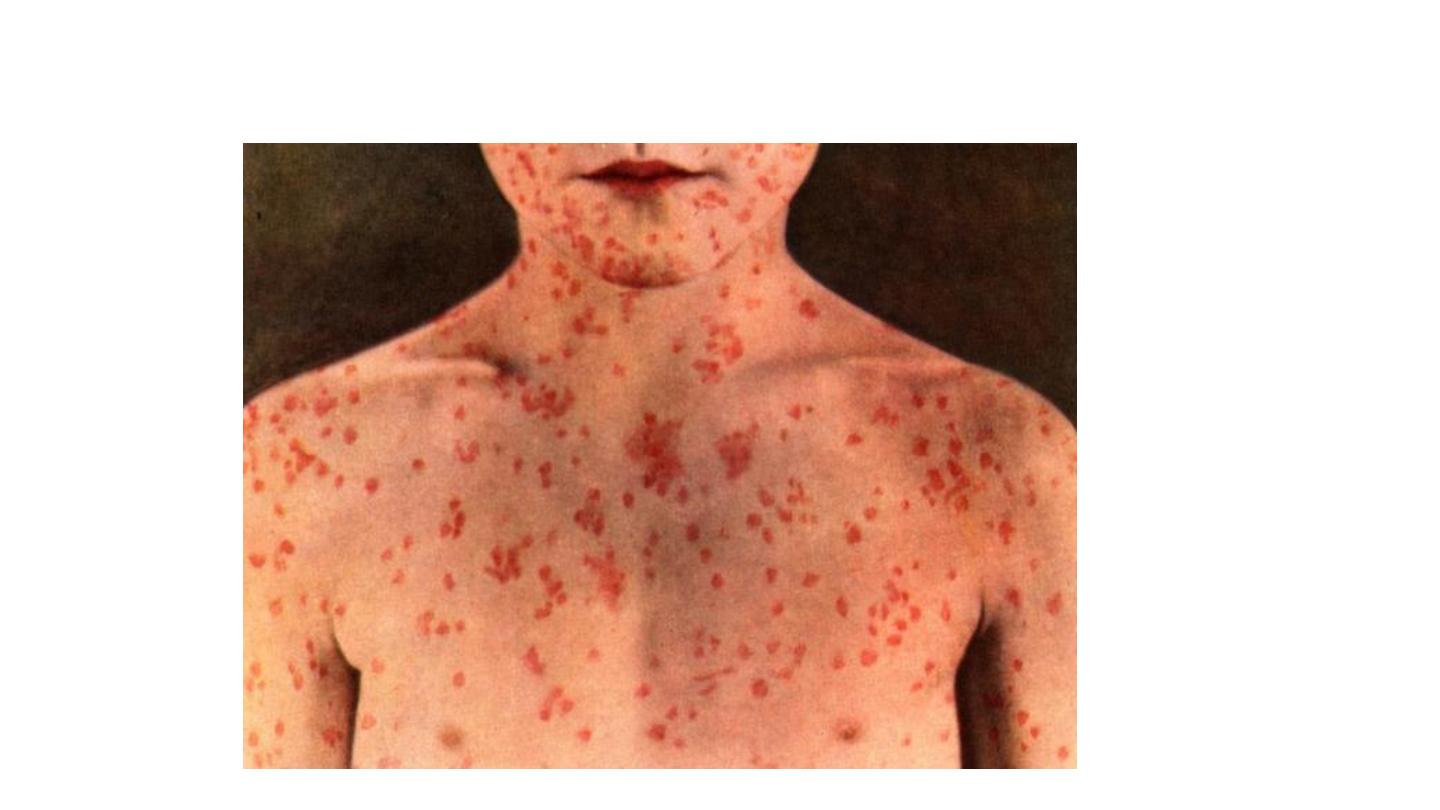
measles

measles
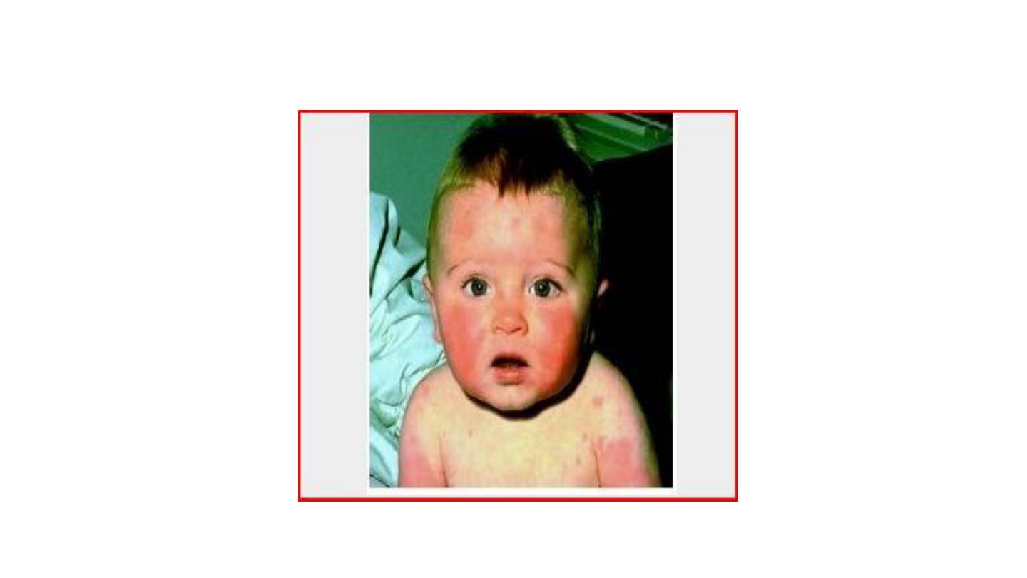
Rosella infantum
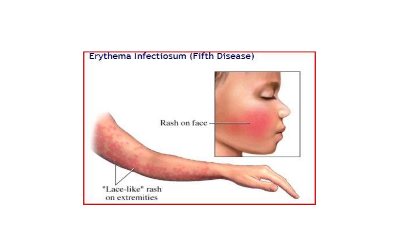
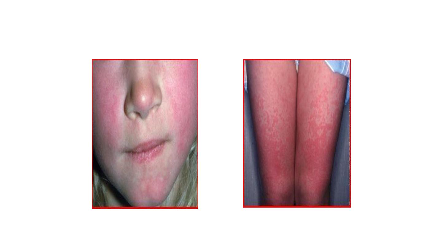
Erythema infectiosum
Erythema infectiosum


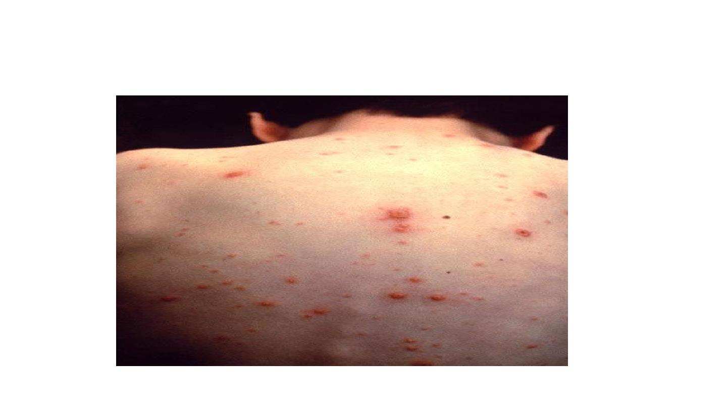
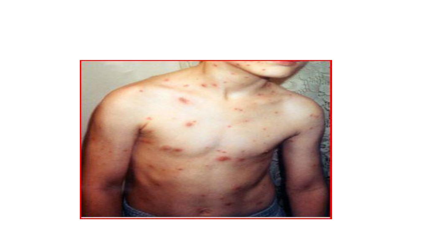
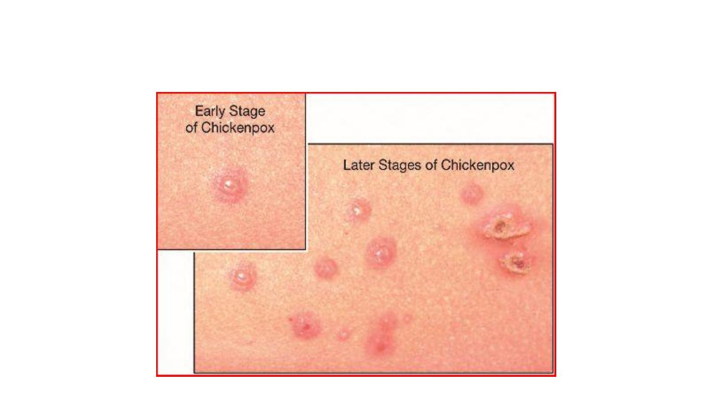
CHICKENPOX

Chicken pox


Infectious mononucleosis
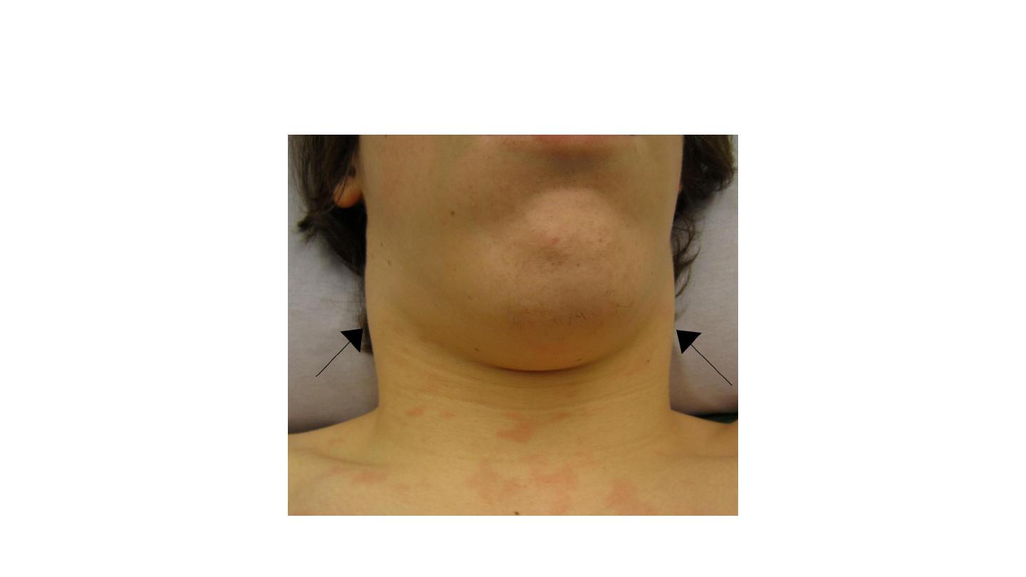
LYMPHADENOPATHY in IMN
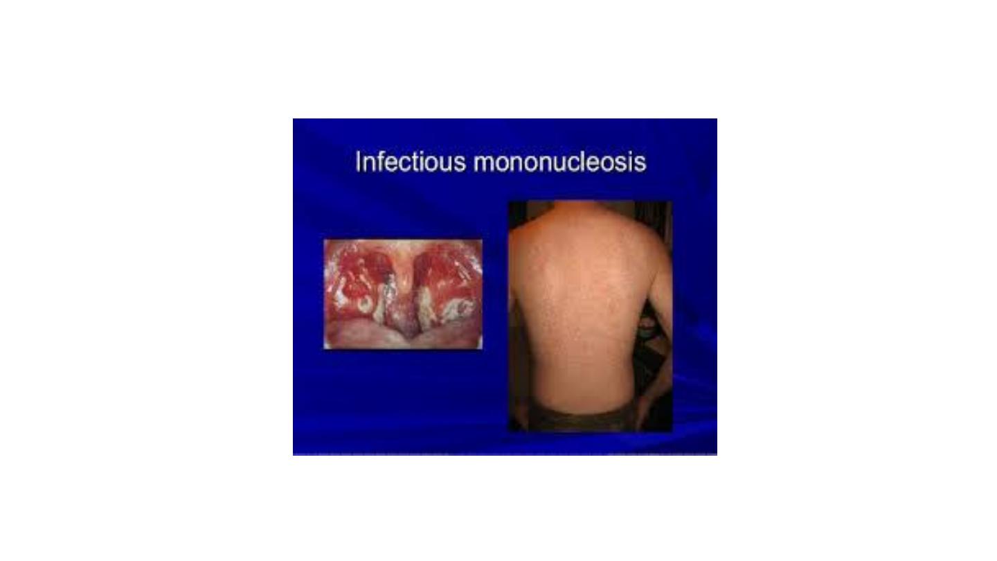
Rash of ampicillin in IMN,
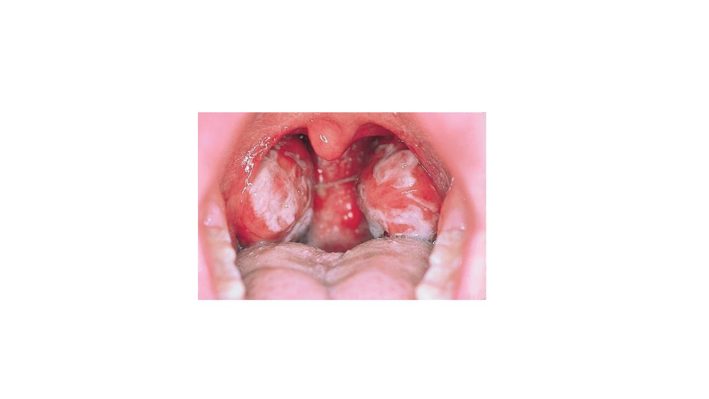
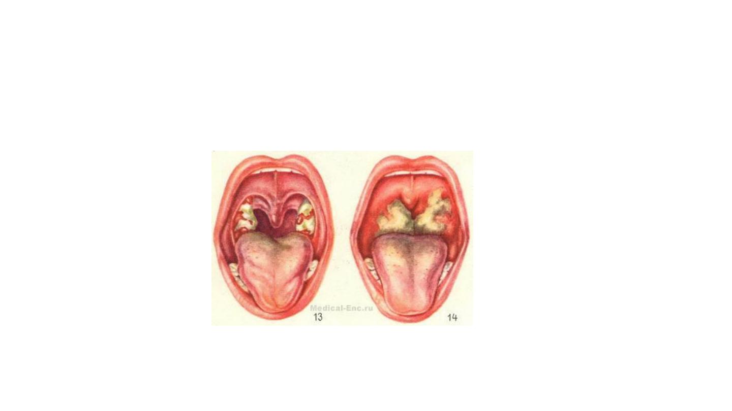
TONSILLITIS
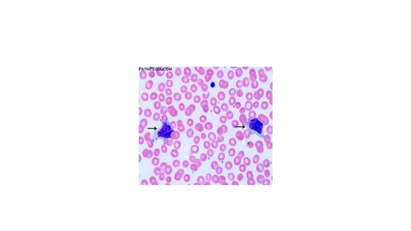
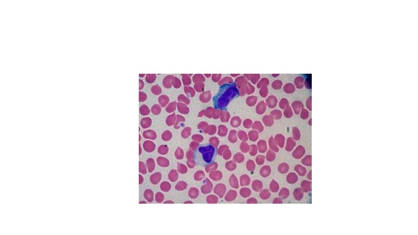
Atypical lymphocytes
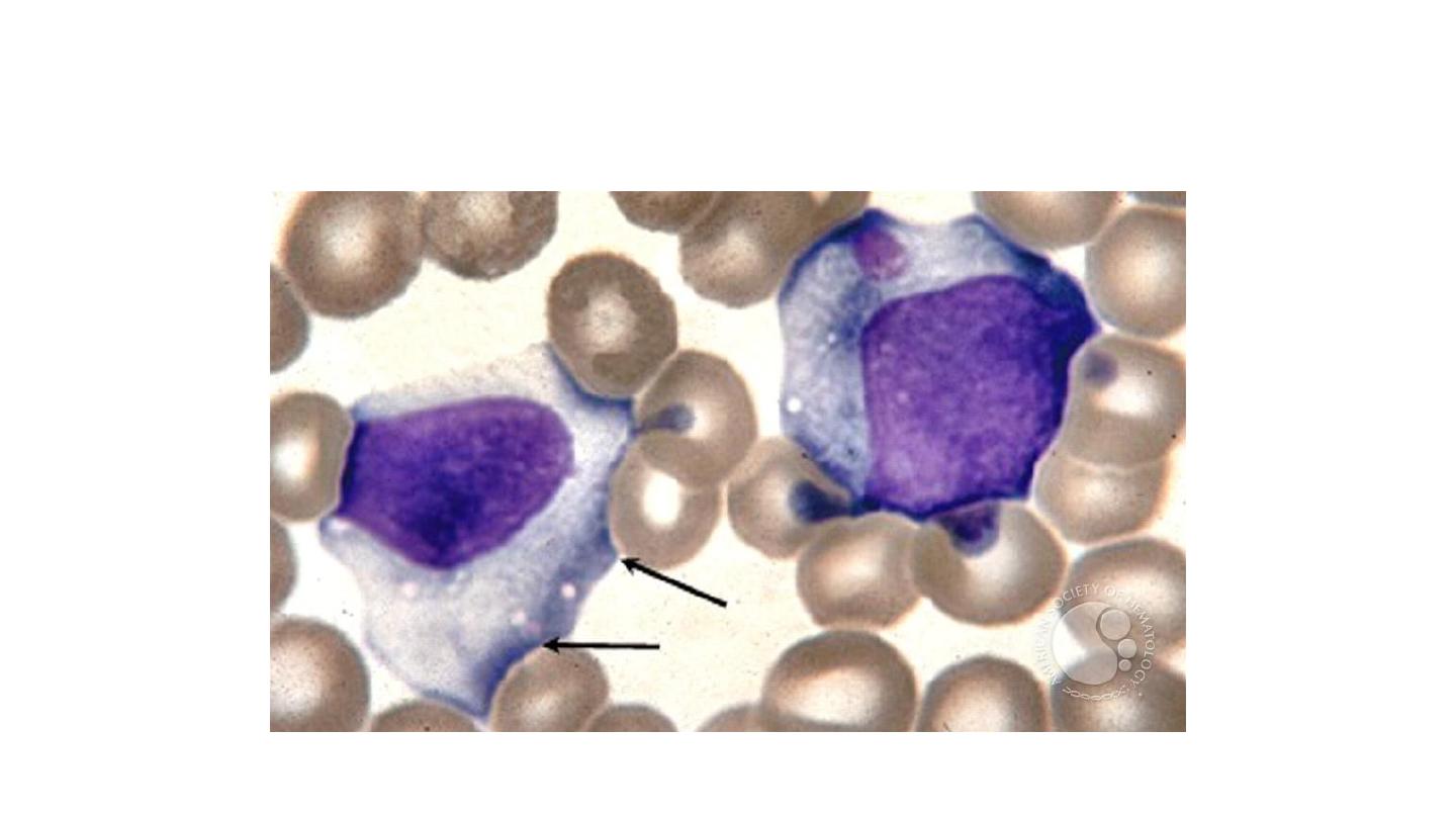
large cytoplasm,nucleoli in the nucleus, indented
by surrounding RBCs

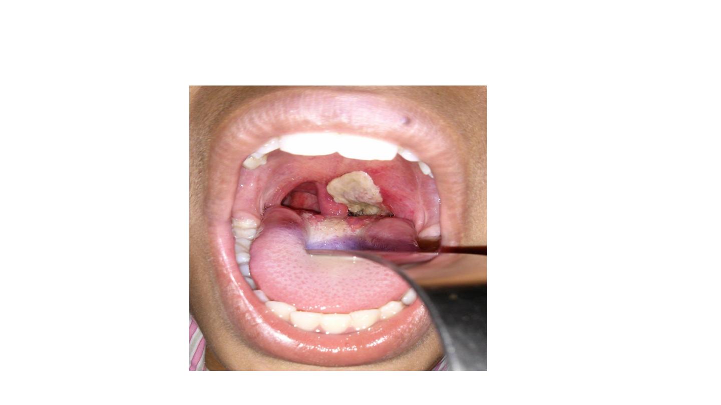
Diphtheria

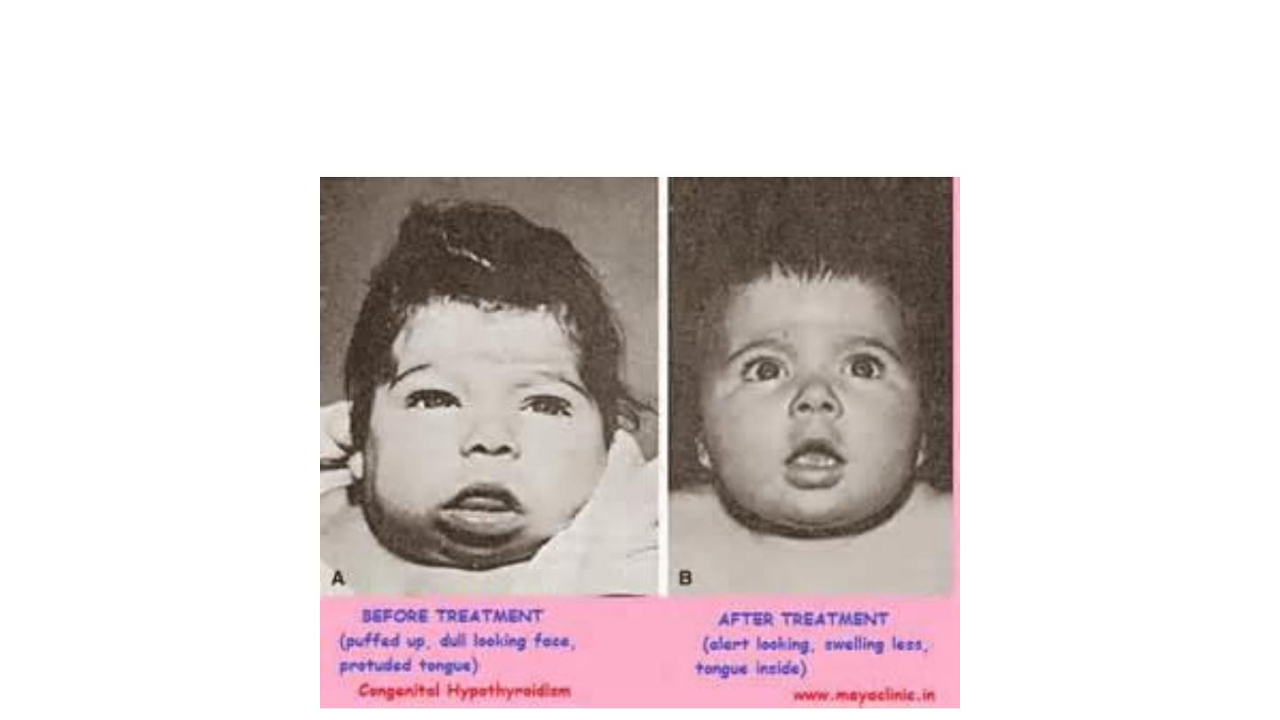
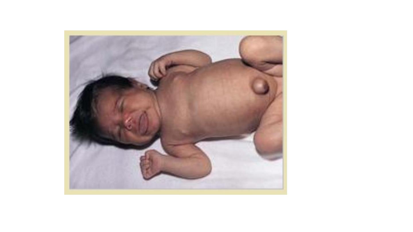
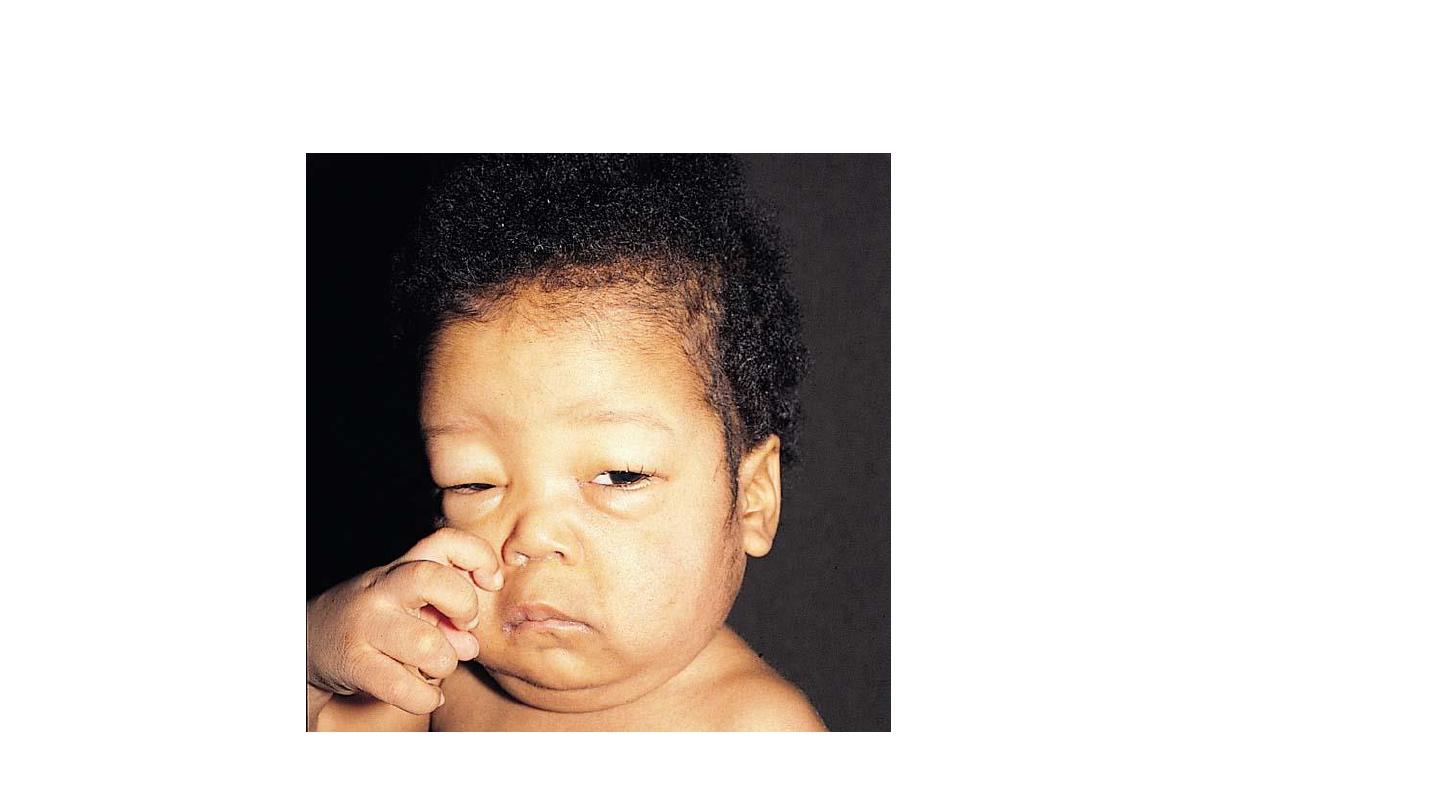
Congenital hypothyroidism

Congenital hypothyroidism
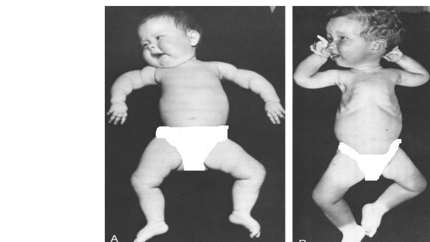
Epiphyseal dysplasia
• Absent femoral epiphysis
Congenital hypothyroidism

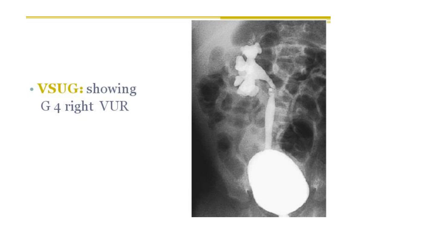
Renal
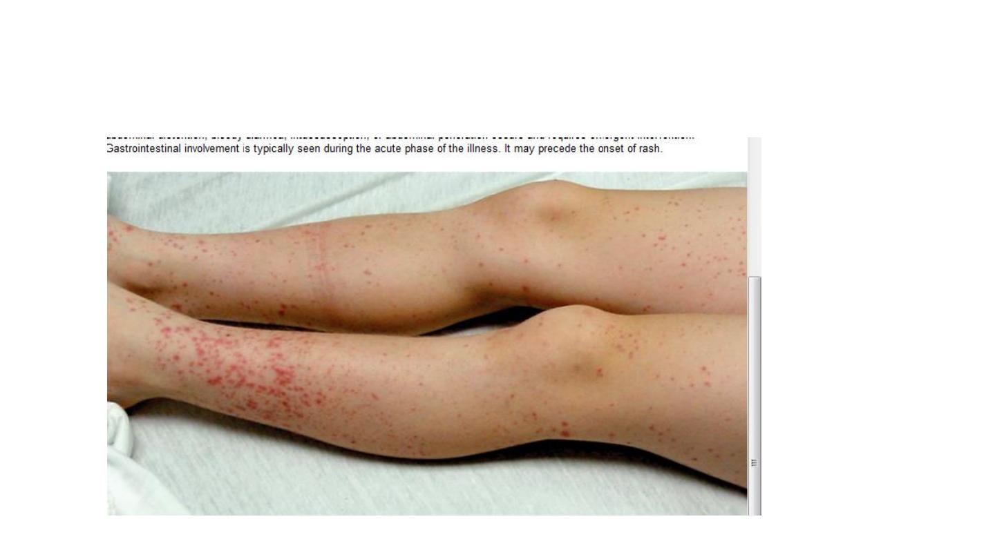
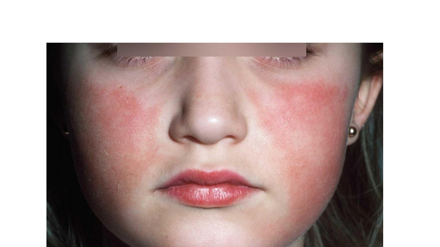
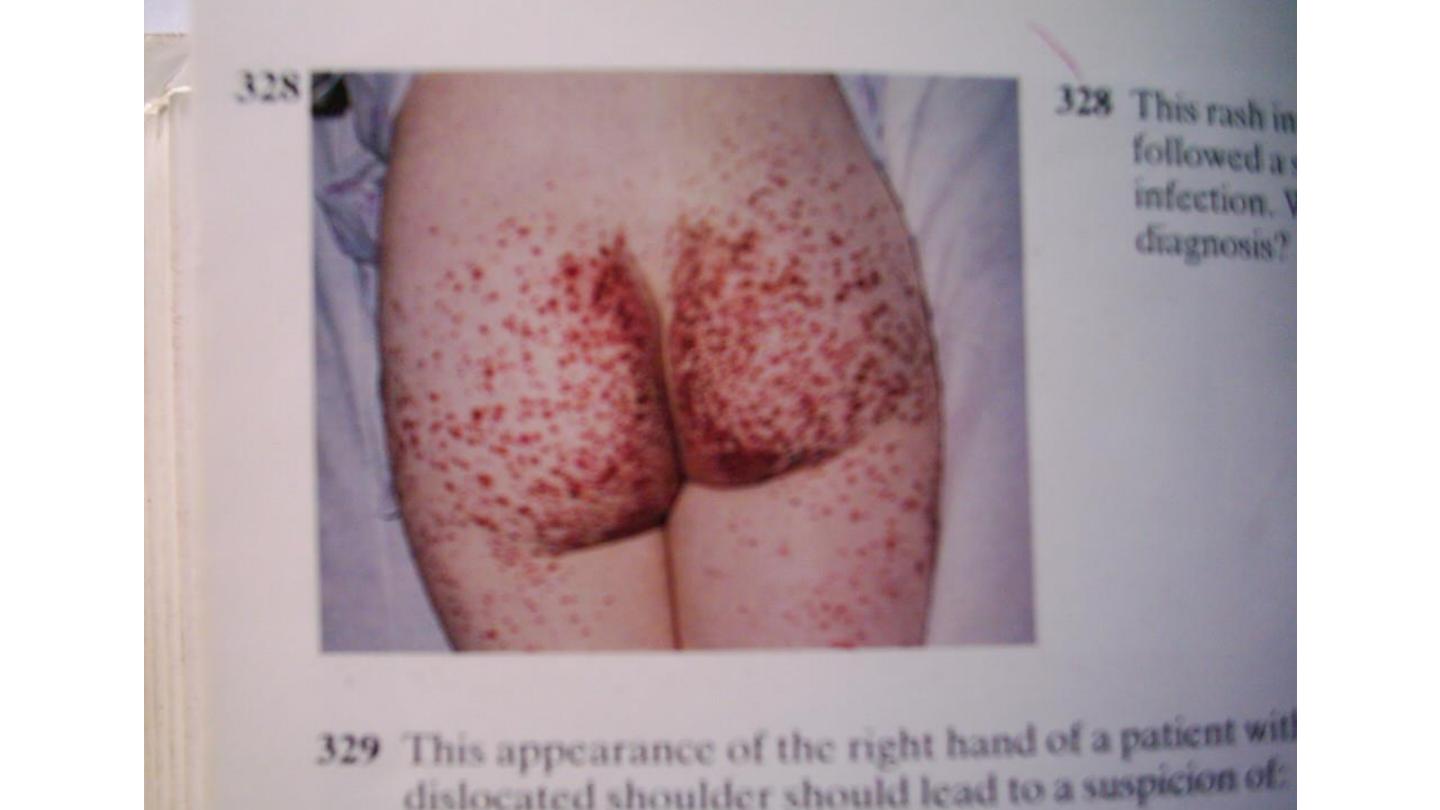
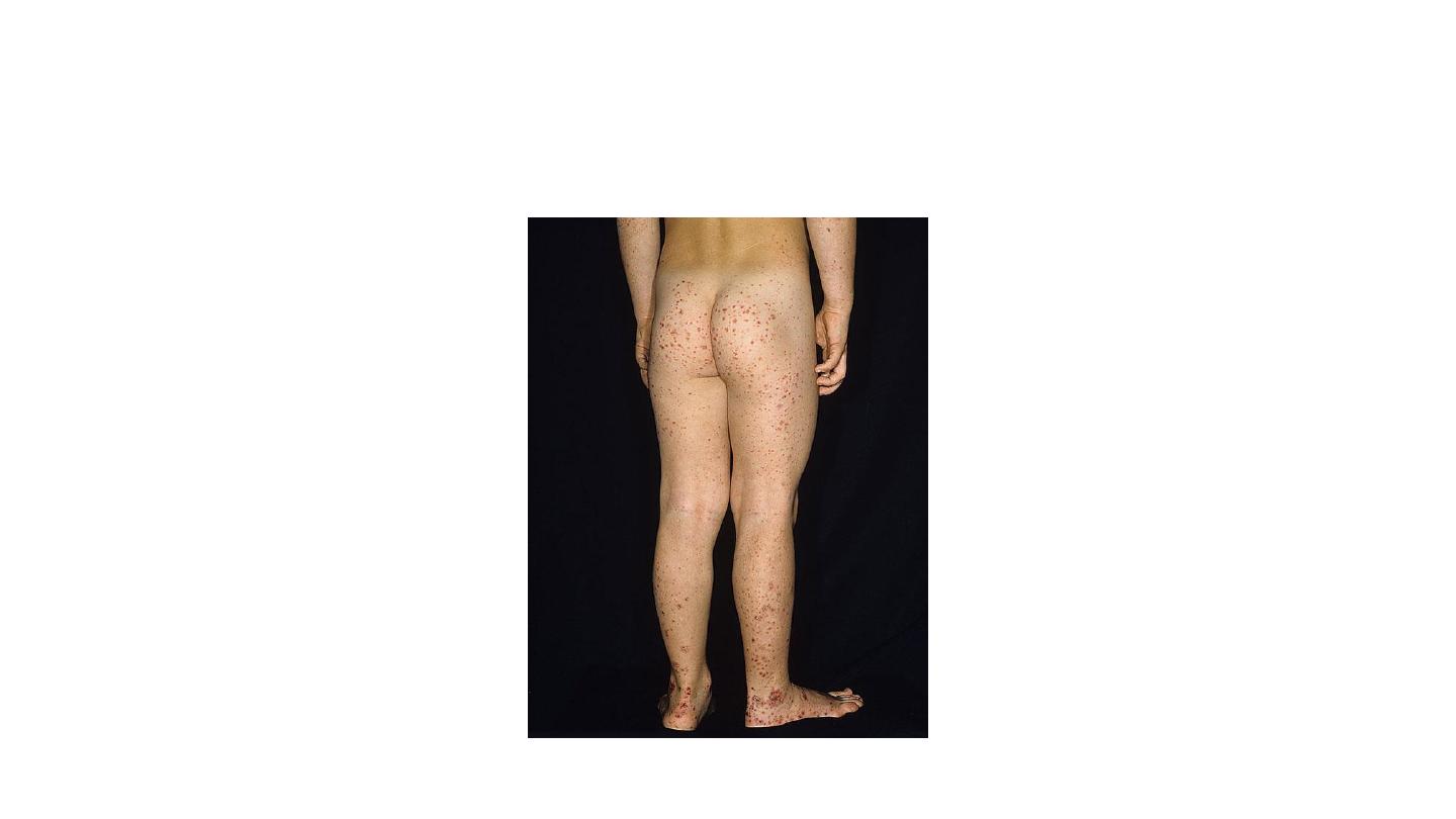
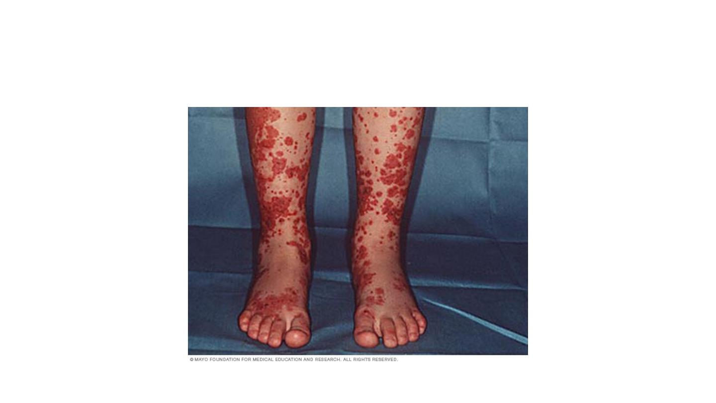
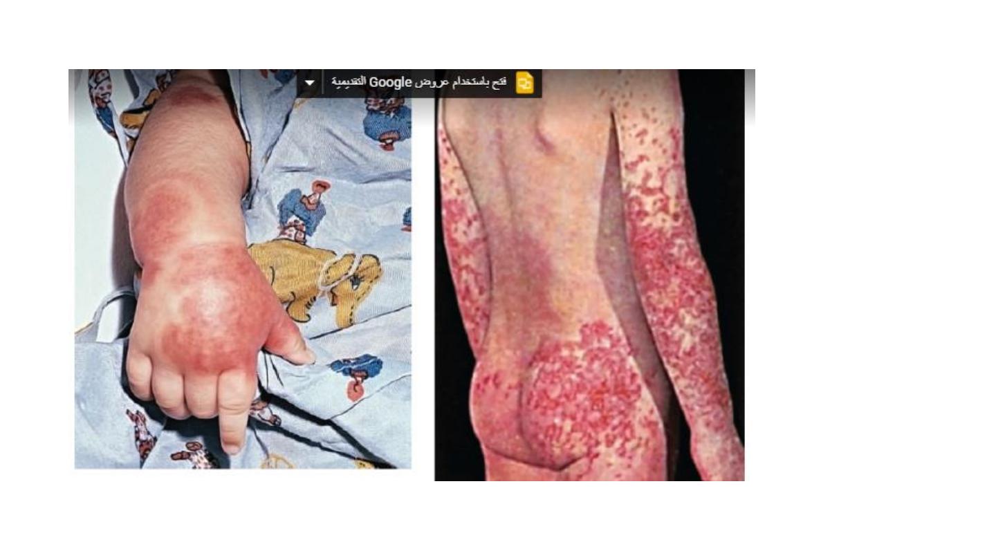
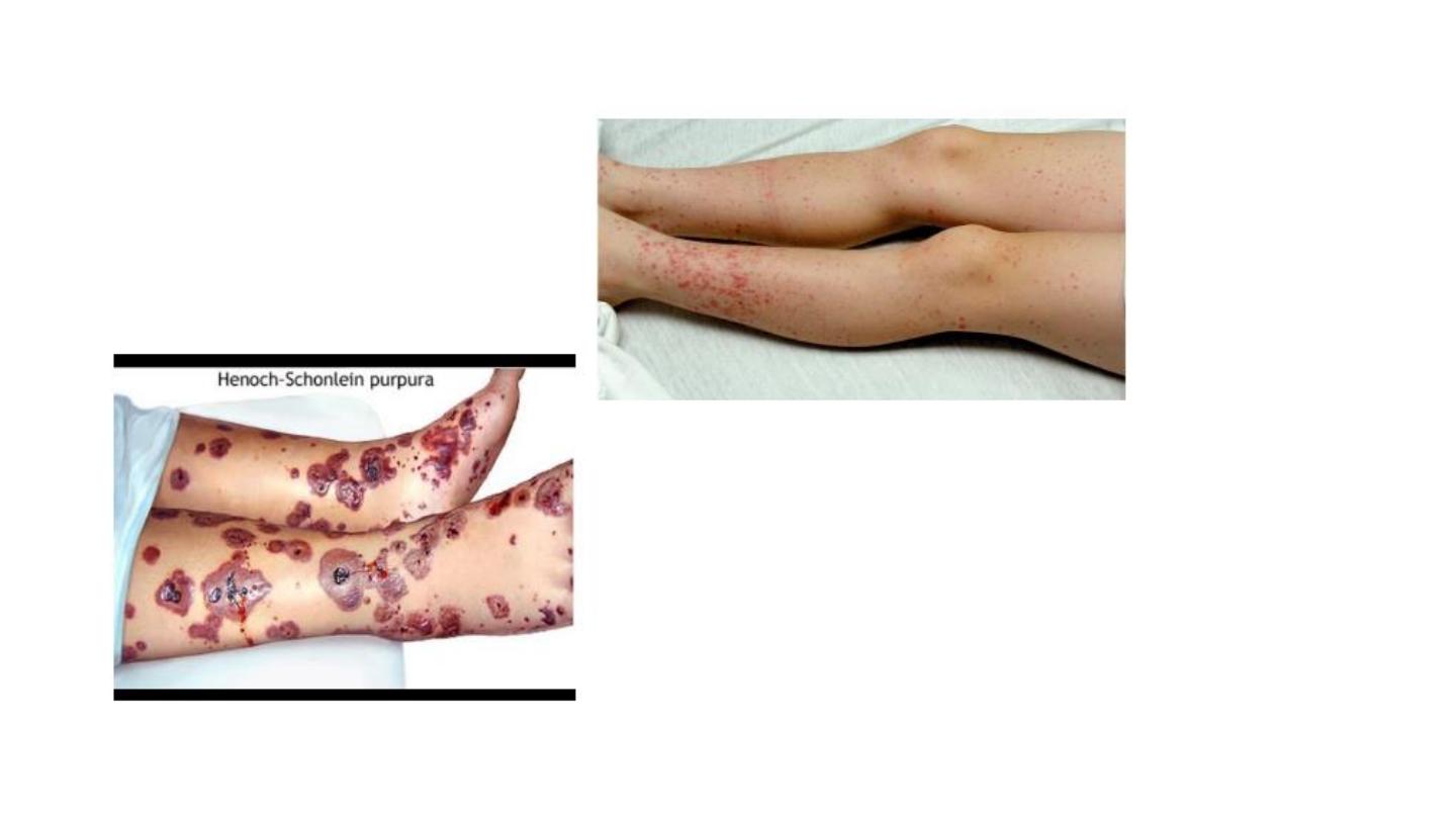
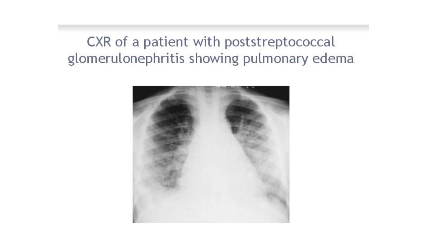
Henoch scoline purpura
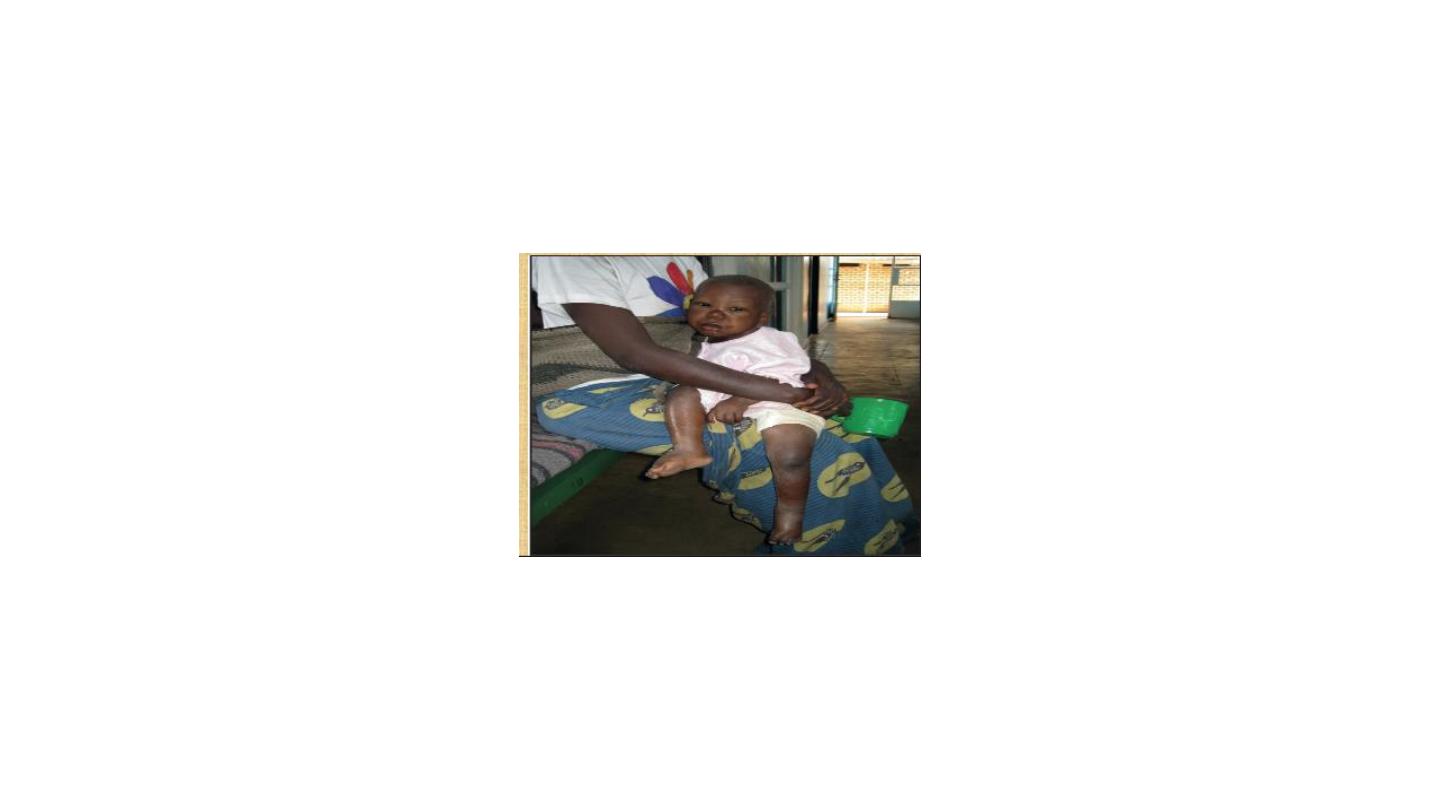
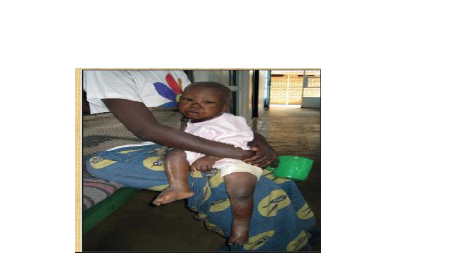
N.S
• Scrotal edema
• In NS
GIT
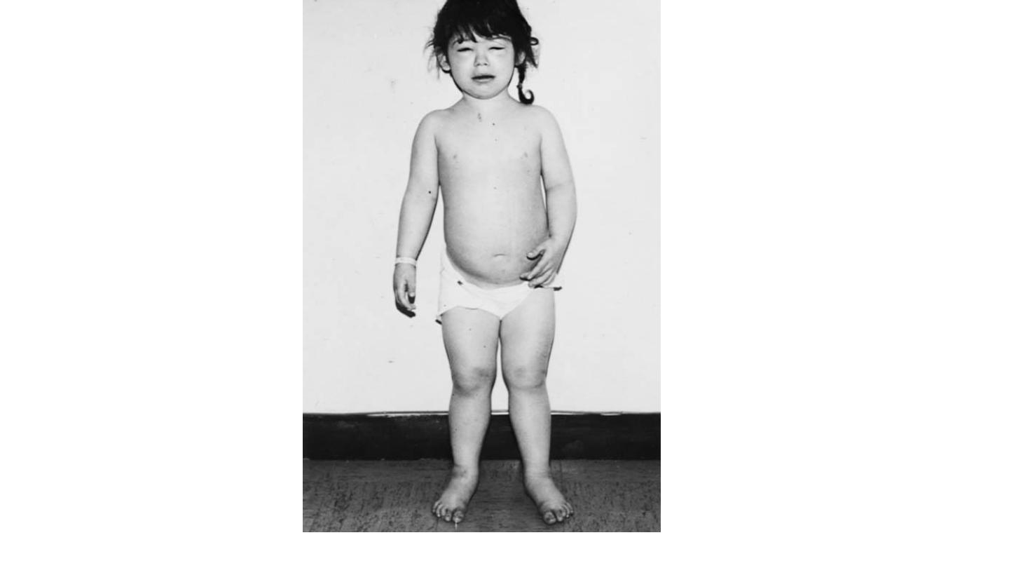
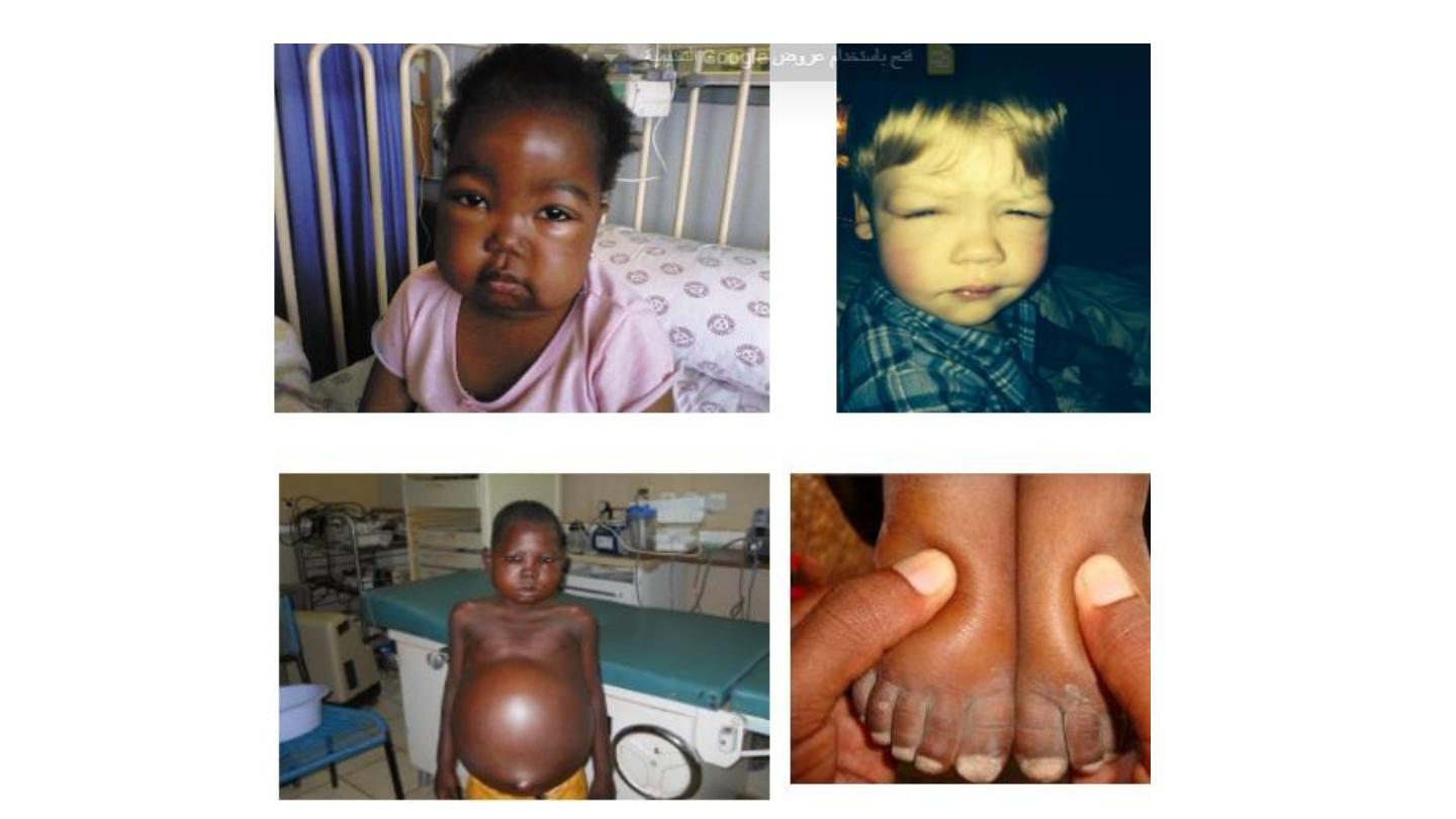
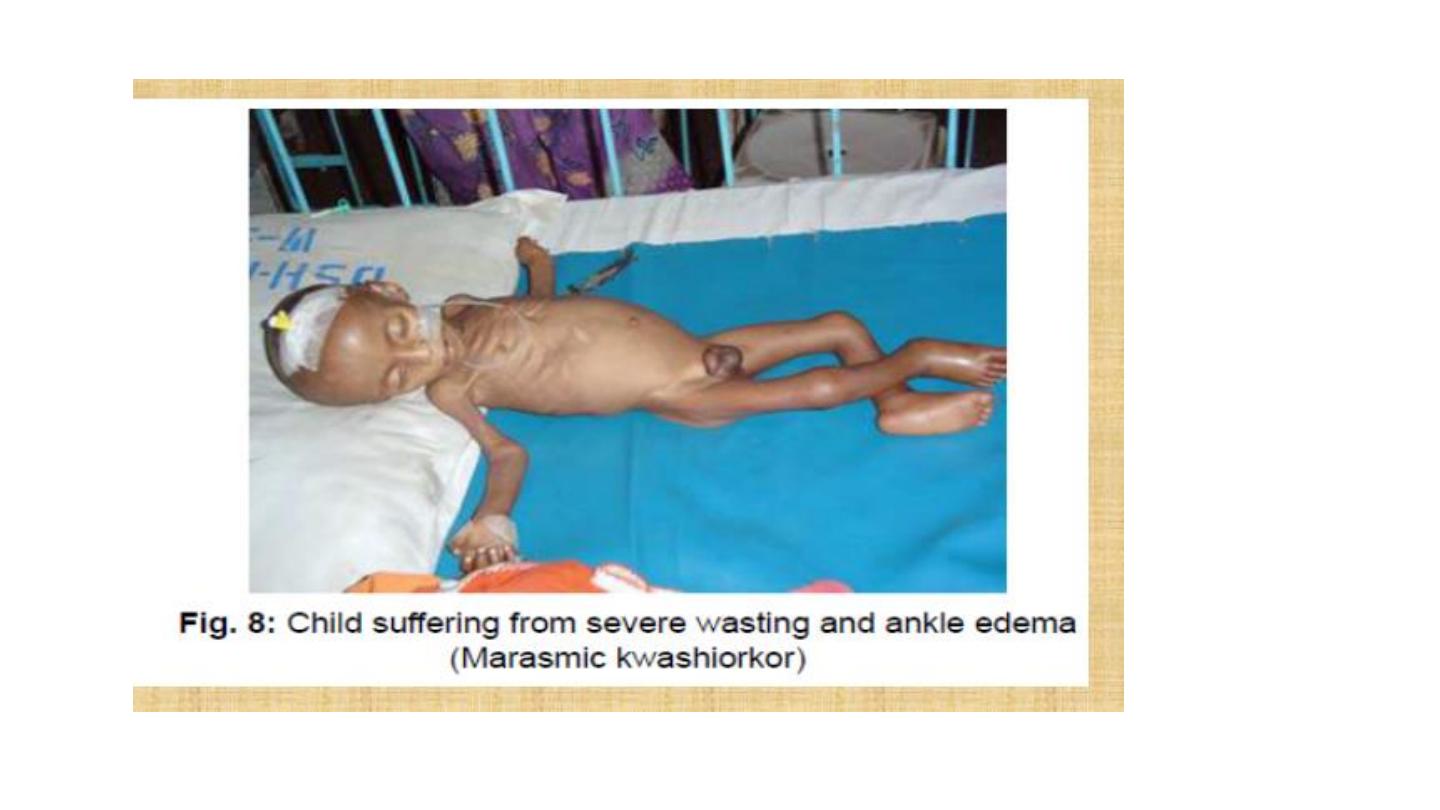
kwashirkor
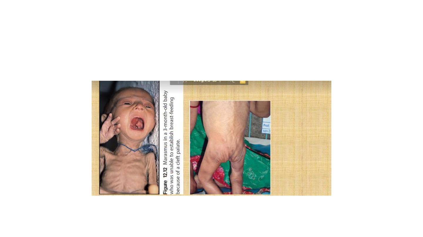
marasmus
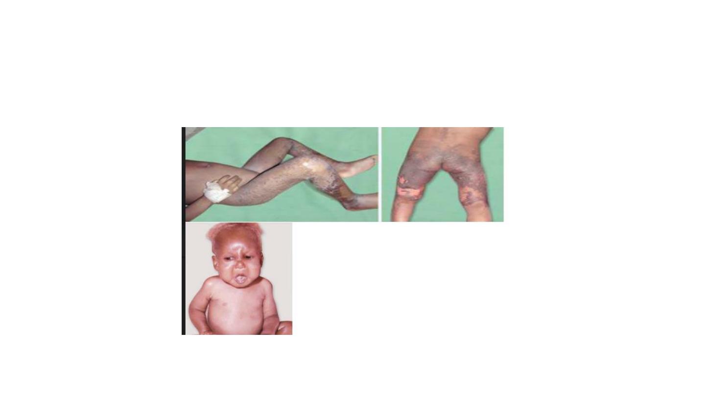
kwashirkor


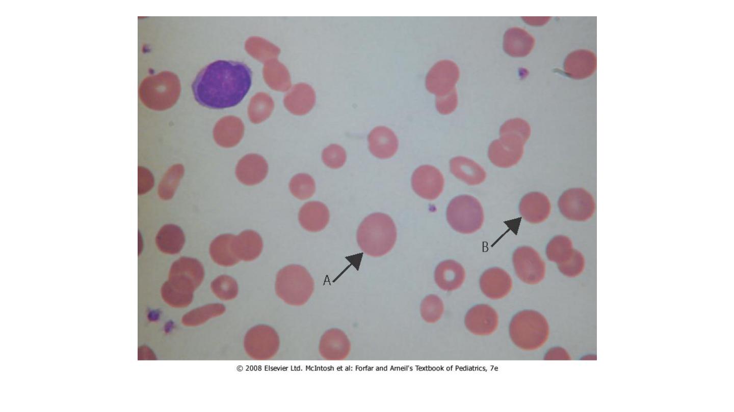
blood
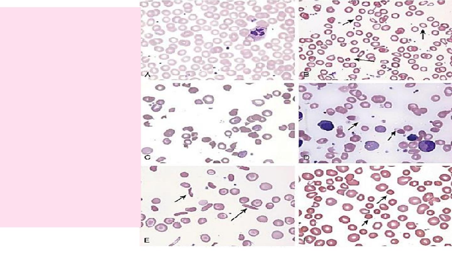
A.
Normal
B.
Hypochromic
microcytes (IDA)
C.
Schistocytes (HUS)
D.
Blister cells (G6PD)
E.
Sickle cells (SCD)
F.
Spherocytes
(autoimmune HA)
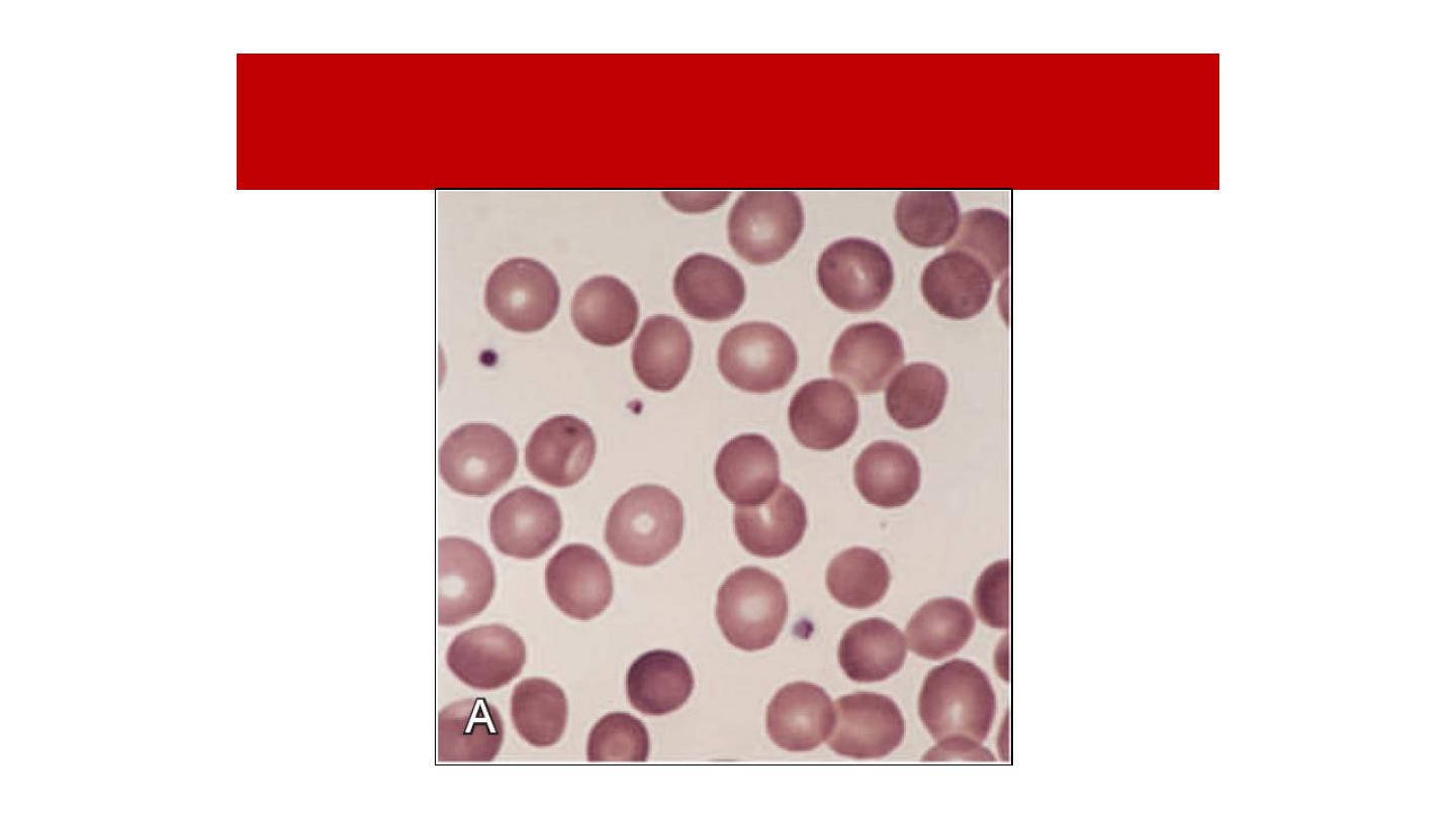
Blood Film of Hereditary Spherocytosis
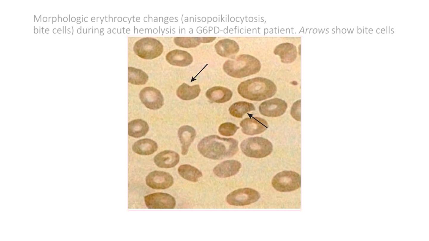
Morphologic erythrocyte changes (anisopoikilocytosis,
bite cells) during acute hemolysis in a G6PD-deficient patient. Arrows show bite cells
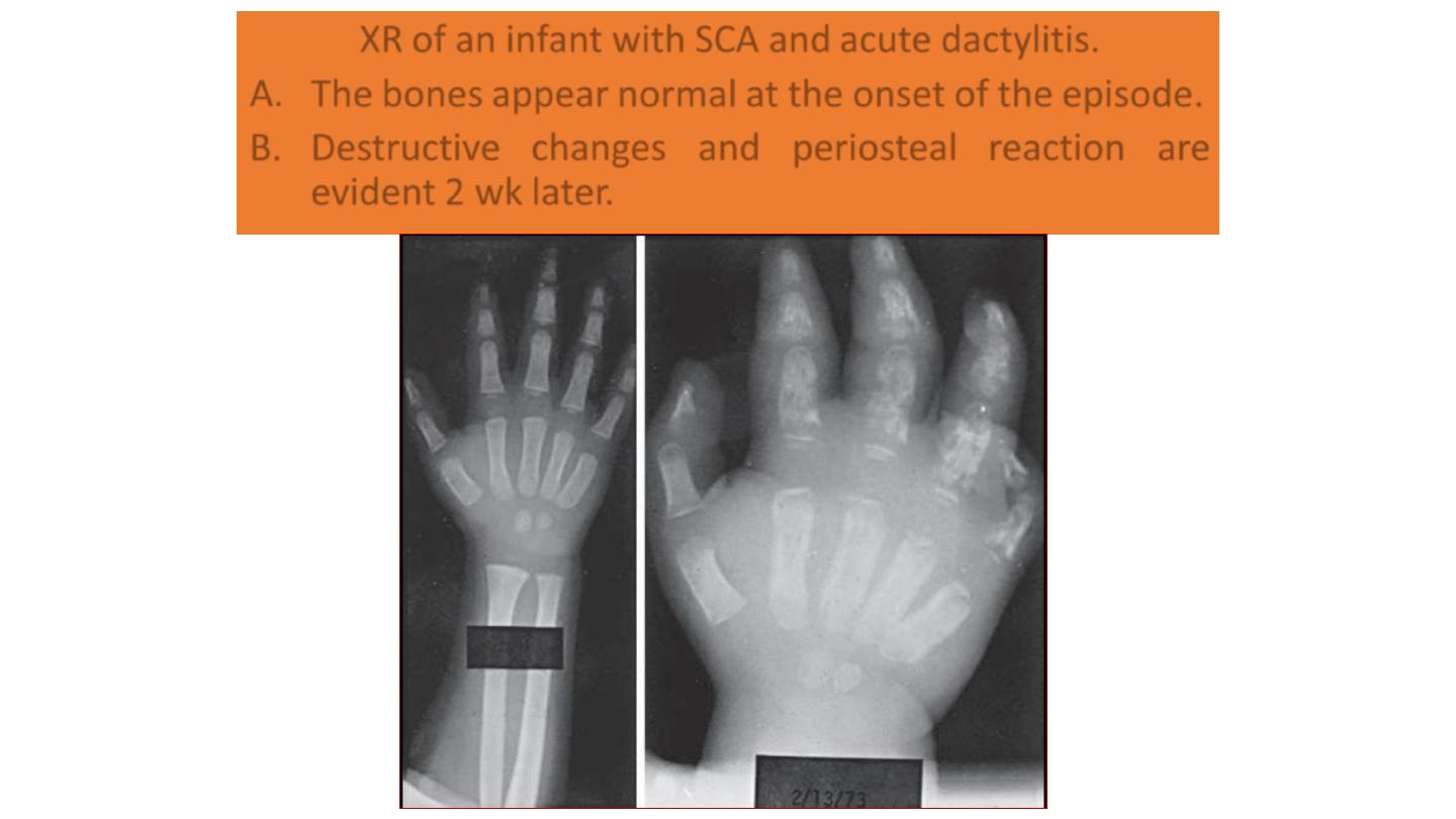
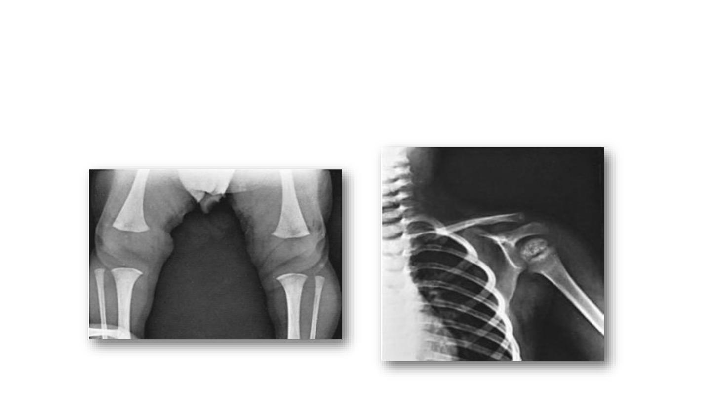
XR of an infant with SCA and acute dactylitis.
A. The bones appear normal at the onset of the episode.
B. Destructive changes and periosteal reaction are
evident 2 wk later.
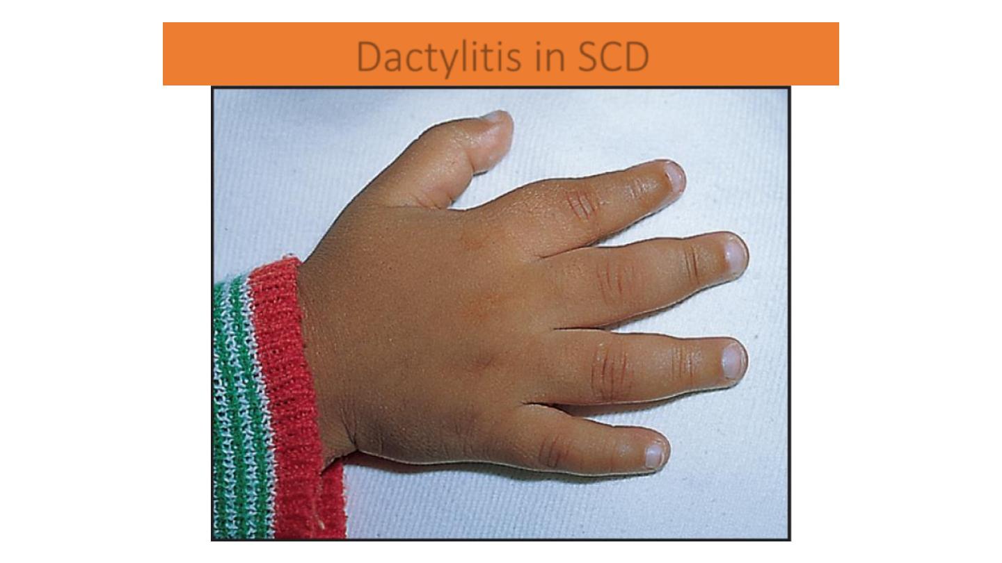
Dactylitis in SCD
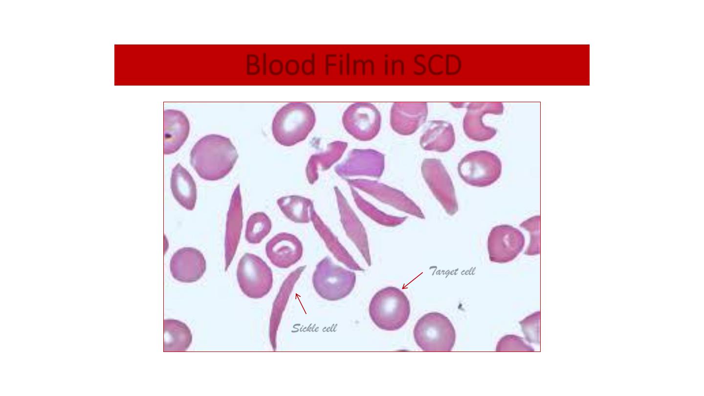
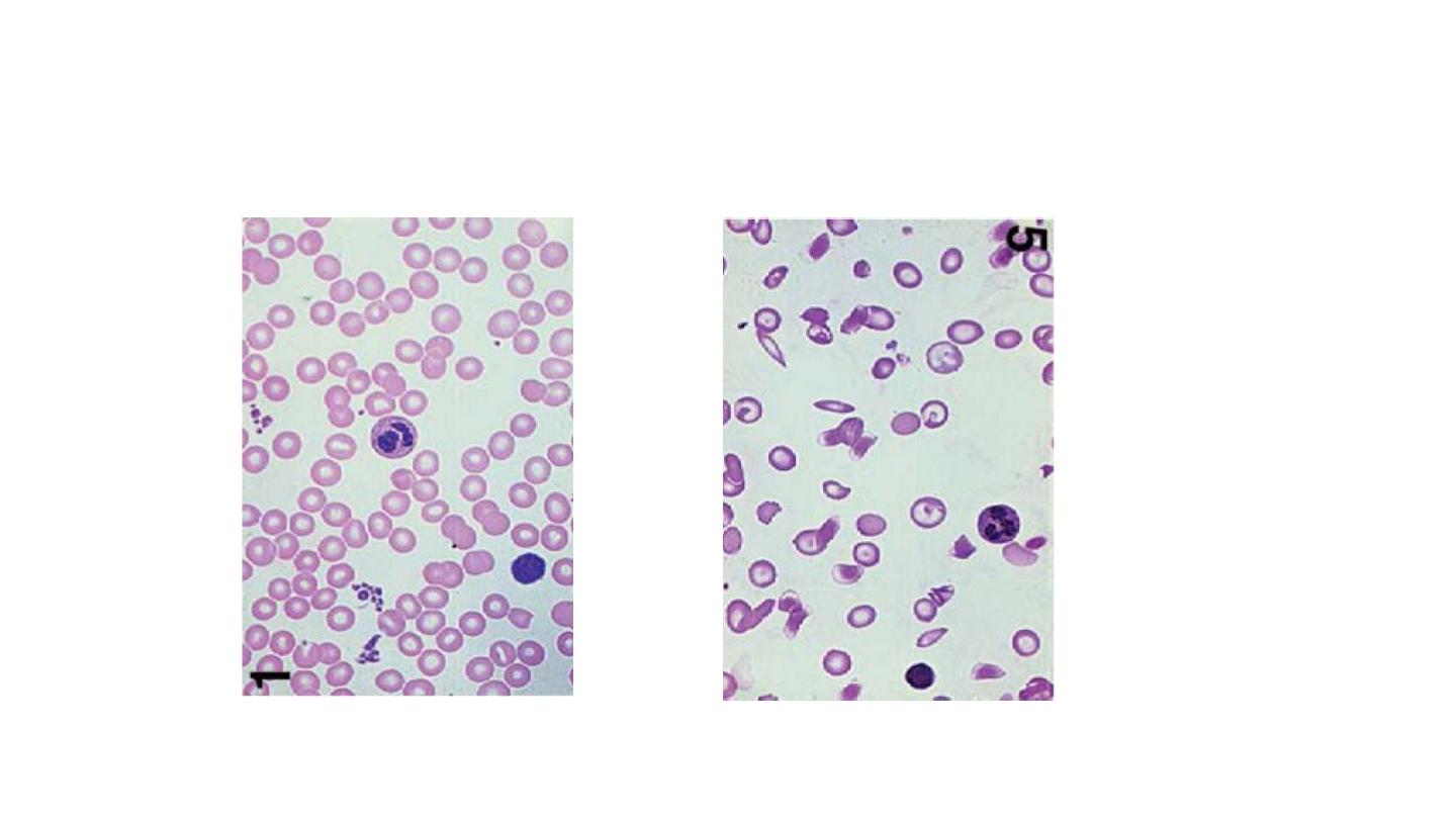
Blood Film in SCD
Target cell
Sickle cell
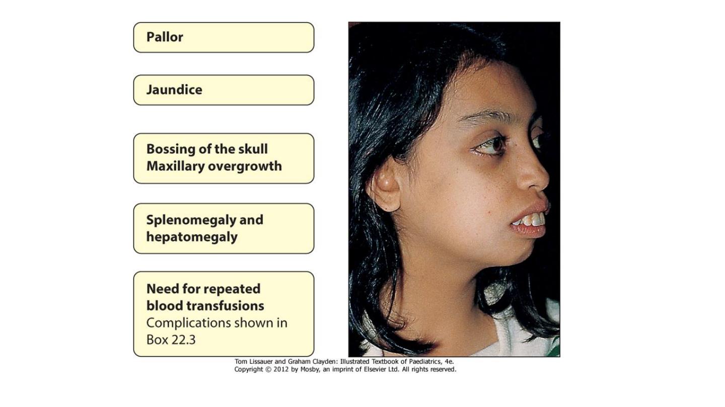
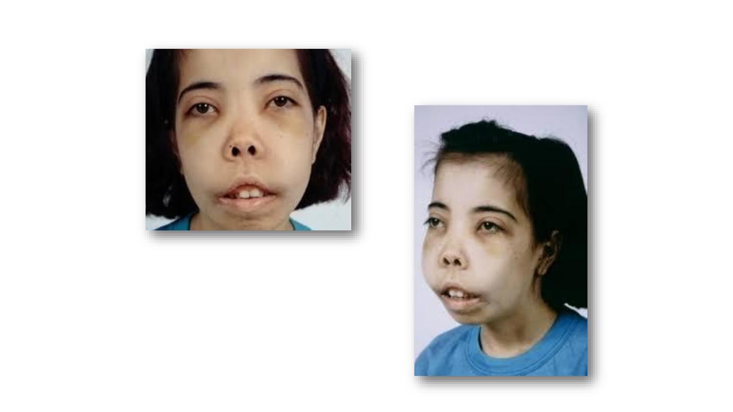
β-Thalassemia
Hair on end appearance SCA

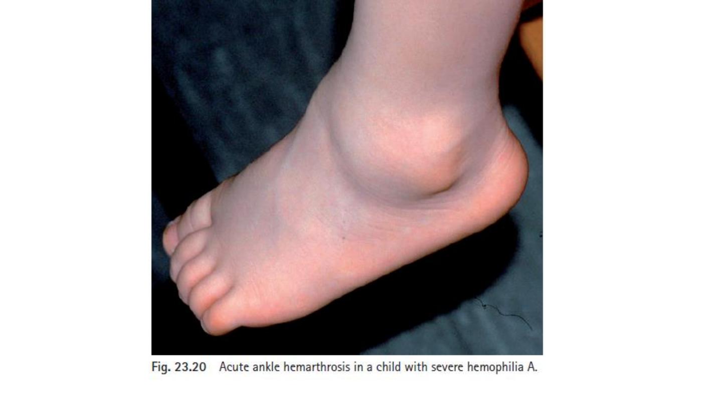
Severe arthropathy from recurrent joint bleeds in haemophilia
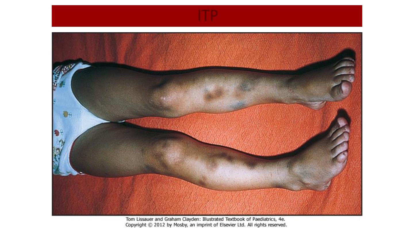
ITP
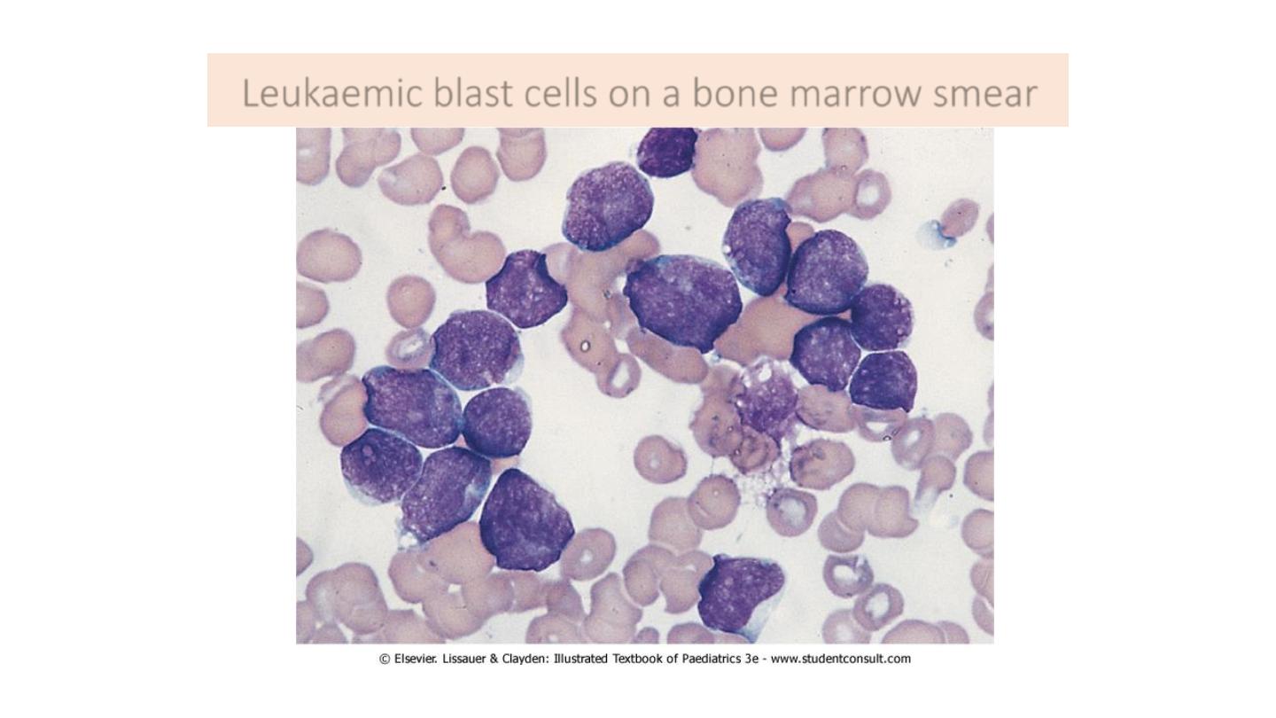
Leukaemic blast cells on a bone marrow smear
HS
A, polychromatic cell; B, microspherocyte
SCA
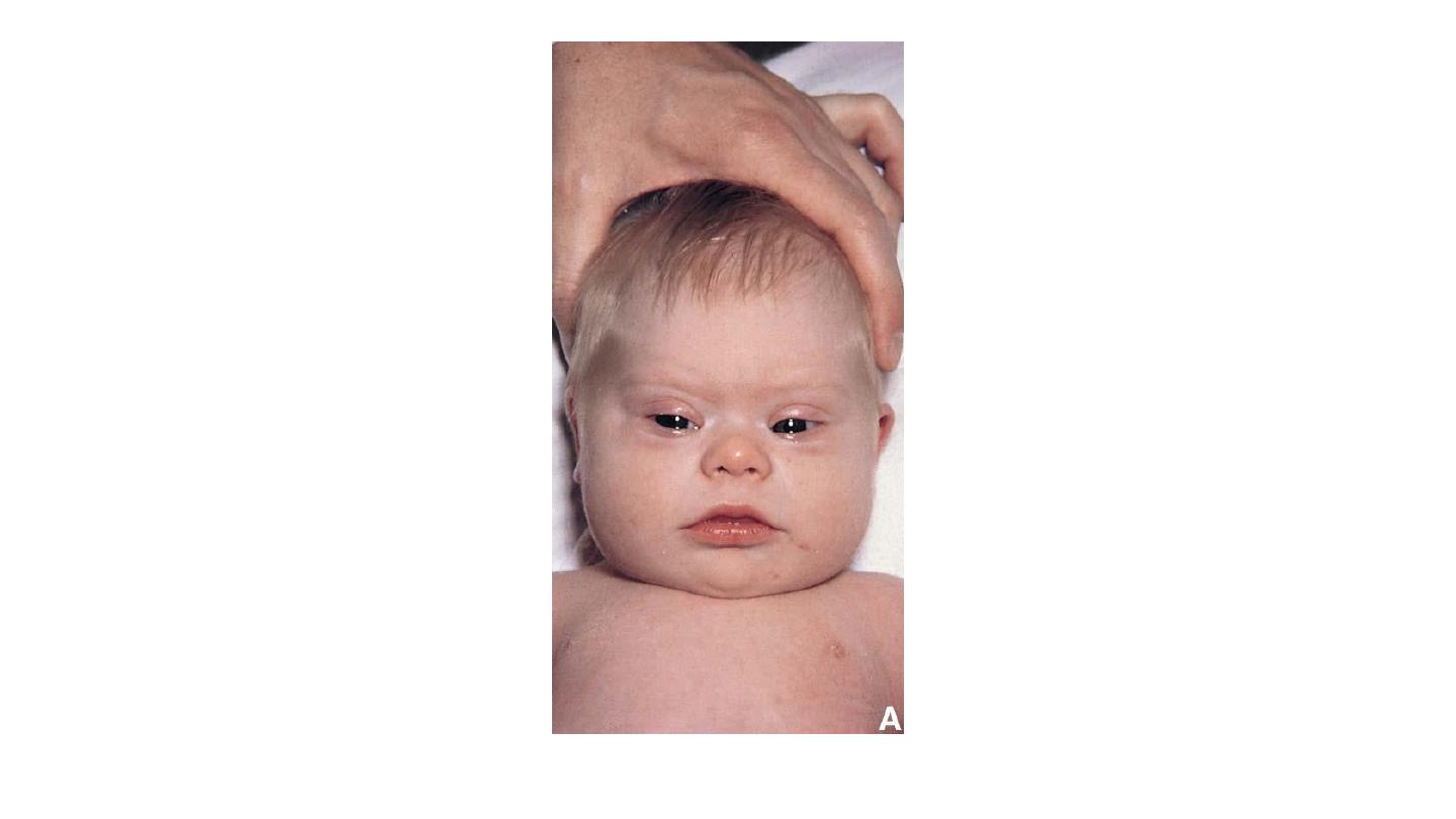
Trisomy 21
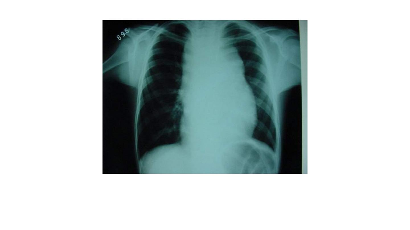
Mediastinal involvement by lymphoblastic lymphoma
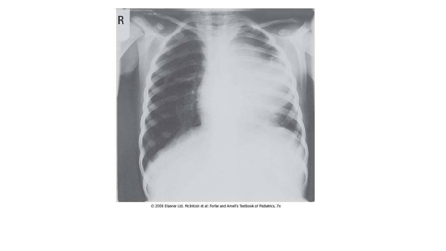
Chest X-ray appearances in acute T cell leukemia with mediastinal
widening and hilar lymphadenopathy causing superior vena cava
obstruction.

Syndroms
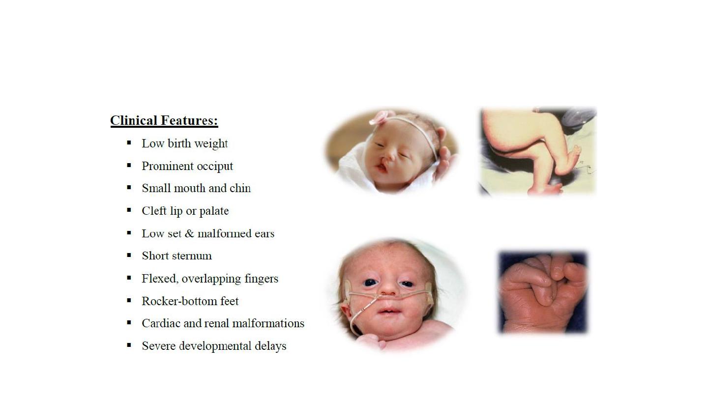
EDWARD trisomy 18
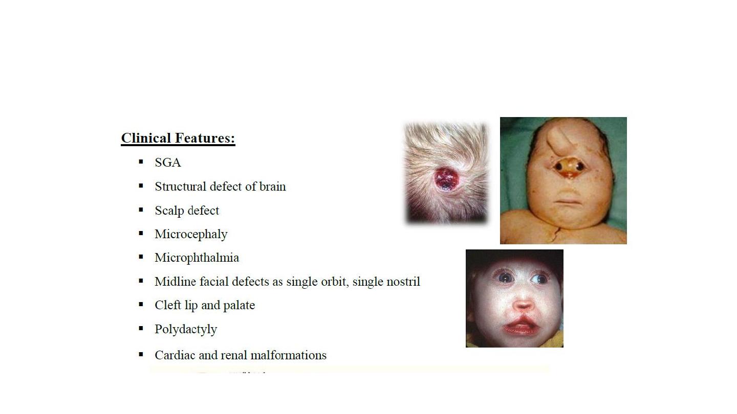
Pataue trisomy 13
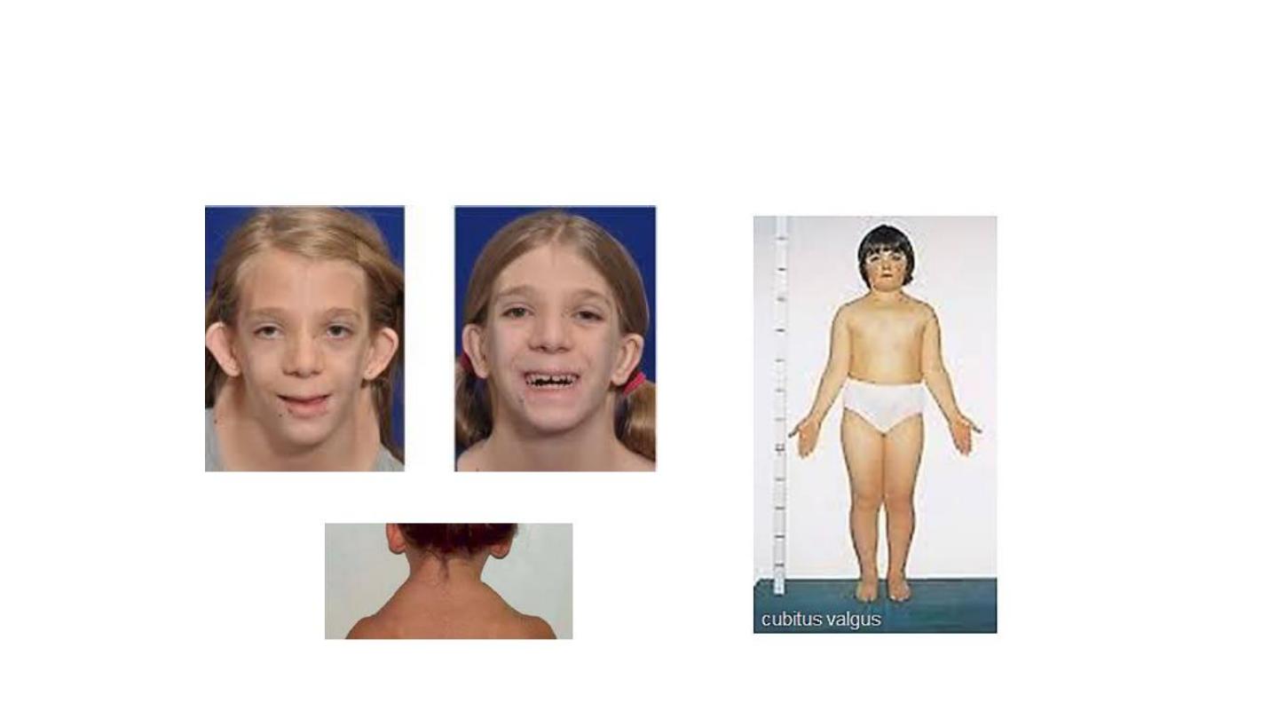
Turner

Turner