Definition:- Ankylosis refers to the partial or complete inability to open the mouth which results in functional and growth deformation of the mandible.
ANATOMY OF TMJ
TMJ is highly specialized, diarthroidal non weight bearing synovial joint of condylar variety
ARTICULAR SURFACES:-
Upper surface:-
a) Articular eminence
b) Anterior part of the mandibular fossaLower surface: - Head of the mandible
BONY COMPONENT:-
Consist of glenoid fossa of the temporal bone & mandibular condyle.
Condyle is roughly elliptical in cross section with mediolateral width being two times the anteroposterior width.
The glenoid fossa is a smooth depression in the temporal bone which is thinnest in its deepest part which separates the joint from the middle cranial fossa.
Articular surface is covered by fibrocartilage.
SOFT TISSUE COMPONENT:-
1) Intraarticular disc or meniscus2) Synovial membrane
3) Lateral pterygoid muscle
4) Capsule of joint
NERVE SUPPLY:-
1) Articulotemporal nerve
2) Masseteric branch of mandibular nerve
BLLOD SUPPLY:-
1) Superficial temporal branch of external carotid artery
2) Middle meningeal artery
APPLIED PHYSIOLOGY
Movement produced by muscle of mastication in the upper joint cavity gliding movements and in the lower joint cavity hinge movements take place.
Movements are:-
– Protrusive -Retraction
-Depression -Elevation
-Lateral or side to side movements
CLASSIFICATION
Kazanjian (1938)
1. True (Intraarticular):- fibrous or bony adhesion between the articular surfaces of TMJ.
2. False (extraarticular):- Results from pathologic condition outside the joint, that results in limited mandibular mobility.
Another classification
A 1. Fibrous
2. Fibroosseous
3. Bony
B 1. Partial
2. Complete
ETIOLOGY
1. TRAUMA- Most common cause of ankylosis. Trauma to the chin forcing the condyle against the glenoid fossa, particularly with bleeding into the joint.
2. INFECTION- From middle ear & mastoid (otitis media, mastoiditis).
3. INFLAMMATORY-
. Primary inflammation of the joint
. Secondary to local inflammatory
Process. (Osteomyelitis)
.Secondary to blood streaminfections
– Septicemia
– Scarlet fever
– Gonorrhea
. Rheumatoid arthritis
4. NEOPLASTIC – (osteochondroma)
5. CONGENITAL
6. MYOGENIC – Myositis ossificans produce limited opening.
7. NEUROGENIC – epilepsy, brain tumor.
CLINIC FEATURES
It includes –
. General features
. Unilateral features
. Bilateral features
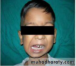
GENERAL FEATURES
AGE- Seen in young age (1 to 10 years)
Symptoms – Trismus (inability to open
mouth)
. Oral problems – poor oral hygiene
carious teeth
periodontal problems
Malocclusion
UNILATERAL FEATURES
Mouth opening is very limited
2. Asymmetry of face with fullness of the affected side & relative flattening of the unaffected side.
3. Face is deviated towards the affected side.
Chin is retracted on the affected side & slightly bypass the midline.
Slight gliding movement towards the affected side.
Cross bite is present.
7. Well defined antegonial notch on affected side.
BILATERAL FEATURES
Bird face appearance/ micrognathia.
No gliding movement neither protrusive nor lateral movement.
Presence of scar on the chin (possibly due to trauma)
Class II malocclusion, protrusive incisors & anterior open bite.
In a long standing case there is atrophy or fibrosis of muscle.
In congenital case-difficulty of introducing the nipple into the mouth of newborn infants.
HISTOPATHOLOGY
1. Atrophy or destructive changes in the cartilaginous component of the joint with loss of meniscus.2. Normal soft tissue is replaced by thick fibrous bands.
3. An overall flattening of articulation.
4. Glenoid fossa & articular eminence become less pronounced.
5. The condyle become enlarged, composed of dense sclerotic bone.
INVESTIGATIONS
For definitive diagnosis & to confirm the extent of bony growth imaging may be required.
1. Lateral oblique view
2. O. P. G. view
3. Cephalometric radiograph
4. Submentovertex view
5. PA view
6. C T Scan
FINDINGS –
1. Decreased ramus height on the affected side.
2. The joint space is completely or partially obliterated with dense sclerotic bone.
3. The condyle can be replaced by shapeless mass of bone.
4. Prominent antegonial notch on the affected side of mandible.
5. Elongation of coronoid process.
6. Sometimes A transverse or oblique dark line crossing the mass of dense bone showing fibrous ankylosis.
MANAGEMENT
Ankylosis can only be treated surgically there is no form of pharmacological management.
The type of surgery depends on the patient & extent of deformity.
Treatment also varies if ankylosis is unilateral or bilateral.
Surgery for ankylosis is done in two stages
1. in first operation only a release of ankylosis is done, Jaw mobility is brought about.
it is belived that growth takes place after ankylosis release.
2. Second procedure, an orthognathic surgery can then be planned to restore facial esthetics. (Especially in children)
GOALS OF TREATMENT
To create mobility in the joint to a satisfactory limit.
To restore vertical height of the ramus.
To restore the mandible & TMJ to normal anatomic & functional state.
To create a new joint space.
To allow the jaw to grow normally in child treated for ankylosis.
To restore the facial esthetics.
To prevent recurrence of ankylosis.
SURGICAL PROCEDURES
It includes
1. Condylectomy
2. Gap Arthroplasty
3. Interpositional Arthroplasty
CONDYLECTOMY
This is a procedure done in ankylosis where the anatomic feature of the joint are not completely changed as in areas of fibrous.
COMPLICATIONS-
1. Loss of vertical height of ramus
2. In bilateral condylectomy it create an anterior open bite.
3. In unilateral condylectomy there is deviation of the jaw on opening.
GAP ARTHRPLASTY
Gap arthroplasty involves creation of an anatomical gap in the ankylosed segment to form an artificial joint space.
The gap is created at a level lower than the original joint space.
Two horizontal bony cuts are made in the most superior aspect of the ramus and the wedge of bone.
A gap of 1-1.5 cm. is created & first interposed with any material.
COMPLICATIONS-
1. Chances of creating excessive gap & reducing vertical height of ramus.
2. Anterior open bite due to excessive bone removal.
3. Reankylosis due to bony contact b/w the cut ends.
INTERPOSITIONAL ARTHROPLASTY
Placing the Interpositional material the two cut ends avoids contact between the bony ends.
It minimizes the chance of reankylosis.
I.P. material may be-
Biological- dermis, facialata, bone & cartilage.
Alloplastic- Vitallium, Tantalum, Silastic
& Acrylic.
COMPLICATIONS-
1. Second surgical site is necessary.
2. Foreign body reaction to alloplastic material.
3. Difficulty in suturing or stablizing the Interpositional material on the medial aspect of joint.
4. Doner site complication such as pleurisy pain, pneumothorax.
SURGICAL APPROACH
Preauricular approach
Risdon’s submandibular approach
Temporal approach
Post auricular approach
Endural approach
Retromandibular approach(post ramus)
OPERATION BY TEMPORAL APPROACH
1. It is the approach through a curvilinear preauricular incision, extending 3 cm. long into the temporal region for the exposure of temporal facia.
2. The dissection of soft tissue is proceeded up to zygomatic arch.
3. The capsule of the joint is opened with a T shaped incision of the facia.
4. Once the joint region is exposed and ankylosed site is identified, aggressive excision of the fibrous & bony mass carried out.
5. Special attention is directed to the medial extension of the callus to ensure adequate excision.
6. Glenoid fossa is recontoured as needed.
7. Coronoidectomy & stripping of muscles are done.
8. It is observed that nearly 1/3 of the height of the ramus is lost following condylectomy & Coronoidectomy.
9. Costochondral graft is removed through inframammary incision.
10. Through submandibular approach ramus is exposed.
11. Masseter & temporalis are stripped from the ramus.
12. Then the surface of the graft and ramus are freshened to produce good bony interface.
13. The graft is placed in position & fixed with stable internal fixation so that 2mm. of posterior open bite is created to compensate for remodelling of the costochondral graft.
14. The length of the graft is determined by the ramus height discrepancy & the facial asymmetry to be corrected.
15. A drain is placed & the wounds are closed in layers.
16. After inter maxillary fixation for a few days, jaw is mobilized vigorously & physiotherapy is continued.
MANAGEMENT SUGGESTED PROTOCOL(KABAN’S)
1. Aggressive total excision of the ankylotic segment in condylar/TMJ region, incomplete removal, particularly medial aspect result in recurrance.
2. Dissection & stripping of muscles of mastication of the ramus & ipsilateral Coronoidectomy are essential.
3. Inter incisial opening should be critically evaluated to ensure that it should be more than 35 mm.
4. If step 1, 2, 3 do not create enough opening opposite side Coronoidectomy is done.
5. A suitable interpositional material must be placed to avoid the risk of reankylosis.
6. Reconstruction of ramus with costochondral graft helps to maintain the ramus height.
7. Rigid fixation of the graft with ramus is necessary for early mobilization of the joint.
8. Creation of open bite on affected side to permit setting of graft for 3-6 months.
9. Early mobilization with minimum IMF less than 3 weeks.
10. Post-operative physiotherapy 6-8 weeks.
11. If no further improvement is noticed postoperatively, joint is stretched under GA. (Brisement Force given.)
12. Later on, if needed, excessive growth of the costochondral graft may have to be recontoured so that no disturbance in occlusion & jaw relationship develop.
REASONS OF RECURRENCE
1. Incomplete excision of callus at the ankylosed site.
2. If the mouth opening during surgery is less than 35 mm & if coronoid process is not excised, recurrence develops.
3. Insufficient & unsatisfactory physiotherapy.
4. Failure to maintain the ramus height leads to narrowing of the inter- fragmentary gap.
5. Failure to provide a satisfactory interpositional material.
6. Heigher osteogenic & periosteal reaction may be responsible for high rate of recurrence in children.
COMPLICATION OF ANKYLOSIS RELEASE SURGERY
A. Preoperative consideration
B. Intra operative consideration
a. complication due to incision
b. complication during osteotomy procedure.
c. Postoperative complications.
COMPLICATIONS DUE TO INCISIONDamage to facial nerve.
Bleeding from superficial temporal artery or facial vessels.
3. Damage to parotid gland.
COMPLICATIONS DURING OSTEOTOMY
1. Inadequate bone removal
2. Profuse bleeding from maxillary artery, pterygoid venous plexus
3. Perforation of middle cranial fossa by damage to glenoid fossa.
POSTOPERATIVE COMPLICATIONS
1. Anterior open bite or deviation of jaw.
2. Infection to surgical site.
3. Reankylosis.
4. Frey’s syndrome.
MANAGEMENT SUMMARY
1. Prior to 1951, it was considered to be incurable.
2. Brisement force (forceful opening under GA) was tried without success.
3. Esmarch suggested- wedge resection of the body of the mandible.
4. Condylectomy in early cases.
5. gap-arthroplasty led to recurrance.
6. Osteoarthrotomy (gap & interpositional arthroplasty) improved the prognosis.
7. Excision of callus & joint reconstruction become the treatment of choice.