Spinal Injuries
ObjectivesThe ability to demonstrate knowledge of the following:
Basic anatomy of the spine.
Initial assessment and treatment of spinal injuries at the field.Principle of spinal stability.
Understanding of neurologic syndromes caused by spinal trauma.
Management of Cauda equina syndrome.
Epidemiology
15-20% multiple non-contiguous levels.10% involving the cervical spine.
90% involving thoracolumbar spine.
25% have neurologic deficit.
Age: mostly between 15-24 years.
Gender: mostly males (4:1).
Mechanism of Injury
High energy trauma such as an MVA or fall from a height or a horse.MVA: 40-55%
Falls: 20-30%Sports: 6-12%
Others: 12-21%
Low energy trauma in a high risk patient (ie a patient with known spinal canal compromise such as ankylosing spondylitis, Osteoporosis or metatstatic vertebral lesions)
Penetrating trauma from gunshot or knives.
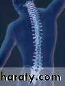
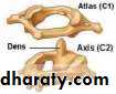
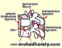
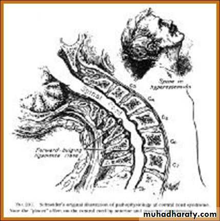
Anatomy
Spinal Column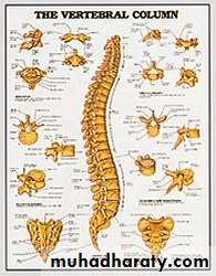
Anatomy
Spinal Column
Anatomy
Spinal Column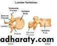
Anatomy
Spinal Column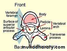
Anatomy
Spinal Column
Anatomy
Spinal Column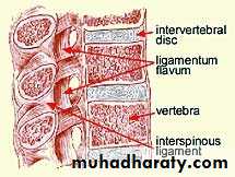
Anatomy
Spinal Column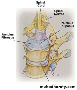
Anatomy
Spinal Column
Anatomy
Spinal Cord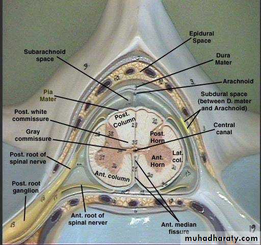
Anatomy
Spinal Cord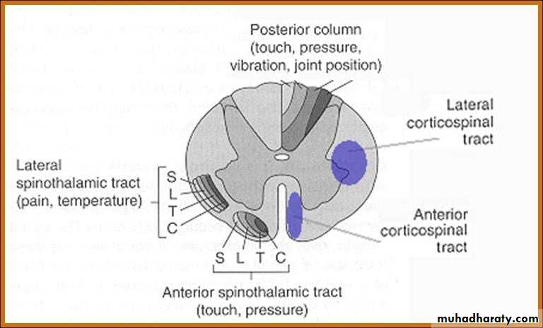
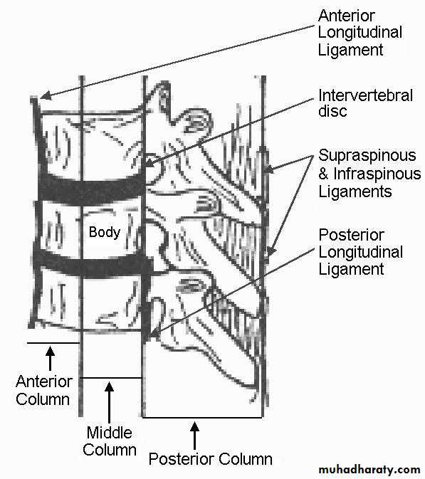
The Three columns
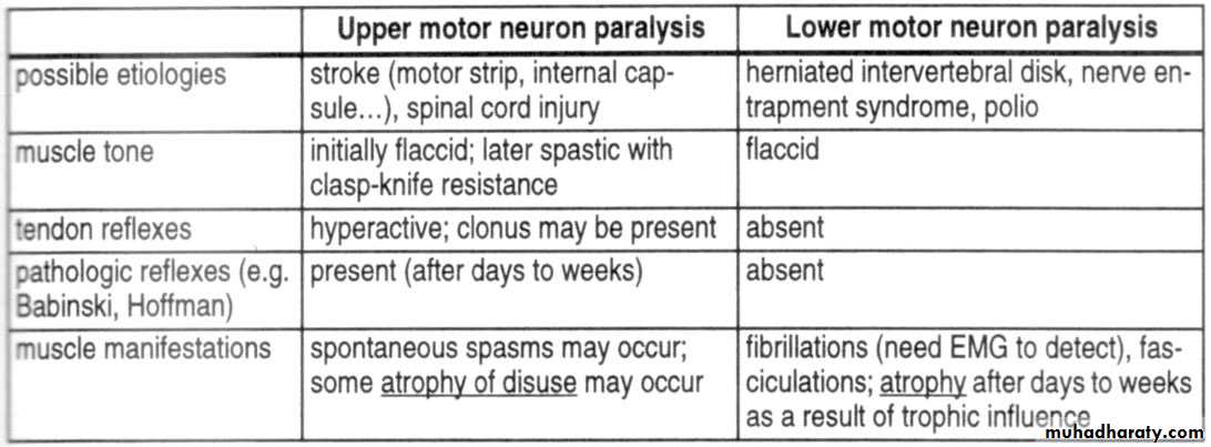
What are the differences between UMN and LMN? (e.g., cauda equina vs. myelopathy)
So to determine the level of injury?
Motor level = the last level with at least 3/5 (against gravity) function
NB: this is the most important for clinical purposesSensory level = the last level with preserved sensation
Radiographic level = the level of fracture on plain XRays / CT scan / MRI
NB: spine level does not correspond to spinal cord level below the cervical region
High cervical injuries (C3 and above)
Motor and sensory deficits involve the entire arms and legsDependent on mechanical ventilation for breathing (diaphragm is innervated by C3-C5 levels)
Midcervical injuries (C3-C5)
Varying degrees of diaphragm dysfunctionUsually need ventilatory assistance in the acute phase
Shock
What is the difference between spinal shock and neurogenic shock?
Neurogenic
Hypovolemic
Etiology
Loss of sympathetic outflow
Loss of blood volume
Blood pressure
Hypotension
Hypotension
Heart rate
Bradycardia
Tachycardia
Skin temperature
Warm
Cold
Urine output
Normal
Low
Low cervical injuries (C6-T1)
Usually able to breathe, although occasionally cord swelling can lead to temporary C3-C5 involvement (need mechanical ventilation)The level can be determined by physical exam
So what do you expect with a cervical lesion?
Quadriplegia or quadriparesis
Bowel/bladder retention (spastic)
Various degrees of breathing difficulties
Neurogenic and/or spinal shock
Thoracic injuries (T2-L1)
Paraparesis or paraplegiaUMN (upper motor neuron) signs
Cauda equina injuries (L2 or below)
Paraparesis or paraplegiaLMN (lower motor neuron) signs
Thigh flexion is almost always preserved to some degree
Assessment
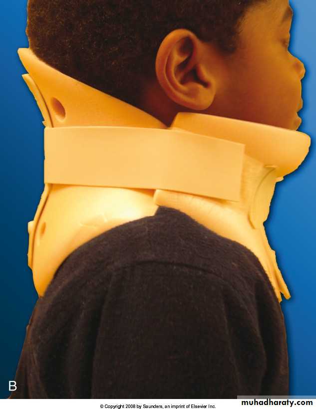
In cases of trauma, ABC….. must be assessed first and treated appropriately.Patients should be examined with spinal collar until spinal pathology is excluded.
Careful log rolling keeping the head, neck and pelvis in line should be done to examine the spine properly.
Assessment
Immobilization.History:
Mechanism of injury:
compression, flexion, extension, distraction
Other injuries.
Seat belt.
Other causalities.
Physical examination:
Inspection, palpation.
Neurologic examination.
Immobilization


Neurologic
Muscle TestSensory exam
light touch, Sharp dull discrimination, Vibration sense, Proprioception and two-point discrimination
Reflexes
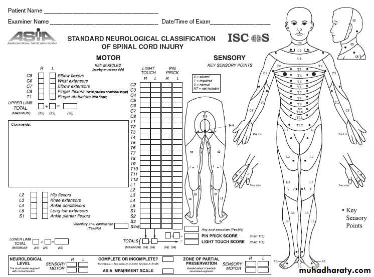
Asia Score: Brief Trauma Neurologic Survey
Level of Cord Injury determines level of function
Prognosis for Recovery of spinal Cord Injury:Poor prognosis for recovery if:
-pt arrives in shock
-pt cannot breath-pt has a complete injury
Assessment
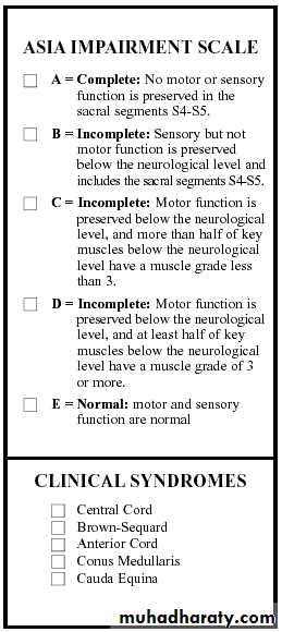
• Severity of neurologic deficit
• CompleteFlaccid paralysis below level of injury.
May involve diaphragm if injury above C5.
Sympathetic tone lost if fracture above T6.
• Incomplete
• ? Any sensation.
• ? Sacral spairing.
Assessment
• Severity of neurologic deficit
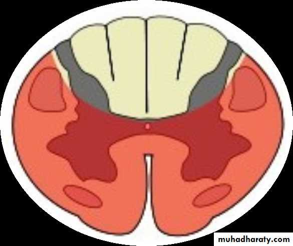
• Incomplete• Central cord syndrome:
• # Characterized by disproportionally (UL>LL).
• # Mechanism: hyper-extension.
• # Occur with or without fractures.
• # Recovery: 50% regaining function.
• # Prognosis is fair.
•
Assessment
• Severity of neurologic deficit
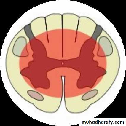
• Incomplete• Anterior cord syndrome:
• # Characterized by loss of corticospinal and spinothalamic tract with preserved posterior column.
• # Mechanism: ischemia or infarction to spinal cord..
• # Common injury.
• # Recovery: 10%.
• # Prognosis is good if progressive recovery within 24hrs, absent SS after 24hrs protends a poor outcome.
•
Assessment
• Severity of neurologic deficit
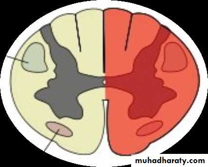
• Incomplete• Brown-Sequard syndrome:
• # Characterized by hemicord injury with ipsilateral paralysis, loss of proprioception and fine touch, and contralateral temperature and pain loss.
• # Prognosis is good, with over 90% regaining of bowel and bladder function and ambulatory capacity.
•
Assessment
• Severity of neurologic deficit
• Incomplete• Conus Medullaris syndrome:
• # Seen in T12-L1 injuries.
• # Loss of voluntary bowel and bladder control with preserved lumbar root function.
• # Uncommon as pure lesion (mixed conus-cauda).
Assessment
• Severity of neurologic deficit
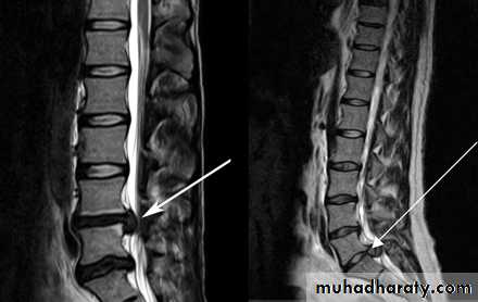
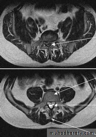
• Incomplete• Cauda Equina syndrome:
• # Saddle anesthesia, urinary retention and stool incontinence.
• # Usually due to large central disc herniation rather than fracture.
• Nerve root deficit: LMN
Spinal Shock
Transient loss of spinal reflexes.Lasts 24-72 hours.
Neurogenic shock
Reduced tissue perfusion due to loss of sympathetic outflow and un-apposed vagal tone.Peripheral vasodilatation (hypotension and bradycardia).
Rx: fluid resuscitation and vasopressors.
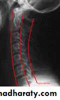
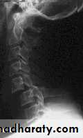
X-rays:
Cervical: 3 views.AP, lateral and open mouth.
Thoraco-lumbar: 2 views.
AP & lateral.
Flexion-Extension views.
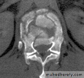
CT: best for bony anatomy.
MRI: best to evaluate soft tissue.
Imaging
Depends on:
Level of injury.Degree and morphology of injury: STABILITY
Presence of neurologic deficit.
Other factors.
Management of Spinal Injuries
Some general rules:
Stable injuries are usually treated conservatively.Unstable injuries usually require surgery.
Neurologic compression requires decompression.
Specific Injuries
Cervical spine fractures
Descriptive: depends on mechanism of injury.Flexion/extension.
Compression/distraction.
Shear.
Presence of subluxation/dislocation
SCI:
high fracture results in quadriplegia.
Low fracture results in paraplegia.
Cervical spine fractures
Thoraco-Lumbar fractures
Spinal cord terminates at L1/2 disc in adultL2/3 in a child
50% of injuries occur at Thoraco-lumbar junction.
Common fractures:
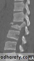
Wedge fracture (flexion/compression).
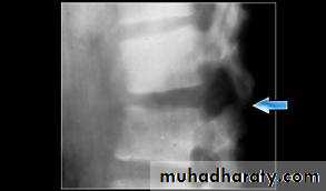
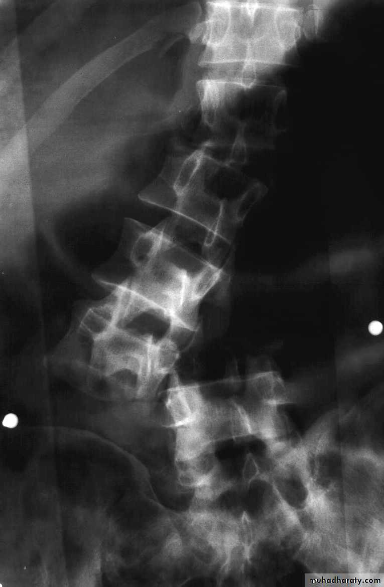
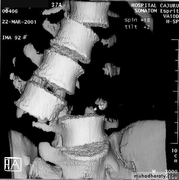
Burst (compression).
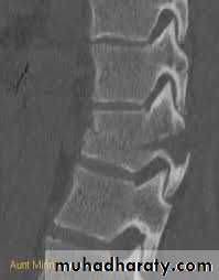
Chance (flexion/distraction).
Wedge fracture
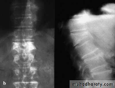
Burst fracture
Chance fracture
Fracture dislocation
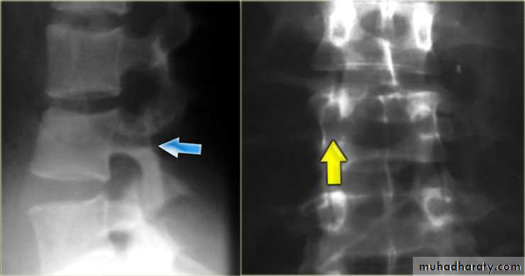
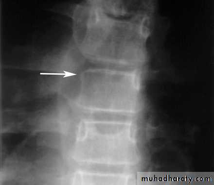
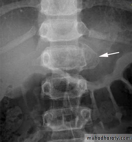
Pathologic fractures
Low-energy fractures.Osteoporotic is common.
Usually due to infection or tumor.
X-rays: “winking owl” sign.
Pathologic fractures

Cauda Equina Syndrome
A surgical emergency.Requires full neurologic examination including rectal examination for anal tone.
Investigations: X-rays initially, but MRI is mandatory as X-rays are usually unremarkable.
Treatment: Emergency decompression-usually discectomy and wide laminectomy within 24 hours.
Cauda Equina Syndrome
Thank you