ا.د اسامه عبيد الخفاجي
MBChBFIBM Cardiothoracic & vascular surgey
MRCS Edn.
PULMONARY HYDATID CYST&BRONCHOGENIC CARCINOMA
Pulmonary Hydatid DiseaseHydatid means cyst full of water
• Epidemiology:
• Hydatid disease is caused by adult and larval stage of Echinococcus granulosus
• The adult worm lives in dogs which is the primary host
• The cystic part of the cycle occur in sheep, ox, pigs, camel, or man which act intermediate host
• The adult worm live in the intestine of dogs for months. The terminal segment proglottidis detach from the worm and release thousands of ova with the stool. These can remain infective for long time.
• Ingestion of the ova by sheep in contaminated water or grass or vegetables lead to infection and hydatid cyst subsequently develop in the viscera
• The parasite goes back into the dog when the sheep is slaughtered and its viscera containing the hydatid cyst are eaten by another dog.
• If a worm gets into a man instead of sheep, the life cycle of the parasite will be broken
The ova hatches in the stomach → hexacanth embryo are released in the stomach and duodenum → pass through the intestinal wall → travel via he portal vein → liver where the major settle and develop hydatid cyst.
Only those embryo which successfully pass through the capillaries of the liver, reach the lung and develop into hydatid cyst and few which get through the lung are then carried to the viscera and tissues supplied by the systemic circulation
Hydatid cyst itself is composed of :
Ectocyst: (the laminated membrane) formed by the germinal layer. It is an acellular white laminated membrane
Endocyst: (the germinal layer) it is the only living part of the cyst which secretes :
Internally: hydatid fluid
Externally: it forms the laminated membrane
Broad capsule: develops from the germinal layer by the process of asexual budding and are attached by pedicles to the inner most wall of the germinal layer
Clinical presentation:
The lesion may be asymptomatic unless complications develop. The patient may present to the thoracic surgical department with incidental CXR findingPatient may experience transient symptoms of : pain, cough and hemoptysis.
The acute onset of : rigor, fever and cough with purulent exudation and pleuritic pain is experienced with all patients with lung abscess.
Fragmentation of the membrane or daughter cysts may be described by the patient as "grape skin" which may be coughed out when the cyst ruptures into the bronchial tree
Severs dyspnea may occur in rare events of rupture into the pleural space → hydropneumothorax or pyopneumothorax.
Rupture of the cyst may cause signs of anaphylaxis
Diagnosis
1-CXRWell defined circular
A pathgnomonic X-ray finding is perivesicular pneumocyst due to presence of air between the adventitia and laminated membrane so appears as a slender crescent "signet ring" in case of adventitial rupture
Double arc sign" appears as a cyst with fluid level on either sided of the dome shaped cyst
"Water lilly sign" or "Camelot sign": cyst with collapsed membrane floating in the fluid seen in cases of rupture of the cyst itself
adventitia calcify → thin shell of egg
Hydropneumothorax is seen
2-Isotope liver scan
3-Ultrasound
4-CT scan
5-Immunological tests:
Casoni intradermal test
Weinberg CFT: best method and it is accurate
Latex slide agglutination test
Indirect hemagglutination test: non-specific
.
6-Blood tests:
Eosinophilia is common but of little value because of multiple parasitic infection in the middle east
Treatment:
Surgical Treatment:
Treatment of hydatid cyst is surgical removal
A-Removal of the cyst:
1-Aspiration – Evacuation technique
2- Enucleation
3- Excision
B-Resection
Medical Treatment:. No drugs have yet been found that can effectively reach and destroy the parasite. Even if drugs capable of destroying the live contents of the cyst become available, the retained membrane will ultimately become infected and produce a lung abscess.
Mebendazole and fluoromebendazole (vermox, fluvermol) are both benzinidazole derivatives and have limited role in treatment
Indications:
Disseminated cases when the cysts are multiple and inaccessible
Surgery contraindicated for reasons of extreme ill health
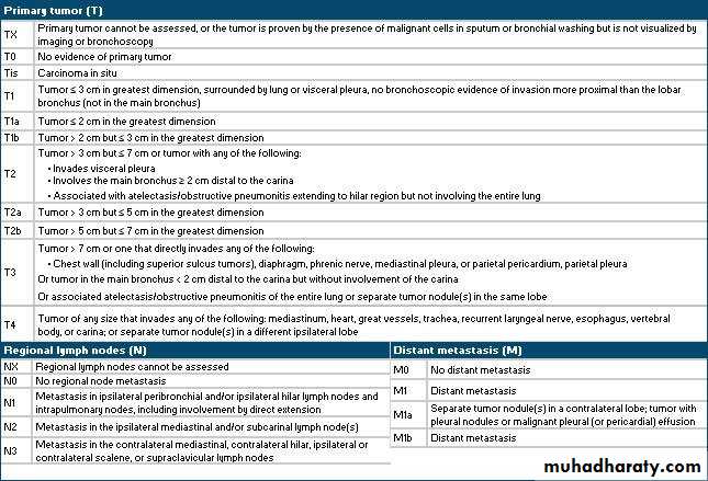
CARCINOMA OF THE LUNG
Lung cancer is the most common cause of death from malignancy in both men and women.
Pathology:
Pulmonary neoplasms arise in the bronchial epithelium
Macroscopically:
Peripheral: 60% of all lung ca. occurring in the lung parenchyma or bronchioles.
Central: 40% of all lung ca. occurring in main stem bronchus, lobar or segmental bronchus
Microscopically:
SCLC: (20%) Small cell lung cancer (SCLC) is characterized by rapid growth, stronger likelihood of metastases being present at the time of diagnosis, and greater responsiveness to chemotherapy and radiation therapy
SCLS is subdivided into:
Oat cell ca
Intermediate
Combined oat cell ca
NSCLC : 80% ,The three major histologic types of NSCLC are
adenocarcinoma,
squamous cell carcinoma,
large cell carcinoma
Pathogenesis:
Ca of the lung shows progressive pathological changes in bronchial epithelium with time (especially squamous cell carcinoma).Basal cell proliferation → hyperplasia of goblet cells → metaplastic stratification of squamous epithelium → atypical metaplesia → Ca in situ → infiltration of the ca through the basement membrane → spread to regional LN → hematogenous dissemination
Etiological factors
• 1-Cigarette smoking: cigarette index: no. of pack multiply by years of habit
• 2-Exposure to carcinogens: arsenic, cadmium, chromium
• 3-Chronic obstructive pulmonary disease (COPD)
• 4-Genetic: involving activation of dominant oncogenes and the inactivation of recessive oncogenes.
Dominant oncogenes: 3 groups associated with lung ca → ras, erb, myc.
Recessive oncogenes: most common p53 and retinoblastoma gene (Rb)
Clinical features:
Asymptomatic
Symptomatic:
I-Bronchopulmonary symptoms: :
Dyspnea ,Hemoptysis, Stridor and wheeze cough Chest pain, Features if lung abscess
II-Extrapulmonary intrathoracic symptoms
Caused by invasion, irritation or compression of the adjacent structures• A- Nerves: Recurrent laryngeal nerve → vocal cord palsy and hoarseness of voice, Phrinic nerve → paralysis of hemidiaphragm
B-SVC syndrome → due to extensive involvement of right mediastinal LN
C-Pleural involvement → pleural effusion
• D-Pericardium and heart → tamponade and arrhythmias
• E-Trachea → dyspnea and stridor
F-Esophagus → dysphagia
G-Ribs and chest wall → chest pain
III-Paraneoplastic syndromes: (extrathoracic non-metastatic symptoms):
Paraneoplastic syndromes are frequently associated with SCLC
• cushing's syndrome
Hypercalcemia
Gynecomastia
Hypertrophic pulmonary osteoarthropathy
Mysthenia gravis
Investigations:
1 CXR: solitary pulmonary nodule, pneumonic infiltrate, pleural effusion 2-CT scan: for evaluation of the primary tumor, ,the assessment of regional lymph node involvement3-Bronchoscopy: Flexible fiberoptic bronchoscopy is an important tools in the diagnosis of carcinoma of the lung. Bronchial biopsy, brushings, washings, and transbronchial aspiration may be used to establish the diagnosis
4-Sputum cytology
5-Mediastinoscopy
6-Thoracoscopy
7-PET (Positron Emission Tomography):
is a noninvasive imaging method that has demonstrated increased glucose metabolism in malignant cells.This technique has demonstrated a sensitivity of 94% and a specificity of 80%
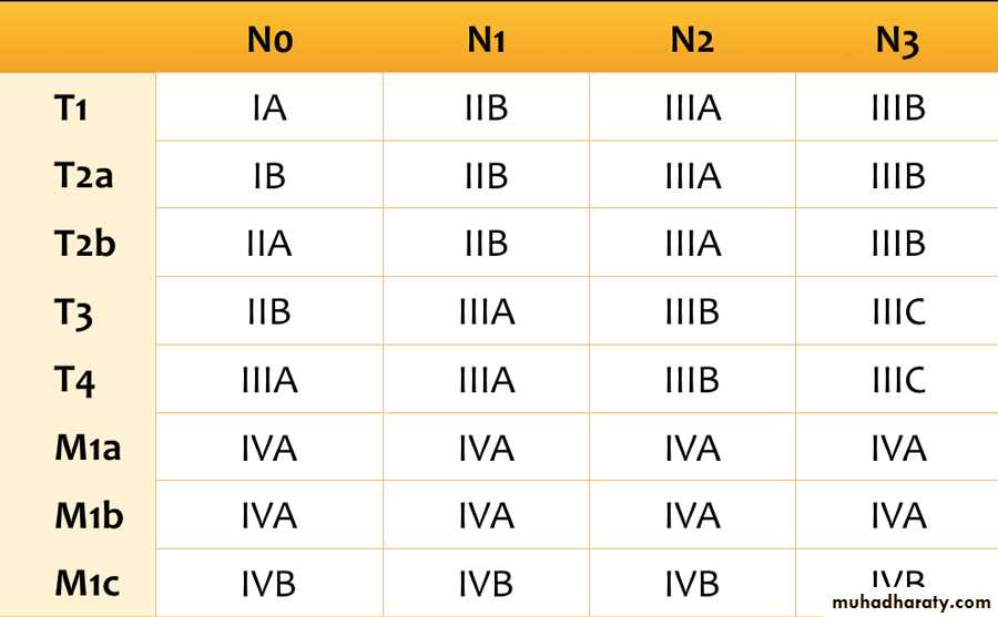
Treatment of lung ca
Surgical :Every solitary pulmonary nodule should be resected unless the lesion is known to be benign or the patient's medical condition contraindicates surgical procedureresection is the standard treatment of patients with Stage I or Stage II disease.
Radiotherapy : pre & post op
Chemotherapy : vincristine,adriomycin,bleomycin
THANKS