Histology
LAB.3
Lymphatic Tissue and the Immune System
Contains of this LAB.
Lymph node (
MH076
)
Tonsil of Human (
MHS276
)
Spleen of human (
MHS212
)
Thymus gland (
MH079
)
Edited by Fahad A.
Mosul Medical College
2018-2019
2th stage
Get it from
www.muhadharaty.com
This copy is used to study tissues with Histology Guide website
to get a better result and a deeper understanding.
Do not hesitate to ask the doctors of the histology branch.
Histology
1
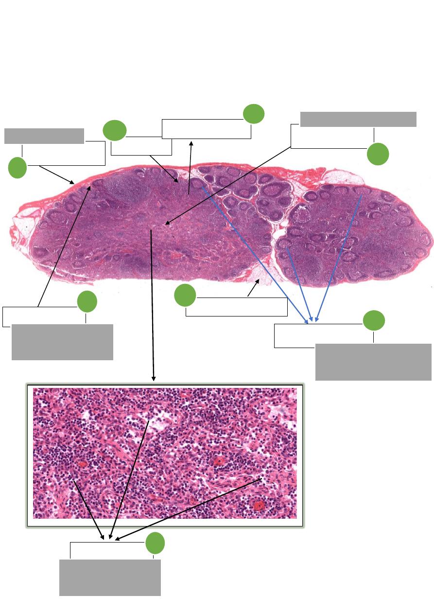
1 - Lymph node
Capsule
enclosing the node
Cortex
Medulla
It's the inner region of the node
Cortical nodules
All the spherical clusters
Remember
lighter center of
nodules called
germinal center
1
3
2
5
Trabeculae
It's connective tissue
that extends inward from
the capsule.
7
Lymphatics at hilus
8
Medullary Sinuses
Intervening spaces in which
lymph flows
4
Paracotical zone
6
صورة مكبرة

2
Notes
Structure
Dense connective tissue enclosing the node.
- contains of :-
1- Subcapsular Sinus
- space underneath the capsule that receives
lymph from afferent lymphatic vessels.
2- Trabeculae
- connective tissue that extends inward from the
capsule.
3- Trabecular Sinuses
- spaces alongside trabeculae in which lymph
flows from the subcapsular sinus into the cortex.
Capsule
Outer region of the node.
- contains of :-
with lighter
Nodules
have
(
region adjacent to the capsule
-
Outer Cortex
-
1
)
germinal center
called
center
2- Inner Cortex
- (paracotex) region between the outer cortex and the medulla
that is free of nodules.
Cortex
Most inner part of the node.
contains of :-
-
1- Medullary Cords
2- Medullary Sinuses
- intervening spaces in which lymph flows
Medulla
MH 076
VS Code
–
.com
guide
istology
H
Lymph Node
تقسيم
ال
odules
N
1
-
Primary nodules
هي
nodules
التي تحتوي
germinal center
2
-
Secondary nodules
هي
nodules
التي ال تحتوي
germinal center
3
-
white pulp
هي
ال
nodules
التي تحتوي على
Central arteries
و
germinal center
وتوجد في
spleen
Remember
lighter center of nodules called
germinal center
3
2- Tonsil
Epithelial lining
TYPE: stratified Squamous
Non-Keratinized Epithelium
Crypts
Nodules
Spherical clusters
الحظ
Germinal center
It's lighter center of nodules
الحظ
صورة مكبرة
Lymphocytes
It's Granules dispersed "lymph cell"
الحظ

4
Notes
Structure
Type of it
Stratified Squamous Non-Keratinized Epithelium
- covers
the numerous nodules that compromise the tonsil.
Epithelial lining
Infoldings of the epithelium into the underlying connective tissue.
Crypts
spherical aggregations of lymphocytes ( have germinal centers )
Lymph Nodules
pass through the epithelium in areas of inflammation.
Lymphocytes
Tonsil on Histologyguide.com
76
2
S
MH
VS Code
–
Histologyguide.com
5
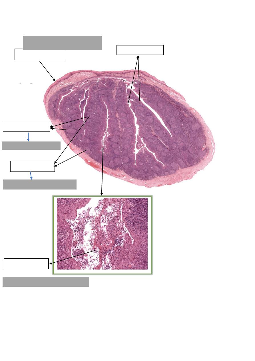
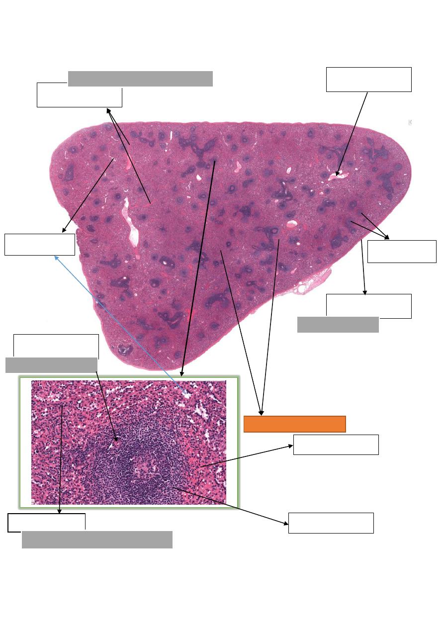
3– Spleen
Capsule
Enclosing the organ
Trabeculae
Connective tissue that extends inward
Blood vessels
Central arteries
Regular circle in nodule
Splenic sinusoids
Lymphocytes
It's Granules dispersed "lymph cell"
الحظ
Red pulp
White pulp
Red pulp
و
هي
ارضية
االساليد
Splenic nodules

6
Notes
Structure
Dense connective tissue enclosing the organ.
Capsule
connective tissue that extends inward from the
Trabeculae
Composed of lymphatic tissue.
It appears
basophilic
due to the large number of nuclei
White Pulp
Clusters of B lymphocytes located on central arterioles.
They usually contain a germinal center of activated B lymphocytes
Splenic Nodules
branches of trabecular arteries.
Central Arterioles
Filters and degrades red blood cells (RBCs).
It appears
eosinophilic
due to the large number of RBCs
Red Pulp
vascular spaces
Splenic Sinusoids
Spleen on Histologyguide.com
212
S
MH
VS Code
–
Histologyguide.com
7
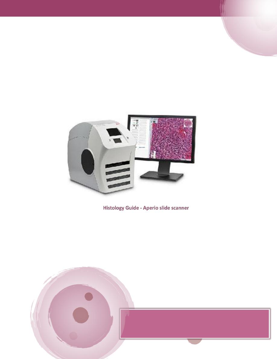
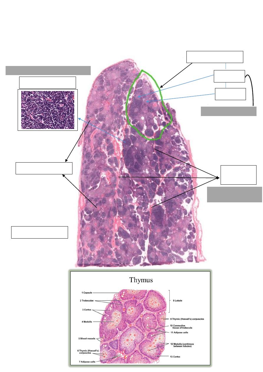
4- Thymus (for adult)
Hassel's Corpuscle
Thymic lobule
Cortex
Medulla
"
nd
& 2
st
It have nodule "1
Lymphocytes
Trabeculae
"septae"
Small nuclei of condensed chromatin.
حواجز
تقسم النسيج لفصوص
Adipose tissue
صورة توضيحية
8
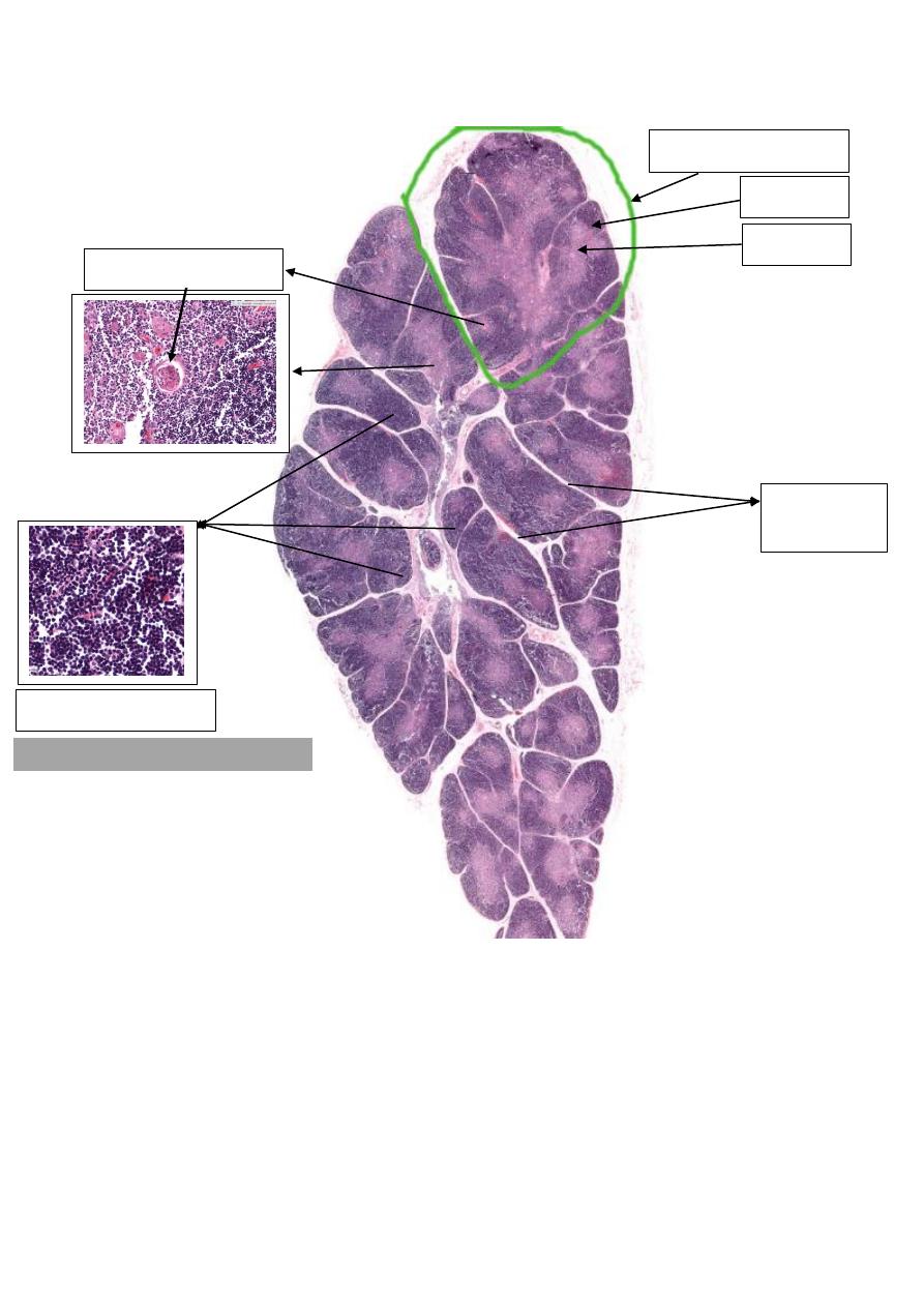
4- Thymus (Neonatal)
Hassel's Corpuscle
Thymic lobule
Cortex
Medulla
Trabeculae
"septae"
Lymphocytes
Small nuclei of condensed chromatin.

9
Notes
Structure
thin connective tissue surrounding the thymus that extends inward
as trabeculae to form incomplete lobules.
Capsule
Adult
= nner, lighter region of large lymphocytes.
Cortex
Neonatal
=
outer darker, region of small lymphocytes.
It have -
1- T-Lymphocytes
2- Epithelial Reticular Cells
inner, lighter region of larger lymphocytes.
Have "Hassel's Corpuscle"
Hassel's Corpuscle = closely packed, concentrically arranged
epithelial reticular cells.
Medulla
Thymus on Histologyguide.com
079
MH
VS Code
–
Histologyguide.com

11
Questions //
Question
: Where else do you find tonsils in the oral cavity?
1 – Palatine tonsils
2 – Pharyngeal tonsils
3 – Lingual tonsil
-------------------------------------------------------------------------
Question
: What is the function of tonsils?
They are the immune system's first line of defense against ingested or inhaled foreign
pathogens.
Play role in immunity of the body because they contain B&T- lymphocytes thus they are
important especially in children.
-------------------------------------------------------------------------
Discuss
the effects of removing the tonsils.
(A very common procedure not so long ago!)
Question
: What is happening to the thymus gland with increasing age?
Is
it still functional in the adult?

11
Task
: List the major immune cells and their functions