Hematuria
Is the presence of at least 5 red blood cells (RBCs) per microliter of urineDefinition:
• Occurs in 0.5-2.0% of school-aged children.
• Presence of 10-50 RBCs/μL by a urinary dipstick may suggest underlying pathology
• Presence of >50 RBCs/μL considered as significant hematuria.
• Presence of formalin (used as a urine preservative)
• False negative results can occur in• High urinary ascorbic acid concentrations (vitamin C intake >2000 mg/day).
• Contamination with oxidizing agents such as hydrogen peroxide used to clean the perineum before obtaining a urinary specimen.
• False-positive results seen in:
• Alkaline urine (pH > 8)
• Pathologic (hemoglobinuria from hemolytic anemia, myoglobinuria from rhabdomyolysis)
Causes of hematuria:
• A-Factitious hematuria:
• Non-pathologic (urate crystals in infants, ingested foods, drugs, dyes)
• Toxin-mediated injury: hemolytic uremic syndrome (HUS)
• B-Glomerular causes:• Immunologic injury: GN e.g., poststreptococcal GN, IgA nephropathy, membranoproliferative GN, systemic diseases.
• Structural disorder: Alport syndrome, thin basement membrane disease
• Structural: cyst rupture, Wilms tumor, urinary tract obstruction, renal trauma
• C-Tubulointerstitial/ Parenchymalcauses:
• Inflammation: interstitial nephritis, pyelonephritis
• Vascular: sickle cell trait/disease
• Hypercalciuria
• D-Lower urinary tract causes:• Inflammation: cystitis, hemorrhagic cystitis, urethritis
• Injury: trauma, stone
• Chloroquine
• Deferoxamine
• Ibuprofen
• Iron
• Metronidazole
• Nitrofurantoin
• Phenothiazines
• Rifampin
• Salicylates
• Sulfasalazine.
**Drug causing red urine:
Acute Poststreptococcal Glomerulonephritis (APSGN)
• It is a postinfectious complication of Group A β-hemolytic streptococcal infections.• It is one of the most common glomerular causes of gross hematuria in children
• It is a classic example of the acute nephritic syndrome characterized by the sudden onset of:
• Gross hematuria
• Edema
• Hypertension
• Renal insufficiency
ETIOLOGY AND EPIDEMIOLOGY:
• It is most commonly sporadic although endemic attacks has been reported.
• APSGN follows infection of “nephritogenic” strains of group A β-hemolytic streptococci which causing pharyngitis during cold-weather months and streptococcal skin infections or pyoderma during warm-weather months.
• 97% of cases occur in less-developed countries.
CLINICAL MANIFESTATIONS:
• APSGN is most common in children 5-12 yr and uncommon before the age of 3 yr.• Typically the acute nephritic syndrome (gross hematuria, edema, hypertension, and renal insufficiency) develop 1-2 wk after a streptococcal pharyngitis or 3-6 wk after a streptococcal pyoderma.
• The severity of kidney involvement varies from asymptomatic microscopic hematuria with normal renal function to gross hematuria with acute renal failure.
• Patients have various degrees of edema, hypertension, and oliguria depending on the severity of renal involvement.
• Hypertensive encephalopathy cause blurred vision, severe headaches, altered mental status, or new seizures.
• Nephrotic syndrome develops in <5% of childhood cases.
• Pulmonary edema and heart failure occur due to hypertension and hypervolemia causing respiratory distress, orthopnea, and cough.• Peripheral edema typically results from salt and water retention and is common.
• The acute phase generally resolves within 6-8 wk. Urinary protein excretion and hypertension usually normalize by 4-6 wk after onset, but microscopic hematuria can persist for 1-2 yr after the initial attack.
• Nonspecific symptoms such as malaise, lethargy, abdominal pain, or flank pain are common.
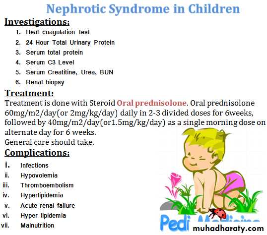
Investigations:
• Urinalysis :RBCs, RBC casts, proteinuria, and polymorphonuclear leukocytes.• Mild normochromic anemia due to hemodilution and low-grade hemolysis.
• Decreased C3 level in >90% of patients and returns to normal after 6-8 wk
• Serum C4 level is normal, or only mildly decreased.
• increased S. creatinine and BUN.
• Positive streptozyme screen (measures multiple Abs to different streptococcal Ags)
• Evidence of a recent streptococcal infection by :
• Positive throat culture in acute infection but it might represent the carrier state.
B. Rising AB titer to streptococcal Ag(s) :
• Antistreptolysin O (ASO) titer is commonly elevated after pharyngitis but rarely after skin infections.
• Antideoxyribonuclease B level is the best single AB titer to document cutaneous streptococcal infection.
• Presence of acute renal failure.
• Presence of nephrotic syndrome.• absence of evidence of streptococcal infection.
• normal complement levels.
• Persistent hematuria and proteinuria, decreased renal function, and/or a low C3 level for more than 2 mo after onset.
• Renal biopsy: indicated in:
• Brain MRI indicated in patients with suspected HT encephalopathy.
• Chest x-ray for patients with signs of heart failure or respiratory distress.• Acute nephritic syndrome (gross hematuria, edema, hypertension, and renal insufficiency).
• Evidence of recent streptococcal infection by culture or serological tests.
• Low serum C3 level.
CLINICAL DIAGNOSIS:
APSGN is Consider in the presence of:
COMPLICATIONS:
• Complication of hypertension : hypertensive encephalopathy which is reversible with appropriate management, but severe prolonged hypertension can lead to intracranial bleeding.
• Complications of acute renal failure: heart failure, hyperkalemia, hyperphosphatemia, hypocalcemia, acidosis, seizures, and uremia.
• Early systemic antibiotic therapy for streptococcal throat and skin Infections, to eradicate the infection but it does not eliminate the risk of GN.
• Family members of patients with acute GN, especially young children, should be cultured for group A β-hemolytic streptococci and treated if positive.
PREVENTION:
• Treatment of acute renal insufficiency and hypertension.
• 10 day course of systemic antibiotic therapy with penicillin to limit the spread of the nephritogenic streptococci, but antibiotic therapy does not affect the natural history of APSGN.TREATMENT:
• Complete recovery in >95% of children.
• Recurrences are extremely rare.• Mortality in the acute stage can be avoided by appropriate management of acute renal failure, cardiac failure, and hypertension.
• Acute phase may be severe and leads to glomerulosclerosis and chronic renal disease in <2% of affected children.
PROGNOSIS:
HEMOLYTIC UREMIC SYNDROME (HUS)
• HUS is characterized by the triad of:• Microangiopathic hemolytic anemia
• Thrombocytopenia
• Renal injury
• It is important cause of acute kidney injury in children.
• HUS is more common in pre-school and school age children but it can occur in adolescents and adults.
ETIOLOGY:
• Typical or Diarrhea-associated HUS:• It’s the most common type of HUS.
• It is associated with a prodromal diarrheal illness (often bloody) then followed by clinical manifestations of HUS.
• Caused by: Verotoxin (VT)-producing E. coli (most commonly E. coli O157:H7)
• Other E. coli strains and other bacteria such as Shigella VT can cause HUS in 5-15% of affected children.
• B. Atypical (non-diarrheal) HUS:
• No preceding diarrhea.• It may occur at any age.
• More severe than diarrhea-associated HUS.
• Infection e. g S. pneumoniae and HIV
• genetic and acquired complement defects.• Drugs: cyclosporine, contraceptive pills.
• Systemic diseases e.g. malignant HT, SLE, malignancy, bone marrow or organ transplantations and pregnancy
• It can be secondary to the following:
• Microvascular injury with endothelial cell damage is characteristic of all forms of HUS.
• In the diarrhea-associated HUS, enteropathic MO produce Shiga toxin or Shiga-like verotoxin causing endothelial cell damage.
• Renal capillary and arteriolar endothelial cell damage leads to localized thrombosis, particularly in glomeruli, causing decrease in GFR.
PATHOGENESIS:
• Progressive platelet aggregation in the areas of microvascular injury results in consumptive thrombocytopenia.
• Microangiopathic hemolytic anemia results from mechanical damage to RBCs as they pass through the damaged and thrombotic microvasculature.
• Typical diarrhea-associated HUS preceded by GE with fever, vomiting, abdominal pain, and diarrhea (often bloody).
• After 5-7 days, patient develop sudden onset of pallor, irritability, weakness, and lethargy due to MAHA.
• Oliguria can be present in early stages but may be masked by ongoing diarrhea.
CLINICAL MANIFESTATIONS:
• Dehydration is often present; however some children have volume overload due to oliguria.
• Hypertension due to volume overload and/or renal injury.
• CNS involvement: seizures, in up to 25% of cases due to focal ischemia secondary to microvascular CNS thrombosis.
• Other manifestations: pancreatitis, cardiac dysfunction, and colonic perforation.
INVESTIGATIONS:
I. Evidence of Microangiopathic HA:• CBC: Anemia and Thrombocytopenia
• Peripheral blood smear: schistocytes, helmet cells, and burr cells
• Increased indirect bilirubin
• Increased reticulocyte count
II. Evidence of renal injury:
• Increased serum creatinine
• Presence of hematuria, proteinuria, pyuria, and casts on urinalysis
• Leukocytosis
• Positive stool culture for E. coli O157:H7 but it might be negative at time of presentation of HUS.
• Positive stool test for shiga-toxin
• Elevated amylase and lipase enzymes due to pancreatitis.
• Negative Coombs test
III. Other potential findings:
• supportive therapy : fluid replacement, control of hypertension, and treatment complications of renal insufficiency, including dialysis if indicated.
• RBC transfusions in severe anemia.
• Platelet transfusions should be avoided because they may add to the thrombotic microangiopathy and are indicated only in active hemorrhage.
• Antibiotics and antidiarrheal agents may increase the risk of developing HUS.
TREATMENT:
• >95% of diarrhea-associated HUS patients survive the acute phase and recover normal renal function.
• Some may have long-term morbidity.
• Poor prognosis associated with non-diarrheal and familial HUS.
PROGNOSIS:
Thank you for your attention