Histology
LAB.5
Gastro-Intestinal System I
Contains of this LAB.
Submandibular Gland (
MH095
)
Parotid Gland (
MH094
)
Sublingual Gland (
MH096
)
Edited by Fahad A.
Mosul Medical College
2018-2019
2th stage
Get it from
www.muhadharaty.com
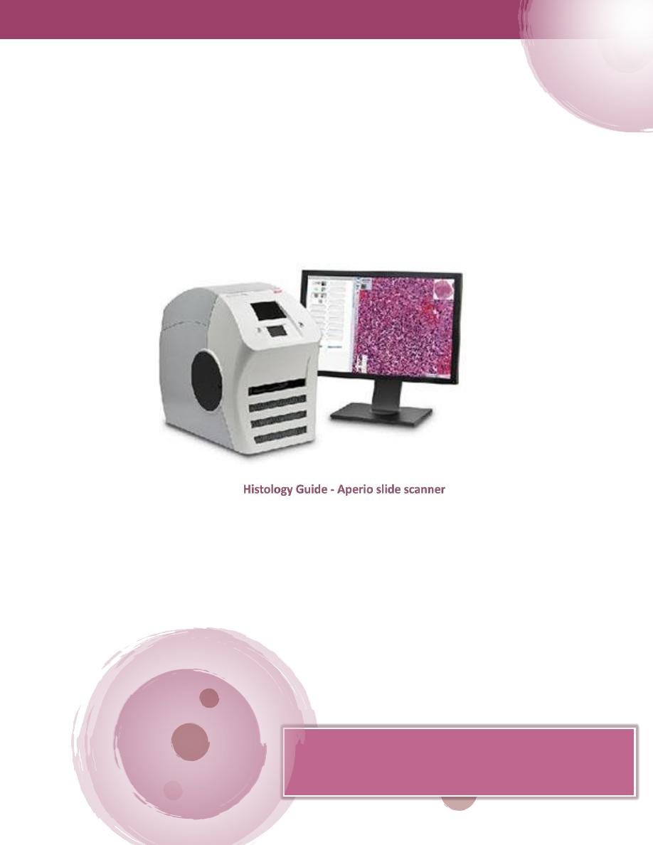
This copy is used to study tissues with Histology Guide website
to get a better result and a deeper understanding.
Do not hesitate to ask the doctors of the histology branch.
Histology
MMC 2019 || By Fahad A.
1
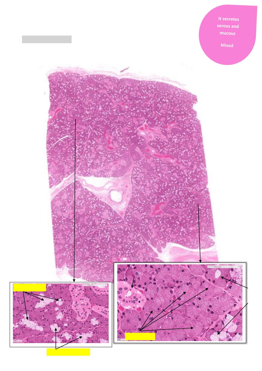
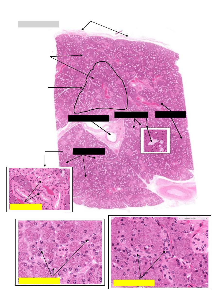
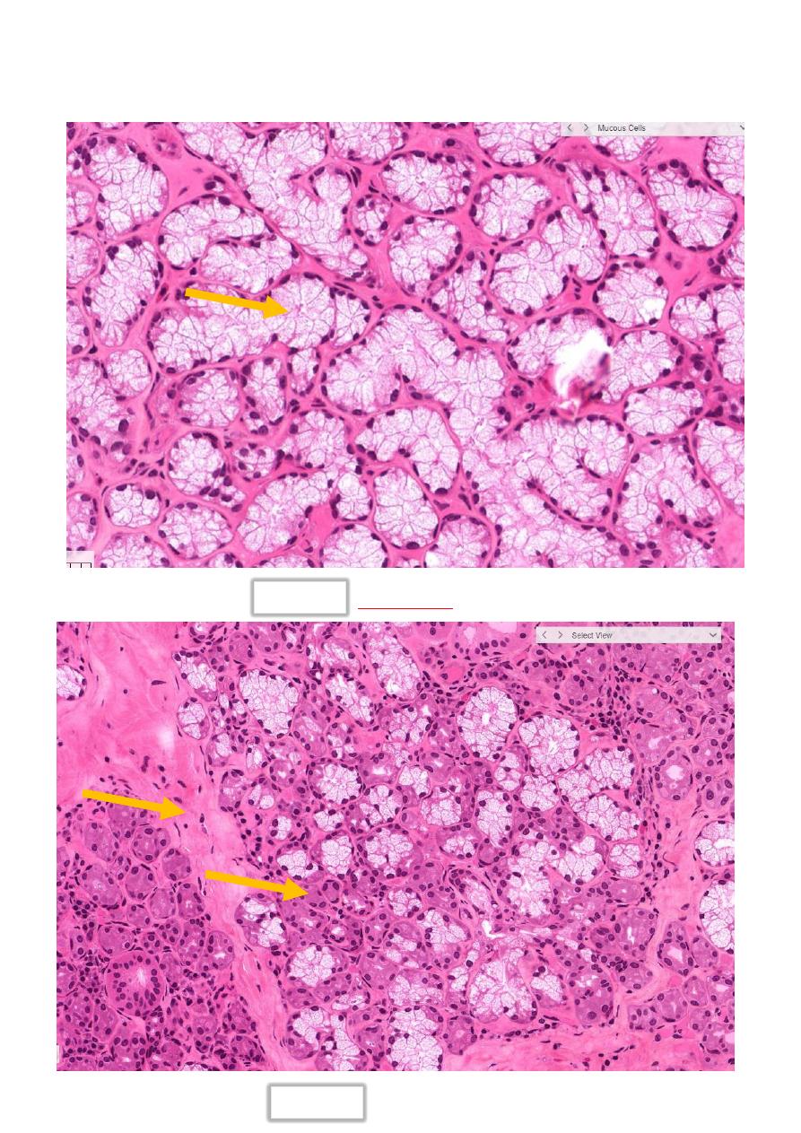
Submandibular Gland
Learning activity 1 :-
Serous acini
Striate Ducts
Serou
s Dem
ilu
ne
Mucous acini
Serous Demilune
MMC 2019 || By Fahad A.
2
ملخص االنواع الثالثة من الخاليا
1
-
النوع االول هو
Serous acini
الشكل
2
-
النوع الثاني هو
Mucous acini
هي خاليا ذا شكل مضيئ وتحتوي على نواة تتخذ
موضع في قاعدة الخلية
3
-
النوع الثالث هي بشكل مب
سط النوع الثاني داخل النوع االول
)(النوع االول كقبعة للنوع الثاني
وهي
Serous demilunes
الشكل
NOTES FREOM GISTOLOGY GUIDE WEBSITE
Serous Cells
- arranged in a tube or acinus. These polarized cells have RER at
their base (basophilic) and secretion granules (eosinophilic) at their apex
Mucous Cells
- polarized cells with flattened nuclei at the bottom of the cells.
They are very lightly stained with a "foamy" appearance (mucous has been
extracted)
Serous Demilune
- serous cells may appear as a cap on mucous cells
Intralobular Ducts - two types are found within lobules
Intralobular Ducts
- two types are found within lobules:
Intercalated Ducts
: the smallest ducts that insert into and drain
individual acini. They are more lightly stained than acini cells and are
low cuboidal
.
Striate Ducts:
arise from intercalated ducts. They are columnar with
basal striations and are surrounded by capillaries
Interlobular (or Extralobular) Duct:
found outside of lobules.
MMC 2019 || By Fahad A.
3
Learning activity 2 :-
Capsule
Serous acini
acini
Mucous
Lobule
* CT Septa
Striate Ducts
intercalated duct
duct
Interlobular
CT Septa = connective tissue Septa
*
Striate Ducts
intercalated duct
MMC 2019 || By Fahad A.
4
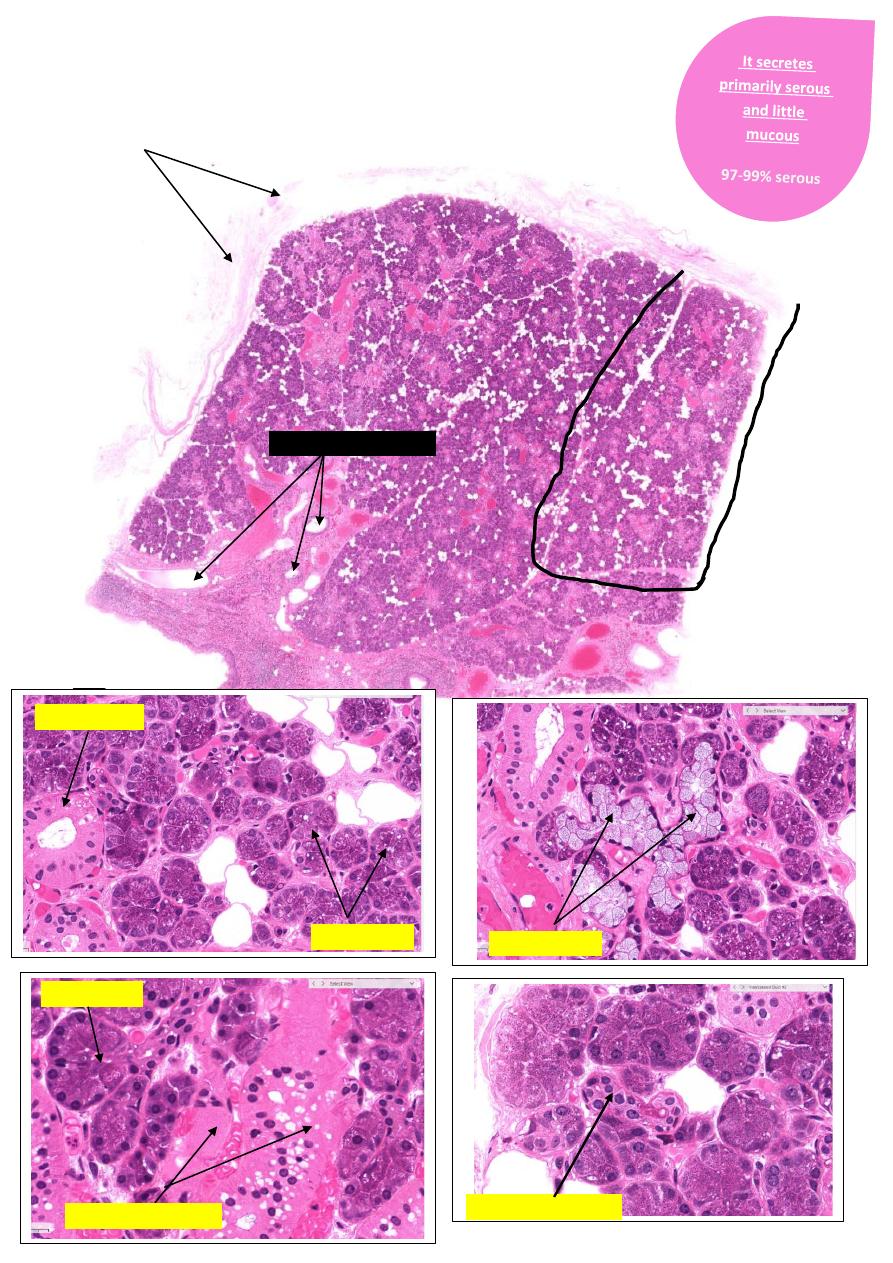
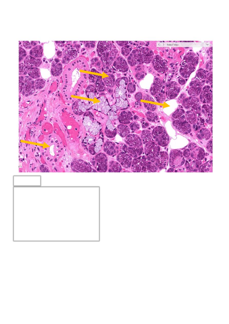
Parotid Gland
Capsule
Lobule
Serous acini
Striate Ducts
Mucous acini
duct
Interlobular
Intercalated Ducts
Intercalated Ducts
Serous acini
MMC 2019 || By Fahad A.
5
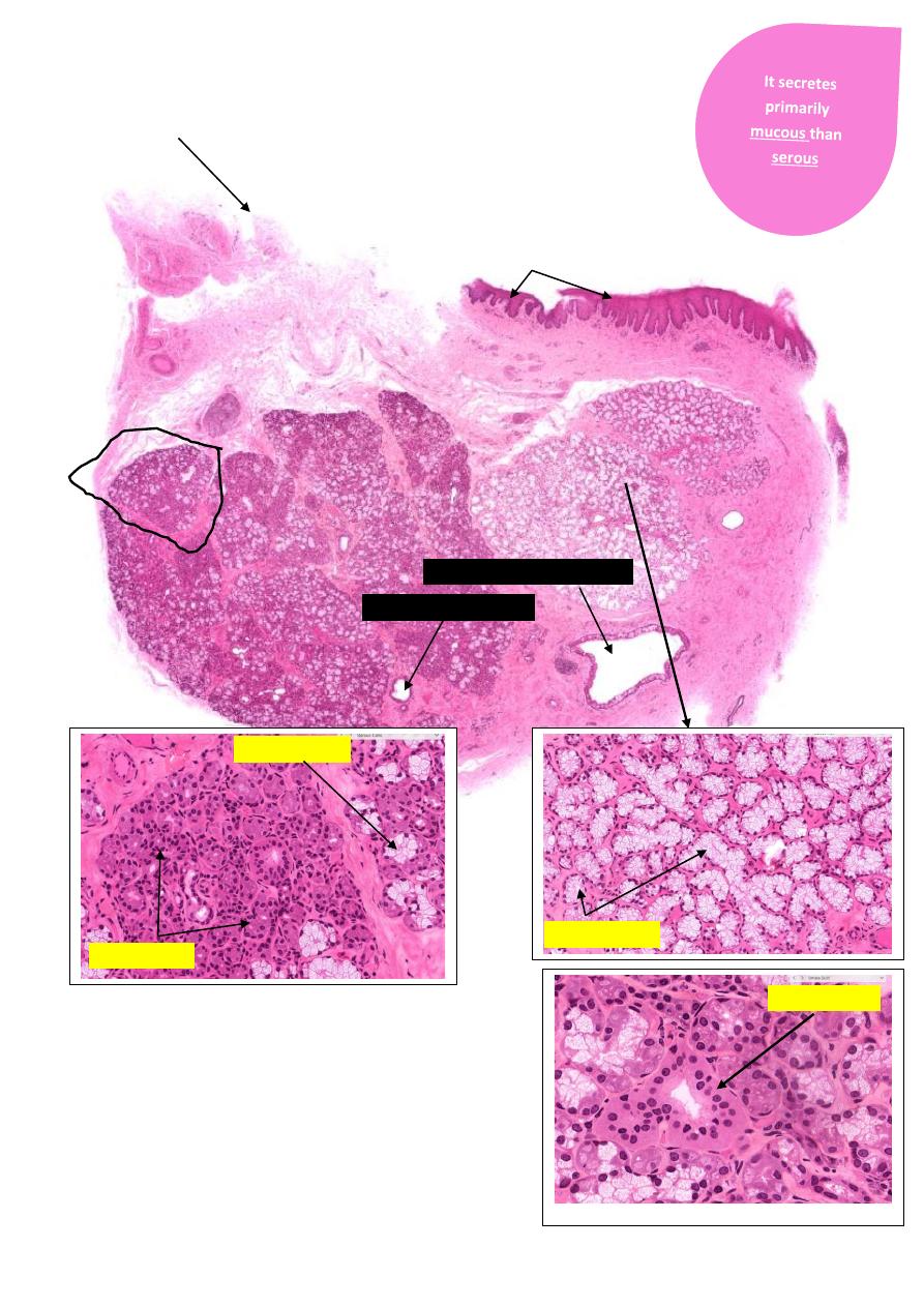
Sublingual Gland
Capsule
Lobule
Mucous acini
Serous acini
Mucous acini
Striate Ducts
duct
Interlobular
Main Excretory Duct
Oral epithelium

MMC 2019 || By Fahad A.
6
-
Questions required for this laboratory:
Question
: What is the function of the saliva?
Question
: Describe the epithelium found in the larger intralobular and interlobular
salivary ducts.

MMC 2019 || By Fahad A.
7
What is the reason behind the basal striation in the striated duct?
Question
: what are histological differences between intercalated and striated ducts
Question
: Compare the distribution of serous and mucous acini in Parotid, sublingual
and submandibular glands.
95
MH 0
VS Code
–
Histologyguide.com
MMC 2019 || By Fahad A.
8
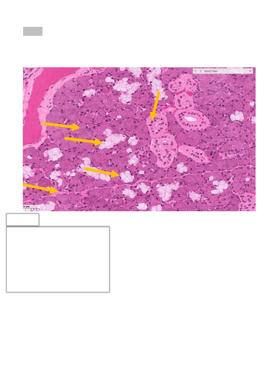
est
T
1- Name the structure pointed.
1
2
4
3
5
1- Serous acini
2- Mucous Cells
3- Serous Demilune
4- Striate Ducts
5- CT septa
The answer
ام
ثلة
عن االسئلة
MMC 2019 || By Fahad A.
9
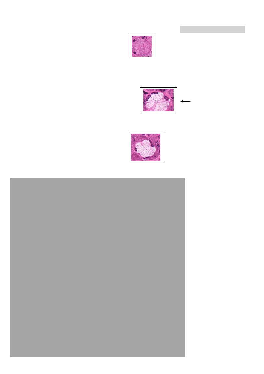
2- Name the structure pointed.
Mucous Cells
The answer
The answer
1- Serous Cells 2- CT septa
1
2
MMC 2019 || By Fahad A.
11
3- Name the structure pointed.
1
1- Serous acini
2- Mucous Cells
3- Adipose cell
4- Striate Ducts
The answer
2
4
3